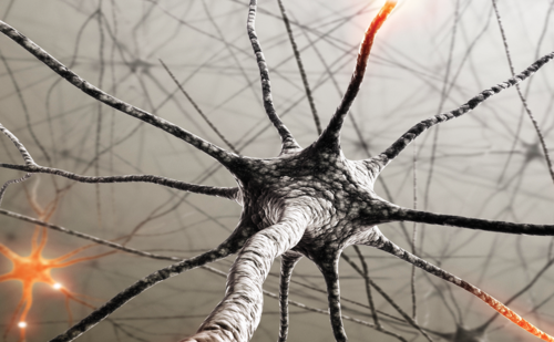
Neuropathic pain (NP) syndromes are chronic pain disorders that develop after a lesion of the peripheral or central nervous structures that are normally involved in signalling pain. It is estimated that about 35% of chronic pain patients suffer from NP, and that up to 5% of the population is affected. The characteristics of NP differ substantially from those of other chronic pain states, i.e. chronic nociceptive pain, which develops while the nervous system that is involved in pain processing is intact. Furthermore, NP states require different therapeutic approaches such as anticonvulsants, which are not effective in nociceptive pain. Many chronic pain states are characterised by a combination of both pain types (see Table 1); the best examples of these so-called mixed pain syndromes are chronic radicular back pain, diabetic foot syndrome or malignant tumour pain. Both categories of pain are characterised by distinct clinical signs and symptoms that can be used to differentiate between them. Therefore, these somatosensory signs and symptoms should always be assessed.
Disease/Anatomy-based Classification
It is common clinical practice to classify NP according to the underlying aetiology of the disorder and the anatomical location of the specific lesion.1 The majority of patients fall into three broad classes: painful peripheral neuropathies (focal, multifocal or generalised, e.g. traumatic, ischaemic, inflammatory, toxic, metabolic, hereditary); central pain syndromes (e.g. stroke, multiple sclerosis, spinal cord injury); and mixed pain syndromes (a combination of nociceptive and neuropathic pain, e.g. chronic low-back pain with radiculopathy) and complex painful neuropathic disorders (complex regional pain syndromes [CRPS]) (see Table 1). One important subgroup of painful polyneuropathies is characterised by a predominant or, in some cases, even isolated affection of small afferent fibres, i.e. unmyelinated C-fibres and small myelinated Aδ-fibres. It is important to realise that conventional electrophysiological techniques assess only the function of myelinated peripheral axonal systems; the affection of small fibres is missed. Therefore, especially in small-fibre neuropathies, alternative diagnostic procedures have to be used).
Signs and Symptoms of Neuropathic Pain
NP is characterised by a variety of sensory symptoms that can be present by themselves or in different combinations. The most typical traits of NP that are often described by patients are burning pain, shooting, electric-shock-like pain and evoked pain (hyperalgesia, allodynia). There is almost always an area of abnormal sensation, and the patient’s maximum pain is often co-extensive with the area of sensory deficit. This is an important diagnostic feature for neuropathic pain. The sensory deficit is usually to noxious and thermal stimuli, which indicates damage to small-diameter afferent fibres or to thespinothalamic tract.
As well as the existence of negative somatosensory signs (deficit in function), there are also positive signs that are characteristic of neuropathic conditions. Paresthesias (ant crawling, tingling) are symptoms typically described by patients that are bothersome but not painful. Painful positive signs are spontaneous (not stimulus-induced) or evoked types of pain (stimulus-induced pain, hypersensitivity). Spontaneous pain is separated into spontaneous ongoing pain, which often has a burning character, and spontaneous shooting, electric-shock- like sensations. Evoked types of pain include mechanical hypersensitivity and hypersensitivity to heat and cold. Two types of hypersensitivity can be distinguished. First, allodynia is defined as pain in response to a non-nociceptive stimulus. The most common example is dynamic mechanical allodynia, which means that even gentle stroking of the skin may cause severe pain. Second, hyperalgesia is defined as increased pain sensitivity to a nociceptive stimulus. Another evoked feature is summation, which is the progressive worsening of pain evoked by slow, repetitive stimulation with mildly noxious stimuli, for example a pinprick. A small percentage of patients with peripheral nerve injury have a nearly pure hypersensitive syndrome in which no sensory deficit is demonstrable. Although all of these characteristics are neither universally present in nor absolutely diagnostic of neuropathic pain, the diagnosis of NP is likely when they are present.
Diagnosis of Neuropathic Pain
Questionnaires for Screening of Neuropathic Pain Components
As described above, there are several pain types and certain qualities of the pain experience that are characteristic for NP states. Therefore, it makes sense to examine the pain qualities as a basis for distinguishing NP from other types of chronic pain. In the last five years, much research has been undertaken to develop screening tools for this purpose.2 One example of such a questionnaire is painDETECT.3
Apparative Assessment of the Afferent System
It is important to realise that conventional electrophysiological techniques such as nerve conduction studies (NCS) assess only the function of myelinated peripheral axonal systems; the affection of small fibres including nociceptors is missed. Similarly, somatosensory evoked potentials (SEP) measure the conduction in the lemniscal system (dorsal columns) and not the spinothalamic tract, which conveys noxious information. Therefore, alternative diagnostic procedures have to be used to analyse nociceptive fibres and tracts in the peripheral or central nervous system, especially to diagnose small-fibre neuropathies and central pain states, which are associated with an isolated or predominant damage to central nociceptive systems.
Quantitative Sensory Testing
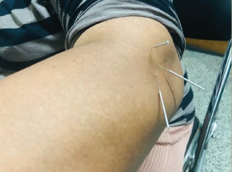
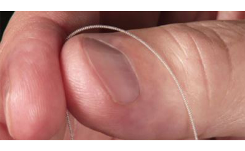
A sophisticated neurophysiological technique to test both the nociceptive and non-nociceptive systems in the periphery and the central nervous system is quantitative sensory testing (QST), which uses standardised mechanical and thermal stimuli (graded von Frey hairs, several pinprick stimuli, pressure algometers, quantitative thermotesting, etc.). Another advantage of QST is that it assesses a loss of function (minus signs) as well as a gain of function (positive signs). For example, allodynia or hyperalgesia can be quantified by measuring intensity, threshold for elicitation, duration and area.
A standardised protocol for QST was recently proposed by the nationwide German Network on Neuropathic Pain (DFNS)4 and includes 13 parameters of sensory testing procedures for the analysis of the exact somatosensory phenotype of NP patients. To evaluate plus or minus signs in patients, an age- and gender-matched database for absolute and relative QST reference data was established for healthy human subjects. At the time of writing, this nationwide multicentre trial holds complete sensory profiles of 180 healthy human subjects and more than 1,200 NP patients of a variety of entities. Thermal detection and pain thresholds including a test for the presence of paradoxical heat sensations, mechanical detection thresholds to von Frey filaments and a 64Hz tuning fork, mechanical pain thresholds to pinprick stimuli and blunt pressure, stimulus/response functions for pinprick and dynamic mechanical allodynia (pain to light touch) and pain summation (wind-up ratio) using repetitive pinprick stimulation are determined. Data of healthy human subjects were analysed for the influence of body side and region, age and gender. For most variables pathological values of positive and negative signs can be detected on the basis of reference data.
Bedside Assessment of Neuropathic Pain
As it is not always possible to perform such an elaborate examination in a clinical routine, we also want to introduce sensory bedside examination, which can be performed more easily. Such an examination should include the following factors: pinprick, touch, pressure, cold, heat, vibration and temporal summation.5,6 To assess either a loss (negative sensory signs) or a gain (positive sensory signs) of somatosensory function, the responses can be qualitatively graded as normal, decreased or increased. The stimulusevoked (positive) pain types are classified as hyperalgesic or allodynic, and according to the dynamic or static character of the stimulus.7
Touch can be assessed by gently applying cotton wool to the skin, pinprick sensation by the response to sharp pinprick stimuli, deep pain by gentle pressure on muscle and joints and cold and heat sensation by measuring the response to a thermal stimulus, for example, thermorollers kept at 20 or 45°C. Cold sensation can also be assessed by the response to acetone spray. Vibration can be assessed by a tuning fork placed at strategic points, e.g. interphalangeal joints. Currently, it is generally agreed that assessment should be carried out in the area of maximum pain using the contralateral area as control.
Therapy
Treatment Guidelines
The number of trials for peripheral NP has expanded greatly in the last few years, and these have been summarised in several recent meta-analyses.8,9 For central NP there are limited data.10 The treatments discussed below have all been demonstrated to provide statistically significant and clinically meaningful treatment benefits compared with placebo in multiple randomised controlled trials. The focus of this article is on the pharmacotherapy of NP, but non-medical treatments such as psychological therapy, physio- and occupational therapy and interventional procedures or neurostimulation techniques are of similar importance. It is important to consider that the suitable drug for each patient should be chosen individually with attention to co-morbid diseases and contraindications. The appropriate dose should be determined through titration and the reported drug-related adverse effects, and the effect on the pain must be borne in mind. It is important to evaluate the efficacy of a drug only after an adequate time-frame of about three to four weeks.
The medical management of NP consists of five main classes of oral medication – serotonin/norepinephrine-modulating antidepressants, sodium-channel blockers, calcium-channel modulators (both anticonvulsants and opioids – and two categories of topical medications, mainly for patients with cutaneous allodynia and hyperalgesia – capsaicin and local anaesthetics. A combination of two or more analgesic agents to cover multiple types of mechanisms will generally produce greater pain relief and fewer side effects. Therefore, in most patients an early combination of two or three compounds out of the different classes is the general practical approach. In fact, in a controlled four-period cross-over trial, combined gabapentin and morphine achieved better analgesia at lower doses of each drug than either as a single agent.11 There are several exceptions from the general treatment recommendations discussed above that should be emphasised. First, treatment guidelines for trigeminal neuralgia are distinct and include carbamazepine and baclofen. Second, the pharmacological therapy may be similar in complex regional pain syndrome, although controlled trials of first-line medications are scarce. However, it is likely that anti-inflammatory strategies in the acute phase in particular may be helpful.
Antidepressants
Tricyclic antidepressants (e.g. amitriptyline) are inhibitors of the re-uptake of monoaminergic transmitters. They are believed to potentiate the effects of biogenic amines in pain-modulating pathways in the central nervous system. In addition, they block voltage-dependent sodium channels and alpha adrenergic receptors. Venlafaxine and duloxetine are newer drugs that block serotonin and norepinephrine re-uptake (serotonin and norepinephrine re-uptake inhibitors [SNRIs]) and have fewer side effects. They have both shown efficacy in the treatment of painful diabetic polyneuropathy.12,13 Selective serotonin re-uptake inhibitors (SSRIs) did not show convincing efficacy in NP states.
Anticonvulsants – Calcium-channel Modulators
There are extensive clinical trials of gabapentin for chronic NP. This substance was examined in patients with peripheral NP syndromes as well as in patients with spinal cord injury. Improvements in sleep, mood and quality of life were also demonstrated. Pregabalin, the successor drug of gabapentin, was also shown to be efficacious in a variety of peripheral NP syndromes and in spinal cord injury. There is growing evidence supporting the mechanism of action on the α2δ-subunit of neuronal calcium channels, which are partly located at the pre-synaptic spinal terminals of primary afferent nociceptors. One advantage of pregabalin over gabapentin is its superior bioavailability, which makes it easier to use without the need for long titration periods. Generally, gabapentin and pregabalin are well tolerated and show minimal drug interactions.
Anticonvulsants – Natrium-channel Blockers
Carbamazepine is effective in trigeminal neuralgia. However, the strength of evidence is much lower for the benefit of these drugs in other types of neuropathic pain. With regards to oxcarbazepine and lamotrigine, there have been contradicting study results: in some studies efficacy has been shown for painful neuropathies, while in other studies a significant decrease of pain in comparison to placebo could not be achieved.14,15
Opioid Analgesics
Tramadol is an SNRI with a major metabolite that is a μ opioid agonist. Sustained efficacy for several weeks has been demonstrated for oral tramadol in patients with peripheral NP syndromes. Concerning strong opioids, their use in NP was highly controversial for many years. However, to date several positive trials of strong oral opioid analgesics in various NP entities have been published. Our experience is that many patients with pain due to central and peripheral nerve injury can be successfully and safely treated on a chronic basis with stable doses of narcotic analgesics. However, the use of opioids requires caution in patients with a history of chemical dependence or pulmonary disease. We recommend using long-acting opioid analgesics (e.g. a sustained-release preparation or transdermal applications) only when alternative approaches to treatment have failed.
Other Medications
A topical medication for NP is topical lidocaine, which blocks voltage-dependent sodium channels. Studies report pain relief with topically applied special formulations of local anaesthetic (lidocaine patches [5%]) in post-herpetic neuralgia and other neuropathies.16 This treatment option is especially interesting for elderly people, who often have contraindications against the use of other drugs or suffer from severe side effects. Capsaicin is another topical agent that acts at the vanilloid receptor (TRPV1), which is present on the sensitive terminals of primary nociceptive afferents. On initial application it produces burning pain and hyperalgesia, but with repeated or prolonged application it inactivates the receptive terminals of nociceptors. Other therapeutic agents include n-methyl d-aspartate receptor (NMDAR) antagonists and cannabinoids, which have been shown to be effective in selected patient groups, but further studies are necessary to evaluate the use of these medications in clinical practice.17
Summary
NP syndromes are frequently encountered disorders and greatly affect the quality of life of patients. It is important to recognise an NP component in chronic pain patients by assessing the somatosensory symptoms a patient presents. A sufficient pain therapy regime including the use of anticonvulsants and antidepressants at an early stage is necessary to prevent chronic stages of the disease. ■