Ultrasound now provides the first medical imaging technique to allow realtime visualisation of stroke. Going beyond new diagnostic applications, this non-invasive modality also offers novel therapeutic opportunities. Emergent treatment of acute ischaemic stroke includes the possibility of using ultrasound to enhance recombinant tissue plasminogen activator (rt-PA) thrombolysis.1,2 Moreover, intravenous microbubbles combined with transcranial ultrasound can open acute intracranial thrombotic occlusions.3
Ultrasound now provides the first medical imaging technique to allow realtime visualisation of stroke. Going beyond new diagnostic applications, this non-invasive modality also offers novel therapeutic opportunities. Emergent treatment of acute ischaemic stroke includes the possibility of using ultrasound to enhance recombinant tissue plasminogen activator (rt-PA) thrombolysis.1,2 Moreover, intravenous microbubbles combined with transcranial ultrasound can open acute intracranial thrombotic occlusions.3
The first clinical studies using microbubbles and ultrasound for treatment of ischaemic stroke have shown encouraging results.4 Further new developments in neurovascular ultrasound involve molecular approaches to diagnostics and therapy. Recent studies document the possibility of using ultrasound to open the blood–brain barrier (BBB) for selective drug therapy, and for targeting gene therapy to the brain.
Realtime Ultrasound Imaging of Stroke
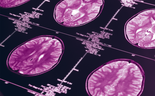
Since perfusion imaging can detect ischaemic lesions earlier than computed tomography (CT) and may distinguish the stroke subtype and severity of cerebral ischaemia, there is growing interest in perfusion imaging for predicting recovery, differentiating stroke pathogenesis and monitoring therapy. Diagnostic tools that are proportional indicators of cerebral blood flow in stroke include (99m)Tc-hexamethylpropylene amine oxime single-photon emission CT (Tc- HMPAO-SPECT), positron emission tomography (PET), Xenon-CT and perfusion-weighted magnetic resonance imaging (MRI). However, these methods can be time-consuming, may require the use of radioactive tracers, are expensive and/or may be intolerable for critically ill or restless patients. Clearly, non-invasive and easily available perfusion studies are needed. Low-mechanical-index ultrasound imaging with contrast agents is a promising new application for bedside assessment of brain perfusion. For the first time, this technique allows realtime visualisation of brain infarctions and cerebral haemorrhages.
Realtime ultrasound perfusion imaging (rt-UPI) combines pulse inversion harmonics and power modulation for extraordinary sensitivity for the detection of ultrasound contrast agents in the human brain. This method allows realtime monitoring of microbubbles flowing through the cerebral microvasculature via insonation through the transtemporal bone window. Parameters that can now be assessed in stroke patients include realtime time-to-peak (rt-TTP) after bolus injection of SonoVue™, microbubble destruction curves of contrast agent on standardised axial planes, realtime microbubble refill kinetics, dynamic microvascular microbubble maps and realtime visualisation of middle cerebral artery infarction.
In combination with contrast agents, ultrasound can highlight cerebral infarctions, as well as demarcated areas of failing or significantly diminished contrast enhancement. Such findings correlate well with perfusion-weighted imaging in MRI. Dynamic microvascular microbubble maps show good demarcation of MCA infarctions and provide impressive displays of low-velocity tissue microbubble refill following destruction of bubbles with high-mechanical-index imaging. In brain regions showing delayed contrast bolus arrival on perfusion-weighted MRI, ultrasound shows decreased or absent microbubble refill kinetics.
Sonothrombolysis
Ultrasonic insonation with frequencies in the diagnostic range has been shown to accelerate thrombolysis.3,4 Indeed, clinical studies using commercial 2MHz ultrasound devices in combination with rt-PA have suggested higher recanalisation rates than with rt-PA alone.5,6 This effect is more pronounced in the presence of microbubbles in experimental models of sonothrombolysis, and the first clinical studies using this combination for acute ischaemic stroke support this concept.7
Dissolution of thrombi without addition of lytic agents has been achieved with ultrasound in combination with microbubbles using frequencies in the low kilohertz range (17kHz)7 and in the megahertz range (1MHz)8 in experimental settings. The mechanism for this effect has been attributed to inertial cavitation resulting from bubble destruction, which then leads to thrombus destabilisation.9 Recent work has also demonstrated the importance of stable microbubble cavitations for thrombolysis, where the bubbles resonate in an ultrasound field without being destroyed.10 Our experimental data on the safety of this approach in a rat model of acute intracerebral haemorrhage has demonstrated no significant effect of 2MHz ultrasound in combination with microbubbles on haemorrhage size, the extent of brain oedema or apoptosis rate.11
Further improvement in thrombolysis may be obtained by selective attachment of microbubbles to the thrombus. Recently, we have developed novel immunobubbles binding abciximab – a humanised fragment of a monoclonal antibody against the glycoprotein IIb/IIIa receptor of platelets – for specific microbubble binding.12 These immunobubbles demonstrate a highly site-specific binding to human clots, allowing in vivo ultrasonographic molecular imaging of the targeted clot.12 In combination with 2MHz ultrasound, the abciximab immunobubbles show pronounced thrombolytic potential compared with non-specific immunobubbles and ultrasound alone in a rat model of acute thrombotic artery occlusion.
Neurovascular Molecular Imaging with Ultrasound
There are several strategies for targeting microbubble contrast agents to specific regions of disease. One takes advantage of inherent chemical or electrostatic properties of the microbubble shell, resulting in the arrest of microbubbles within the microcirculation. This method, ‘passive targeting’, relies on the disease-related upregulation of receptors that bind non-specifically to either albumin or lipid components of the microbubble shell. The second approach is referred to as ‘active targeting’. This involves the attachment of specific antibodies or other ligands to the microbubble surface, leading to the accumulation of targeted contrast agents at a specific site. A number of adhesive ligands have been explored and include antibodies, peptides and polysaccharides. Most targeted ultrasound contrast agents are microbubbles, but other vehicles can be used such as acoustically active liposomes and perfluorocarbon emulsions.
The addition of targeted ligands to microbubbles opens new avenues for the identification of vascular injury. Adhesion molecules such as the integrin αvβ3, intercellular adhesion molecule-1 (ICAM-1) and fibrinogen receptor GPIIb/IIIa are overexpressed in regions of angiogenesis, inflammation or thrombus, respectively. These molecular signatures can be used to localise ultrasound contrast agents through the use of complementary receptor ligands. Recently, this approach has been demonstrated for imaging of angiogenesis using microbubbles targeted to αv-integrins.5 Likewise, lipid-based perfluorobutane-filled microbubbles have been synthesised with various densities of anti-ICAM-1 monoclonal antibodies conjugated to the bubble shell to investigate early stages of atherosclerosis.6
As mentioned above, we have recently developed immunobubbles directed to the GPIIb/IIIa receptor of activated platelets for in vivo visualisation of human thrombus.13 Likewise, leukocyte-targeted microbubbles can be used to characterise inflammation14 and to identify inflamed plaques.15 Although application of targeted contrast-enhanced ultrasound for molecular imaging is in the early stages of development, it is potentially easily translatable to routine clinical practice.
Ultrasound-mediated Gene Therapy
Although most conventional therapeutic agents are relatively stable in the blood, genetic materials are rapidly metabolised by serum esterases and therefore are not stable to intravenous administration unless the genetic material is stabilised in some fashion. Moreover, genes are usually too large to pass across the capillary fenestrations of blood vessels unless they are assisted by some mechanism. After genes reach the brain tissue, they must pass across cell membranes and enter the cell nucleus. This is not easy, since cells have designed efficient mechanisms for processing exogenous molecules. Once cells take up macromolecules, these are generally digested by lysosomes within the cells. Therefore, successful gene therapy requires effective gene delivery. Recent work suggests that ultrasound may play an important role in the development of new approaches for gene delivery to the brain.
Simple exposure to ultrasound increases transgene expression in vascular cells by up to 10-fold after naked DNA transfection. The mechanism of this transfection is mostly likely cavitation, which in turn increases microvascular permeability. This effect can be dramatically increased in the presence of ultrasound contrast agents.7 As microbubbles are cavitated by ultrasound, local shock waves increase capillary permeability. This process increases transcapillary passage of macromolecules or nanospheres co-delivered with the microbubbles.8 Cavitation probably opens micropores in small blood vessel walls, making the vessels more passable to molecules and nanoparticles. Therefore, microvascular permeability caused by cavitation of microbubbles can be used to increase local delivery of therapeutic materials such as genes.
Not only can microbubbles be used to enhance the effects of ultrasound on gene expression, they may also serve as carriers of gene therapeutic agents.9,10 There are a number of ways to entrap drugs with microbubbles. One technique is to incorporate them into the membrane that stabilises microbubbles. Charged drugs can be stabilised in or onto the surfaces of microbubbles by virtue of electrostatic interactions. In this way, cationic lipid-coated microbubbles can bind DNA. This is because DNA is a polyanion and binds avidly to cationic (positively charged) microbubbles.
Several in vitro and in vivo animal experiments have suggested the feasibility of microbubble-ultrasound enhanced gene therapy to the brain. After intracisternal injection of microbubbles and plasmid DNA, the reporter gene was detected in meningeal cells exposed to ultrasound.16 Likewise, following intrastriatal injection of microbubbles and naked plasmid DNA, significantly increased gene transfer was demonstrated in glial cells.16 In another study, 210kHz ultrasound and 5.0W/cm2 of insonation for five seconds effectively transfected plasmid DNA into culture slices of mouse brain (nearly 150-fold increase). The effect was reinforced five-fold by using a combination with an echo contrast agent. When DNA was intracranially injected, the contrast agent also enhanced gene transfection in newborn mice.17 Thus, ultrasound can enhance gene expression.
In the presence of microbubbles a synergistic effect is attained, and cavitation is a likely mechanism. Acoustically active materials that bind or entrap genetic materials have a potential role for gene delivery. These materials can be injected intravenously, and targeted gene delivery is attained within the tissue exposed to ultrasound. The ability to focus ultrasound and cause local cavitation with these gene carriers may provide a new tool for gene therapy of a variety of brain diseases. However, as opposed to other organs, gene therapy to the brain using high-molecular-weight complexes of plasmid-DNA and microbubbles must overcome the BBB. Fortunately, ultrasound can also be used to facilitate drug delivery across the BBB.
Permeating the Blood–Brain Barrier with Ultrasound
In addition to the physiological barrier at the level of basal lamina, the BBB is formed by the endothelial cells of the cerebral microvessels that connect to each other by means of intracellular attachments known as tight junctions. The factors that determine penetration of substances from the blood to the central nervous system are lipid solubility, molecular size and charge. The BBB prevents penetration of ionised water-soluble materials with a molecular weight greater than 180 Daltons. Chemical modification of drugs to make them lipophilic or the use of other carriers, such as amino acid and peptide carriers, are ways to support propagation through the barrier. Another possibility is to diffusely alter the function of the BBB by temporarily opening the tight junctions, which is now possible with an increasing number of chemicals.
High-intensity focused ultrasound has been shown to allow selective and non-destructive disruption of the BBB in rats.17 If microbubbles are introduced into the bloodstream prior to ultrasound exposure, the BBB can be transiently opened at the ultrasound focus without acute neuronal damage.18 The introduction of cavitation sites into the bloodstream concentrates the ultrasound effects to the vasculature and reduces the power needed to produce BBB opening by a factor of two orders of magnitude. This diminishes the risk of tissue damage and makes this technique more easily applied through the intact skull.
Conclusion
In summary, the intact BBB, which is a major limitation in using genes for the therapy of brain disease, can be opened with ultrasound.19 This allows a localised, transient and reversible opening to provide anatomically selective targeted drug delivery. This approach will likely be used in conjunction with ultrasound-mediated gene delivery to the brain. Future developments in neurovascular imaging will include improvements in technologies for ligand attachment to microbubbles or ultrasound-sensitive nanoparticles, better methods for imaging targeted ultrasound agents in the brain and optimisation of ultrasound-mediated gene delivery. ■


