Although placement of particles and other agents, including platinum coils, were subsequently attempted, it was not until the late 1980s, when detachable platinum coils were introduced, that endovascular techniques finally came of age.6 These coils were placed using a steel-wire delivery system via a microcatheter positioned at the aneurysmal sac. When the coils were in place, an electrical current was used to detach the platinum coil inside the aneurysm.7 The presence of the coil inside the aneurysm provided protection, likely from immediate hemodynamic changes and redirection of blood flow through the proper vessel. A thrombus eventually forms inside the aneurysm that protects the weaker aneurysmal neck and leads to the aneurysm being obliterated with connective tissue.8 Most endovascular treatments are performed under general anesthesia, although a very select group of patients can tolerate the procedure while awake.9 The current procedure involves a coil being placed in the aneurysmal sac with the assistance of a balloon,10 stent,11 or microcatheter.12
Improvements in coil design have led to improved obliteration of aneurysmal sacs.13 Complex aneurysms are also treatable with the use of liquid embolic agents; for example, ethyl vinyl alcohol polymer in conjunction with intravascular stents also leads to obliteration of the aneurysm.13 In the early stages, endovascular procedures were limited to patients who were considered poor candidates for neurosurgical clipping, including: elderly patients,14 those with poor Hunt-Hess Scales (IV or V),15 posterior circulation aneurysms,16 or cavernous segment internal carotid artery aneurysms17 with active vasospasm.18 As the technology matured, with improvement in operator technique and coil design, embolization has been attempted in patients who traditionally have been good candidates for neurosurgical clipping.19 Finnish investigators compared both clipping and endovascular techniques in ruptured aneurysms, and confirmed the safety and effectiveness of coil embolization.20
Mortality in both coiling and clipping was found to be comparable, with no difference in short-term clinical outcome. In a phase II trial with 57 patients in the clipping arm and 52 patients in the embolization arm, the angiographic outcome in anterior circulation aneurysm repair was better in the surgical group, while endovascular treatment had better angiographic outcome in posterior circulation patients. There was a technique-related mortality rate of 4% in the surgical group and 2% in the endovascular group.20 Another recently concluded, larger randomized multicenter phase III trial—the International Subarachnoid Aneurysm Trial (ISAT)21—compared the safety and efficacy of embolization versus neurosurgical clipping. A total of 2,143 patients with ruptured aneurysms who were considered suitable for either treatment were enrolled. There were 1,070 patients enrolled in the clipping group, while the group receiving endovascular treatment by detachable platinum coils had 1,073 patients. Clinical outcomes were assessed at both two months and one year, with interim updates on re-bleeds and death. Out of 801 patients allocated to the endovascular arm, 190 (23.7%) became dependent or died at one year compared with 243 (30.6%) of the 793 allocated to the neurosurgical clipping arm (p=0.0019). Exmination of the risk of re-bleeding at one year showed that from endovascular treatment to be two per 1,276 patients, while risk of re-bleeding from clipping was 0 per 1,081 patients. The conclusion was that one-year outcomes in disability-free survival were significantly better with endovascular coiling.21 Further follow-up of these patients will hopefully add more information about the durability of coiling versus clipping.22 Figure 1 summarizes the results of the ISAT trial.
The outcome of endovascular treatment of unruptured aneurysms has improved following the introduction of platinum detachable coils. In a 2005 article, Taha et al. showed that endovascular embolization of cerebral aneurysms is a safe alternative to surgical clipping in the treatment of both ruptured and unruptured cerebral aneurysms; however, long-term outcome needs to be evaluated.23 A retrospective comparative analysis of endovascular versus surgical treatment of unruptured aneurysms was published by Johnston et al. in 1999.24 In this review of data from 60 universities, factors such as age, sex, race, mode of admission, and year of treatment were all considered. When they looked at rates of in-hospital mortality, adverse outcomes (discharge to rehabilitation centers versus nursing homes) and length of stay were all greater for clipping compared with endovascular treatment.24 More than one study examining noncontrolled patient series has been published that supports the safety and effectiveness of cerebral aneurysm embolization.25–27 New coil designs, including 2-D and 3-D configurations, have led to improved radiographic obliteration of aneurysms.28,29The focus of treatment on aneurysmal anatomy, especially on the neck:dome ratio and the method of coil packing in a stepwise fashion, has led to remarkable success in aneurysms previously thought of as difficult. Murayama et al. in 2001 demonstrated that treating experimental porcine aneurysms with a bioabsorbable-polymer-coated coils led to improved formation of neointimal proliferation across aneurysm necks compared with standard platinum coils.30 The bioabsorbable-polymer-coated coils may decrease the chances of coil compaction and aneurysm recanalization in future years.30
Newer technologies have been developed to keep the colis from prolapsing out of an aneurysm with a large neck. A temporary balloon10 or a permanent stent designed for intracranial vessels (e.g. the Neuroform® stent11 by SMART Therapeutics, Inc., San Leandro, CA) could be used to provide coil stabilization within intracranial aneurysms.31 It is evident that the future can only hold more promise in improved treatment techniques and device design. Figure 2 illustrates an example of coiling in an anterior cerebral artery aneurysm.
Cerebral Vasospasm
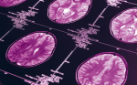
The management of the complications of aSAH can be a daunting, if not challenging, task. Aneurysmal re-bleeding, seizures, hydrocephalus, and hyponatremia, as well as cardiac and pulmonary concerns, can at any time confront the treatment team. However, vasospasm, which can lead to delayed cerebral ischemia, is the leading cause of disability following aSAH and remains a continuous challenge to the neuro-intensivist.32 Angiographic vasospasm occurs in up to 75% of patients following SAH.33 Delayed ischemic neurological event, described as a decline in neurological status during the first two weeks that is not attributed to other causes, occurs in up to 40% of patients.34,35 These values have remained consistent with contemporary retrospective reviews.36,37 Angiographic confirmation is not always necessary with the increasing use of non-invasive methods employed to monitor patients with suspected cerebral vasospasm.
Gradual or stepwise evolution of neurological deficits most often heralded by onset of headache or worsening headache, changes in level of consciousness, lower extremity diplegia, visual field deficits, or any constellation of focal neurological deficits are common neurological manifestations of cerebral vasospasm.38 Because vasospasm occurs in a delayed fashion and is often predictable, it is an obvious target for therapeutic strategies.
Cerebral vasospasm usually develops 3–5 days after the initial SAH insult, peaks at 6–10 days, and gradual resolution will occur within 2–4 weeks. The combination of induced hypertension, hypervolemia, and hemodilution (triple H therapy) is commonly used as prophylaxis and treatment of cerebral vasospasm. Endovascular therapy offers an additional treatment for patients who continue to exhibit neuro-radiographic and/or clinical signs and symptoms of delayed ischemic neurological deficits despite optimal medical management. Currently, the most commonly employed endovascular treatments are percutaneous transluminal angioplasty with or without intra-arterial vasodilation using papaverine, nicardipine, verapamil, nimodapine, amrinone, or milrinone.39
Percutaneous Transluminal Angioplasty
Since the successful development of arterial angioplasty in 1977, significant advances have allowed for the evolution of neuro-endovascular applications. This, coupled with the advancement of smaller balloons and eloquently flexible catheters, allows almost all vessels to be treated with percutaneous transluminal angioplasty (PTA). At the cellular level, the results of balloon angioplasty are thought to create a fragmentation of collagen fibers and myocytes resulting in restoration of vessel diameter.37 The effects of PTA seem to be sustained and long-lasting and have thus made this approach a favorable treatment.37 PTA does have its limitations to treating larger and more proximal vessel segments.40 Also, the procedure carries some complications including aneurysmal rupture, vessel rupture, thrombus, and reperfusion hemorrhage.41 Early efficacy of PTA has been shown in smaller series, such as the one using prophylactic PTA revealing that there may be benefit for patients with Fisher grade three SAH.42 This evidence has led to designing of a larger, multicenter National Institutes of Health (NIH)-funded trial that may be able to answer this very important question in future.
Intra-arterial Injections
Neuroendovascular catheterization provides a direct means for introducing agents that will facilitate the attenuation of spastic vessels. These medications are injected using small repeated doses through the avenue of a superselected microcatheter positioned proximal to the affected vessel. Papaverine and other calcium channel blockers are utilized in this fashion. Papaverine hydrochloride, a potent vasodilator, is a synthetic alkaloid that inhibits cyclic adenosine monophosphate (cAMP) and cyclic guanosine monophosphate (cGMP) on smooth muscle cells, and is also thought to block calcium channels. The results of endovascular papaverine therapy are derived from cases series.40 It appears to have particular utility in severely vasospastic vessels.43 The effect of papaverine depends on appropriate dosing and adequate delivery to a vasospastic vessel rather than into a normal artery. Complications include increased intracranial pressure, tachycardia, arrhythmias, and sedation.
Other Pharmacological Applications
The calcium channel antagonist nimodipine has been used extensively in the treatment of the prevention of vasospasm related to SAH. Several randomized controlled trials have been conducted. In a meta-analysis of several published randomized trials, the evidence in favor of routine prophylactic use of nimodipine is strong.44 Nicardipine has also been the subject of several controlled trials. Although the largest trial showed a considerably lower rate of ischemic deficits secondary to cerebral vasospasm, the overall outcome was not improved.44 Other calcium antagonists have also been used from time to time. A recent series showed a 20% incidence of delayed ischemic deficit when oral diltiazem was used and favorable outcome in 75% of 123 consecutive cases.45 Other treatment options—for example the phosphodiesteraseinhibitor milrinone,46 the platelet-activating factor receptor antagonist E588047, or the protein kinase inhibitor fasudil hydrochloride48—are under continuing investigation. Of considerable initial interest was the modified steroid free radical scavenger tirilazad mesylate, which was used in four large, controlled trials in the 1990s.49 In the first trial, it appeared likely to be effective in reducing spasm, but later meta-analysis showed only a trend, whereas outcome was improved only in patients categorized in clinical grades IV and V.50 Improvements in neuro-intensive care monitoring and diagnostic studies have allowed rapid identification of patients who reach critical reductions in cerebral blood flow.
The introduction of endovascular treatment has allowed minimally invasive methods of treating intracranial aneurysms. Similarly, a major late cause of morbidity in patients with aSAH is vasospasm, which is now being treated more effectively with interventional neurology techniques. The future for the interventional techniques is exciting and promises minimally invasive therapeutic options for patients with aSAH. A more rigorous assessment of the efficacy of the interventional approach with aSAH requires further trials.51