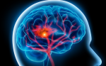
Primary intracerebral haemorrhage (PICH) originates from the spontaneous rupture of small arteries as a result of chronic degenerative changes due to chronic hypertension or amyloid angiopathy.1 Although environmental factors are important, there is accumulating evidence that genetic elements also contribute to the pathogenesis of PICH.2,3 In an epidemiological study, familial clustering of PICH was noticed, especially when involving deep brain structures, indicating genetic predisposition to cerebral haemorrhage.4 Increased incidence of intracerebral haemorrhage in specific animal models also provided additional evidence for the existence of susceptibility genes.5 The importance of genetic factors was unequivocally demonstrated with the identification of causative mutations in monogenic cases of familial intracerebral haemorrhage. Furthermore, several association studies have suggested the presence of susceptibility genes that predispose to PICH (see Table 1). In this article we briefly discuss the current state of knowledge regarding the known major and susceptibility genes that have been implicated in PICH.
Familial Cases
Familial Cerebral Amyloid Angiopathy
Cerebral amyloid angiopathy (CAA) is caused by the deposition of amyloid in the small and medium-sized cortical and leptomeningeal arteries leading to intracerebral haemorrhage, ischaemic infarction or dementia. Amyloid is caused by the aggregation of β-amyloid peptide (Aβ) and other proteins, promoting vasculopathic changes such as fibrinoid necrosis and microaneurysms. Aβ peptide is formed by the proteolytic fragmentation of amyloid precursor protein. Amyloid formation has also been reported in familial cases of CAA caused by mutations in the cystatin C gene, 6,7 the transthyretin gene8–12 or the BRI gene. 13,14 The clinical presentation of these familial cases includes dementia, vascular cognitive decline and PICH. PICH has also been reported in a member of a Volga-German family with Alzheimer’s disease and a mutation in the presenilin-2 gene. 15 Recently, a novel mutation in presenilin-1 gene was also associated with early-onset dementia of Alzheimer type and lobar PICH. 16 However, most familial cases of CAA and PICH are caused by mutations in the amyloid precursor protein. Of note, these mutations are located in the Aβ segment of the amyloid precursor protein, whereas mutations in the flanking regions cause Alzheimer’s disease or ischaemic stroke. PICH has been documented in Flemish,17 Dutch,18 Arctic,19 Iowan20 and Italian21 CAA families. Recently, duplication of the amyloid precursor protein gene was reported to be the cause of familial CAA presenting with dementia and PICH.22,23
Type IV Collagen a1 Chain
Type IV collagen a1 chain (COL4A1) is an integral component of the basement membrane in the brain vasculature and other tissues. A few families and a sporadic case with PICH and mutations in COL4A1 have been reported so far.24–27 Mutations in COL4A1 seem to compromise vascular wall integrity and blood supply, leading to small-vessel diseases including PICH, microbleeds, lacunar strokes or leukoaraiosis.24–26,28 Electron microscopy of the vascular wall in patients with COL4A1 mutations reveals structural defects of the basement membrane such as interruptions, variable thickening and inconsistent density.28
Cerebral Autosomal-dominant Arteriopathy with Subcortical Infarcts and Leucoencephalopathy
Cerebral autosomal-dominant arteriopathy with subcortical infarcts and leucoencephalopathy (CADASIL) is a monogenic disorder caused by a variety of mutations in the Notch3 gene, which is responsible for cell signalling and vascular development.29 The clinical manifestations of this disorder include migraines, transient ischaemic attacks, lacunar strokes and subcortical dementia. Magnetic resonance imaging reveals extensive peri-ventricular white matter leucoencephalopathy and the presence of microbleeds, predominantly in subcortical areas and the thalamus, detected on T2-weighted gradient echo imaging. Microbleeds can be present in 31–69% of patients with CADASIL.30 It was found that PICH can occur in 25% of symptomatic patients with CADASIL, and this is closely related to the number of cerebral microbleeds.31
Genetic Association Studies
Apolipoprotein E
Apolipoprotein E (ApoE) is a glycoprotein involved in cholesterol transport and has three isoforms: ε2, ε3 and ε4. Accumulating evidence implicates ApoE ε2/ε3/ε4 polymorphism with CAA-related PICH.32–34 The e4 allele increases Aβ deposition in the cerebral vasculature in a dose-dependent manner.35,36 The ε2 allele is associated with vasculopathic changes in amyloid-laden vessels and rupture.33 It has also been documented that ε2 and ε4 alleles of the ApoE gene are risk factors for the occurrence of lobar PICH, probably due to the presence of cerebral amyloid angiopathy in the carriers of these alleles.2 In addition, the e4 allele was associated with earlier age at onset of CAA-related PICH37 and with warfarin-related PICH.38 ε2 and ε4 allele carriers are also at increased risk of recurrent haemorrhage compared with ε3 carriers.39 Moreover, the presence of the e4 allele was linked to poor outcome of PICH patients.40 However, other studies did not find any association between ApoE polymorphism and PICH.41–45 In a recent meta-analysis, the ε2 allele was found to be an independent risk factor for PICH (odds ratio [OR] 1.32, 95% confidence interval [CI] 1.01–1.74), whereas ε4 genotypes were not (OR 1.16, 95% CI 0.93–1.44).46
VKORC1 Gene
An interesting association between a haplotype in the vitamin K epoxidase reductase complex subunit 1 (VKORC1) gene and arterial vascular diseases including PICH (OR 1.53, 95% CI 1.09–2.16; p<0.05) has been reported.47 VKORC1 is implicated in haemostatic processes through γ-carboxylation of vitamin-K-dependent proteins. Common polymorphisms of VKORC1 gene have also been found to affect interindividual differences in warfarin sensitivity.
Interleukin-6 Gene
In a large-scale association study, 282 Japanese patients with PICH and 2,010 controls were genotyped for 202 polymorphisms of 152 genes that were implicated in vascular biology, platelet function, leukocyte biology, coagulation processes, regulation of the circulation, blood pressure or endocrine function and various metabolic factors, as well as lipid, glucose and homocystein metabolism. It was found that the C allele of the interleukin-6 (IL-6) gene -572G/C polymorphism increased the risk of PICH (OR 1.57, 95% CI 1.21–2.07; p<0.001). It was suggested that IL-6 may damage the vascular wall through induction of matrix metalloproteinases, which degrade the extracellular matrix around blood vessels and thus weaken the vascular wall.48 However, recently, in a small group of patients IL-6 -174G/C gene polymorphism was not found to be an independent risk factor for PICH.49
Engoglin Gene
Endoglin is a glycoprotein in the surface of endothelial cells that interacts with transforming growth factor-β. Endoglin is important for vascular development and structural integrity. Variable mutations in the endoglin gene were found to cause hereditary haemorrhagic telangiectasia. A homozygous 6bp insertion in the endoglin gene was found in 8.7% of PICH patients compared with 2% of controls (OR 4.76, 95% CI 1.28–21.6; p=0.012).50 The same polymorphism was also associated with increased frequency of intracranial aneurysms.
Angiotensin-converting Enzyme Gene
Angiotensin-converting enzyme (ACE) plays an important role in regulating both the production of angiotensin II and the degradation of bradykinin at the endothelial surface. Angiotensin II, which is the main active product of the renin–angiotensin system, has been linked to vascular remodelling, inflammation and endothelial dysfunction. It was reported that the DD genotype of ACE insertion/deletion (I/D) polymorphism in intron 16 was over-represented in Polish patients with non-lobar PICH (OR 2.13, 95% CI 1.10–4.14; p=0.02). However, after excluding the individuals who were receiving ACE inhibitors and adjusting for other variables, the association was no longer statistically significant.51 In a previous study, the distribution of ACE genotypes and alleles was the same among the controls and patients.52 It was shown that ACE I/D polymorphism only partially determines the variation in plasma ACE levels, and it is uncertain whether it represents a functional polymorphism; this may explain the inconsistency between the two studies.
Alpha-1 Antichymotrypsin Gene
Alpha-1 antichymotrypsin (ACT) is an acute-phase protein member of the serine proteinase inhibitors that has been implicated in vascular pathology. ACT has anti-inflammatory properties as it strongly inhibits neutrophil cathepsin G, but it is also known to interact with Aβ peptide, promoting amyloid plaque formation. The TT genotype of ACT A/T signal peptide polymorphism was associated with PICH in Spanish patients (OR 2.80), especially those with normal blood pressure (OR 3.40).53 By contrast, a study from China reported a more robust association in hypertensive patients.54 However, these associations were not replicated in a study from Poland55 and in a group of 147 Greek patients from our department.56 In our group we observed only a marginal association in the non-hypertensive group (p=0.05). It is possible that in non-hypertensive patients the absence of hypertension unmasks the relatively minor effects of ACT A/T signal peptide polymorphism on the cerebral vasculature, making it more susceptible to haemorrhage.
Lipoprotein a Gene
Elevated lipoprotein a (Lp(a)) levels have been associated with increased risk of cardiovascular diseases, possibly by being implicated in atherosclerotic arterial damage. In a large multicentre study in a Chinese population, low numbers of TTTTA repeats (PNTR polymorphisms) of the Lp(a) gene were found in patients with PICH.57
Apolipoprotein H Gene
Apolipoprotein H (ApoH) has been implicated in several physiological pathways including lipid metabolism, coagulation and increased blood pressure. In a study in a Chinese population it was found that the Ser88Asn (G341A) polymorphism was associated with increased risk of PICH.58
Platelet Glycoproteins
Glycoproteins Ia, IbA and IIIa are platelet surface receptors for fibrinogen, von Willebrand factor and collagen, playing an important role in platelet adhesion and aggregation. However, genetic polymorphisms of these factors were not found to increase the risk of PICH.59
PAF-H Gene
Platelet-activating factor acetylhydrolase (PAF-H) is implicated in thrombosis. A Val2793Phe substitution in the PAF-H gene has been associated with ischaemic stroke, possibly through increased thrombotic processes. The same mutation was also found to be a risk factor for PICH.60
Factor XIII Gene
Blood coagulation factor XIII plays an important role in clot stabilisation by cross-linking fibrin chains. A point mutation in codon 34 (Val34Leu) of XIII gene was known to be protective against thrombotic diseases including myocardial infarction and ischaemic stroke. However, a potential association of Val34Leu and PICH was reported.61 The authors suggested that the Leu34 allele might cause the formation of weaker fibrin structures that predispose to PICH. Subsequently, this polymorphism was extensively investigated in various populations but with contradictory results.62–66
Other Association Studies
Other genetic variants that have been associated with PICH are polymorphisms in the oestrogen receptor alpha gene,67 the osteoprotegerin gene,49 factor V-leiden,63 factor VII63 and b1-tubulin.68 Genetic variants not associated with PICH are polymorphisms in the paraoxonase 2 gene, 69 the protein Z gene,70 the interleukin-1a gene,71 the methylenetetrahydrofolate reductase (MTHFR) gene,72 prothrombin,63 factor VII,73 the growth-arrest- specific gene74 and plasminogen activator inhibitor-1 (PAI-1).66
Conclusions
PICH is a complex multifactorial disorder that probably results from an interaction between various environmental factors and the genetic background of the patient. Linkage analyses in familial cases of PICH have identified chromosomal loci linked to PICH. In addition, several association studies of sporadic cases have revealed a number of genetic variants that possibly confer susceptibility to PICH. Whole-genome association studies are now feasible via current technology. Implementation of genome-wide scans may provide substantial benefits, including the development of genetic markers for determination of specific molecular profiles in individuals and assessment of disease risk. In the future, this may offer the prospect of early diagnosis, personalised risk assessment and novel genomic-based preventative therapies.3 ■