Sports-related injuries account for 10 % of head and spinal cord injuries,1 and yearly 1.5 million Americans suffer traumatic brain injury (TBI) without loss of consciousness or need for hospitalization. Further evidence that a growing public health dilemma is growing comes from the Centers for Disease Control and Prevention (CDC), who attributed 207,830 emergency room visits per year between 2001 and 2005 to non-fatal sports-related head injuries.2 A study of high school athletes found that all sports were at risk for concussions, with boys’ football the highest at 63 % of the total, followed by wrestling (10 %), soccer (6 %), and the rest in basketball, softball, baseball, field hockey, and volleyball.
Concussion is generally defined as a mild injury to the brain caused by traumatic biomechanical forces without radiographic evidence of damage. It may be caused by a direct blow to the head or an impulsive force that leads to acceleration of the head without direct trauma. Loss of consciousness is frequently absent, in fact 90 % of sports-related concussions present this way.3 The most common signs and symptoms are headaches, dizziness, confusion, disorientation, and blurred vision. Although once thought to be benign, there is increasing evidence that concussions or mild traumatic brain injury (MTBI) have more serious complications, particularly when they occur repeatedly. Sports-related chronic traumatic encephalopathy (CTE) manifests as a progressive worsening of cerebral neurologic symptoms, initiated and maintained from repetitive concussions. Recent epidemiologic data has shown that 17 % of the individuals with repetitive MTBI develop CTE, however, the severity and the frequency of repetitive injury needed to cause CTE is still unknown.4–8
Early Concepts of Chronic Traumatic Encephalopathy
In 1928, Martland introduced the term ‘punch drunk’ to describe neurologic symptoms such as confusion, tremors, slowed speech and gait seen in boxers suffering from repeated blows to the head.9 This came to be known as ‘dementia pugilistica’ by Millspaugh,10 and then the term ‘psychopathic deterioration of pugilists’ was introduced by Courville.11 The cause of these symptoms became clear once the neuropathology of CTE was described by Brandenburg and Hallevorden and later Corsellis who found several characteristic areas of damage—septum pellucidum, adjacent periventricular gray, frontal and temporal lobes, substantial nigra, cerebellar scarring, and diffuse neuronal loss.12,13,14 Table 1 lists the four components of punch drunk syndrome with their suspected anatomical substrate. Although the terms ‘punch drunk’ and ‘dementia pugilistica’ came from the original descriptions found in boxers, later reports showed that CTE was also found in athletes practicing other sports besides boxing where there is a risk of repetitive brain injury (football, wrestling, hockey, and soccer).12,13
Pathology
The gross pathology of CTE includes a reduction in brain weight, enlargement of the ventricular system, especially lateral and third ventricle, thinning of the corpus callosum, atrophy of cerebral hemispheres, mesial temporal lobes, thalamus, mammillary bodies, olfactory bulbs and brainstem, pallor in the substantia nigra and locus coeruleus, a cavum septum pellucidum, and scarring of the cerebellar tonsils.
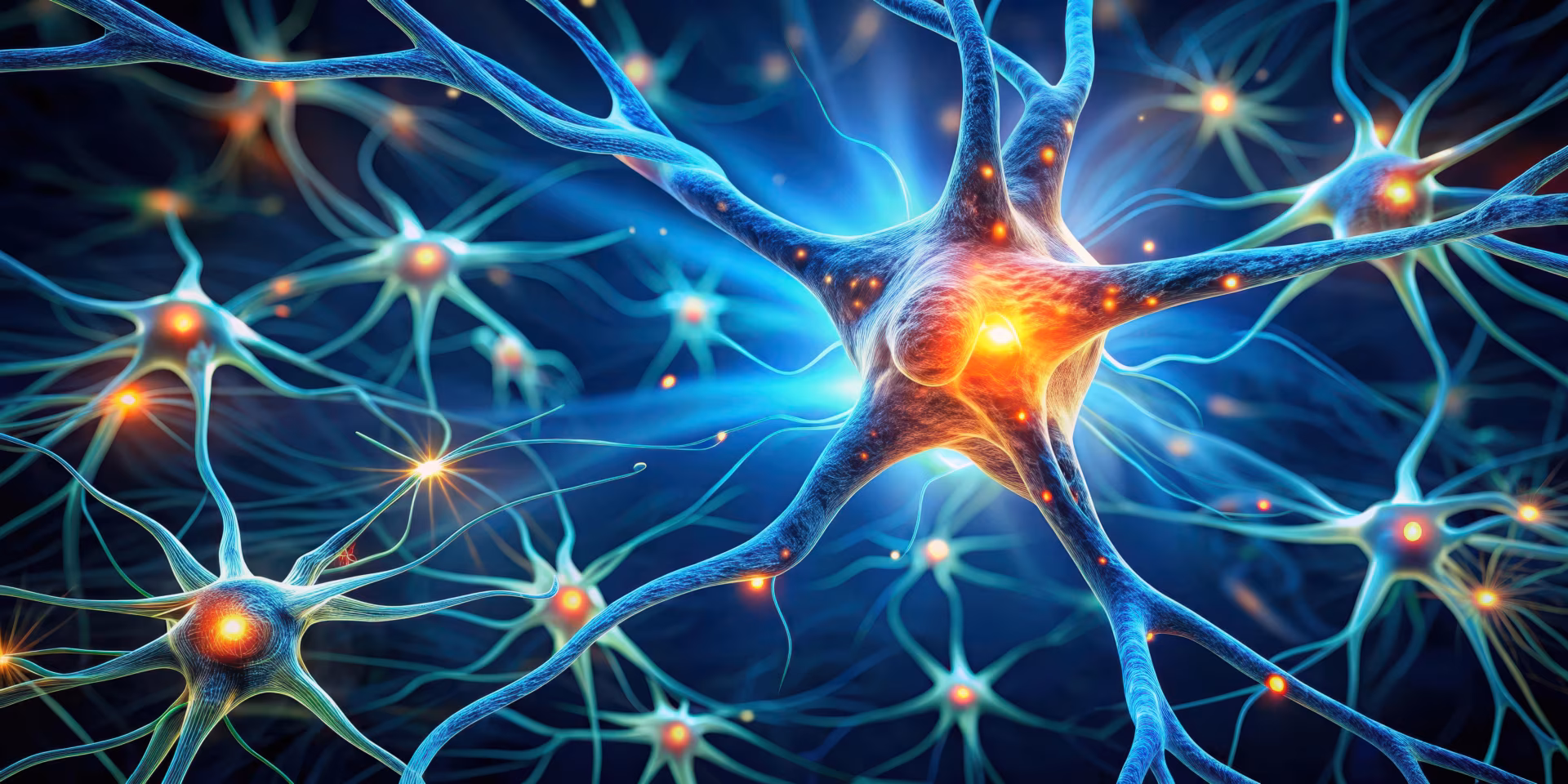
Neuronal loss and gliosis are the most common microscopic findings in CTE and this is usually associated with neurofibrillary degeneration, especially in the hippocampus, subiculum, entorhinal cortex amygdala, subcallosal and insular cortex, frontal and temporal cortex mamillary bodies, substantia nigra, locus coeruleus, nucleus accumbens medial thalamus, pars compacta, and pars reticulata.4,15,16,17 The pathognomonic neuropathologic signs of CTE are tau-immunoreactive neurofibrillary tangles (NFTs) with preferential involvement of superficial cortical layers, irregular and patchy distribution in the frontal and temporal cortex, a propensity for sulcal depths, and a perivascular, periventricular, and subpial distribution (see Figure 1). Unlike Alzheimer’s disease (AD), which shares a similar neuropathology, tau deposition in CTE is denser and preferentially found in neocortical layers II and III. Also unlike AD, the deposition of beta amyloid in CTE is not consistently present.4
According to Buee, the shear forces occurring with repetitive head trauma can cause vascular damage followed by a perivascular NFT formation and neuropil neurites (NN).18 Damage of the blood–brain barrier and release of local neurotoxins may then contribute to the perivascular formation of tau immunoreactive NFT, tau-positive glia, and NN. Although beta amyloid deposition is not found consistently in CTE, it presents diffusely in frontal, parietal, and temporal cortex without signs of vascular amyloid and without correlation to areas of injury. As with AD, inheritance of an Apo E e4 allele is a risk factor for the development of CTE.4
In 2005, Omalu et al. confirmed what had long been suspected—pathology consistent with CTE in a National Football League (NFL) player who had died of atherosclerotic heart disease 12 years after a long career in the NFL.19 Interestingly, family members noticed that he had suffered from memory deficits and Parkinsonian symptoms. A second case of CTE was reported in 2006 by Omalu, again in a player with a long history in the NFL who had been diagnosed with severe depression and had multiple suicide attempts.20 Since then, additional autopsy-confirmed cases of CTE have been reported, including a football player as young as 18 years old and athletes in hockey, soccer, and wrestling.4 Although the significance is unclear, football players compared to boxers were diagnosed at a younger age and had a shorter duration of illness.
Pathophysiology
CTE likely results from progressive neuronal loss, but the exact mechanisms are unclear. In acute TBI there is direct physical damage caused by acceleration and deceleration forces. The severity of injury is proportional to the severity of the generating forces, and depends on the direction in which these forces are applied. For example, rotational forces may cause more damage than linear translational forces of the same degree. Other mechanisms such as release of excitatory transmitters (glutamate), focal ischemia, breakdown of the blood–brain barrier, and inflammatory mediators likely play a role in initiating and maintaining a necrotic and apoptotic death cascades that can cause neuronal cell death.4 CTE caused by repetitive MTBI continues to progress decades after the injuries have stopped indicating that once the cascades are initiated, they continue to execute their effects, and the longer the individual lives, the worse the symptoms.4,21 The severity threshold of the brain injury that can initiate these progressive chronic neurodegenerative changes, as well as the frequency, is still unknown.
The patchy cortical distribution of NFTs suggests that their location is correlated with areas of direct mechanical trauma from blows to the side or top of the head. An ischemic basis of injury comes from the observation that tau deposition tends to be in the depths of cortical sulci. The associated breakdown of the blood–brain barrier following brain injury with release of local neurotoxins potentially explains the perivascular clustering of NFTs. Buee et al. examined the microvasculature in patients with dementia pugilistica and found decreased microvasculature density and tortuosity, which corresponded to the laminar distribution of tau-positive NFTs.18 In addition to tau,other protein inclusions may play an important role in CTE. The DNA binding protein TDP-43 along with tau has been observed in brains and spinal cords of athletes presenting simultaneously with CTE and motor neuron disease, suggesting a common link between CTE and sporadic amyotrophic lateral sclerosis.22 While the exact function of TDP-43 is unknown, its overexpression causes neuronal degeneration and cell death in animal models.23 One possible hypothesis is that MTBI causes axonal shear with cytoskeletal disruption. As part of the injury response, TDP-43 is upregulated and binds to neurofilament mRNA in order to stabilize the transcript. Because this protein is prone to aggregation, pathologic TDP-43 deposits form, resulting in neurotoxicity and cell death.
Clinical Manifestations and Diagnosis
Patients with CTE present with headaches, dizziness, an unsteady gait, fatigue, and dysarthria. In addition, they may have cognitive and psychosocial symptoms such as memory loss, attention deficit, difficulty in concentration, slow information processing, confusion, loss of judgment, irritability, emotional distress, and show an inability to stay employed. In severe cases there is a progressive slowing of movement, a propulsive gait, tremor, masked facies, deafness, dysarthria, dysphagia, ptosis, and other ocular abnormalities.4 Clinical deterioration in CTE typically occurs in three stages.4,13
- Stage 1—affective disturbances and psychotic symptoms.
- Stage 2—social instability, erratic behavior, memory loss, and initial symptoms and signs of Parkinsonism.
- Stage 3—general cognitive dysfunction, progression of dementia, speech and gait abnormalities, or full-blown Parkinsonism.
Early in the disease there is deposition of tau protein in the limbic system, which explains behavioral symptoms such as emotional lability, aggression, and violence. Hippocampal involvement as well as medial thalamus and entorhinal cortex account for the memory disturbances. Disinhibition occurs as more frontal lobe involvement develops and degeneration in temporoparietal and occipital cortex results in visiospatial symptoms. Finally, in the late stages, degeneration of the substantia nigra causes Parkinsonian symptoms.4,21,24 The diagnosis of the CTE related to sports is based in the history of repetitive MTBI, the presentation of the disease progression, and the detailed neurologic examination of the patient.
CT and MRI can be helpful for excluding other causes of neurologic symptoms such as chronic subdural hematoma or brain tumor. Recently, diffusion tensor imaging (DTI) has been used to look for changes that correlate with concussion. A study by Zhang et al. in 2006 looked at MRI compared with DTI in 49 professional boxers. While conventional MRI showed nonspecific white matter changes, diffusion anisotropy analysis revealed a decrease in the average diffusion constant and whole brain diffusion.25 Changes in regional anisotropy may indicate an early response to concussion in athletes and could become a useful tool both for detecting early damage and monitoring long-term neurologic deficits.
Prevention of Chronic Traumatic Encephalopathy
Because there are no known treatments for CTE, prevention of concussion and particularly avoiding repeated concussions is the onlyoption. An important part of this is the recognition of concussion on the field. It has been shown that suffering a first concussion increases the likelihood of a second concussion up to threefold.26 Standardized concussion tools have been developed that assist physicians and trainers in this assessment. Concussion grading scales, such as the Cantu system, use loss of consciousness, post-traumatic amnesia length, and post-concussion signs and symptoms to determine severity (see Table 2).
There has been tremendous research and development in designing new helmets, but their effectiveness in preventing concussions has not been proven. Mouth guards can reduce dental injuries but also have little impact on preventing concussions. There is some concern that protective equipment results in more aggressive play and may paradoxically increase the incidence of injuries.27
Much of the focus on preventing CTE centers on avoiding repeat brain injuries that occur within a short time frame. Return to play (RTP) guidelines have been developed that will hopefully decrease the risk of catastrophic injuries such as second impact syndrome as well as CTE. The RTP guidelines are based on concussion severity and require that the athlete be symptom free for at least one week before being considered ready to return (see Table 3). When a second concussion is suffered, the time period that the athlete must wait varies from two to four weeks depending on concussion severity. After three concussions (two if severe), the player should terminate the season and return the next year. For purposes of the RTP guidelines, an asymptomatic patient is one that is free of symptoms both at rest and exertion.
Conclusions
CTE is a growing public health concern and recent evidence suggests that younger athletes (particularly girls) are at greater risk of concussion due to less developed neck muscles, which allow a greater force to be exerted on the brain from trauma. The pathologic changes are similar to AD, but there are key differences in tau protein and amyloid deposition. As our sophistication around concussion detection and prevention improves, hopefully the incidence of CTE will decline.