Multifocal motor neuropathy (MMN) is a rare, immune-mediated neuromuscular condition characterized by impairment of the peripheral motor nerves, leading to muscle weakness, affecting the arms more than the legs. The disease was formerly known as MMN with conduction block (MMNCB), since a predominant feature is CB at multiple sites along the motor nerves, although at early stages the axons are not affected.1–3Entrapment neuropathy (EN) occurs when single nerves in various body locations become chronically compressed or mechanically injured as a result of various factors, including anatomical structures, fibro-osseous tunnels, ligaments, local edema, tumors, diabetes, medications, arthritis, hypothyroidism, obesity, or repetitive movements. Nerve compression causes sensory symptoms, such as paresthesias, numbness, dysesthesias, muscle weakness, and atrophy (if untreated), in the affected anatomical area. The involvement of a single or more than one nerve can resemble some of the motor symptoms of MMN, and consequently, in its early stages, it is possible to confuse the two conditions, which can have serious clinical consequences.4
Given its clinical presentation and topography, that is, distal predilection, MMN can simulate various entrapment mononeuropathies. These include median nerve compression at the wrist secondary to carpal tunnel syndrome (CTS), ulnar neuropathy at the elbow, distal ulnar neuropathy at the wrist, radial mononeuropathy at the spiral groove, radial tunnel syndrome, posterior interosseous mononeuropathy, proximal median mononeuropathy, or peroneal compressive neuropathy at the fibula.5–8The estimated incidence of MMN is 0.3–3/100,000, which is equivalent to 1,000–10,000 cases in the US, unlike EN, which is highly prevalent.
MMN usually presents with pure motor symptoms only, and that while it is unlikely to be misdiagnosed as EN by an experienced neurologist or neuromuscular specialist, it occasionally presents with vague, unrelated sensory symptoms. General physicians and even certain neurologists may have seen very few cases and may mistake some MMN presentations, especially early on and when focal in distribution, for EN. MMN commonly affects dexterity and walking


ability but most patients maintain autonomy9 and a high percentage remain employed.10However, if untreated, its clinical course tends to be chronic and protracted, and significant functional impairment may result.11–13 If MMN is misdiagnosed as EN, the subsequent delay in making the correct diagnosis may result in progression in the condition and significant motor disability.14This article aims to discuss the comparative signs and the diagnoses of MMN and EN and identify the issues in diagnosing the conditions correctly.
Diagnosis of entrapment neuropathy and multifocal motor neuropathy
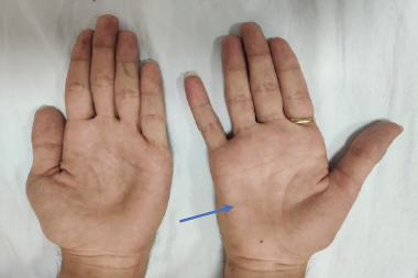
MMN is a demyelinating neuropathy, characterized by CB of myelinated motor nerves that can lead to axonal loss as the disease progresses, resulting in increasing weakness and muscle atrophy. Figures 1 and 2 illustrate these symptoms, as well as the impact of misdiagnosis, showing scars (at the elbow and at the wrist) representing sites of previous surgeries for presumed diagnosis of entrapment neuropathies. The main clinical features of MMN are discussed in a number of reviews: the most notable finding is asymmetric weakness in the distribution of peripheral nerves that is out of proportion to muscle atrophy. Weakness usually starts in a distribution of a single peripheral nerve with unilateral wrist drop, foot drop, or grip weakness.1–3,15It can differentially involve branches of a peripheral nerve; a common clinical finding noted in some patients is the presence of differential weakness of finger extensors.16
The European Federation of Neurological Societies (EFNS) has defined the diagnostic criteria for MMN (see Table 1).15,17The condition usually presents with slowly progressive focal weakness and atrophy without pain or sensory complaints (see Figure 2). There is often focal or multifocal involvement of the upper-extremity motor nerves in the forearm or upper arm. The disease course is slowly or stepwise progressive and typically shows asymmetric involvement of two or more nerves. Sensory symptoms or signs are usually absent in the distribution of the affected nerve. Recently, the extent of sensory signs and symptoms in MMN has been added to the criteria.15CB may not be identified in all patients;16 the EFNS define CB as definite or probable based on electrodiagnostic findings.15,17
A common EN is CTS, the presentation of which is pain or paresthesias in an area that includes the median nerve distribution. Symptoms are characteristically relieved by hand movements.18 Bilateral CTS is commonly seen at initial presentation.19Recommended diagnostic procedures include collecting accurate patient history, conducting a physical examination that includes personal characteristics, performing a sensory examination, performing manual muscle testing of the upper extremity, performing provocative tests, and/or performing discriminatory tests for alternative diagnoses. The most widely used provocative physical tests are Phalen’s sign, which involves flexion of the wrist to 90 degrees for 60 seconds, and Tinel’s sign, which involves tapping over the carpal tunnel.20However, these tests have limited sensitivity and specificity.21Electrodiagnostic study assists significantly in validating the diagnosis, determining the pathology and prognosis, as well as ruling out other mimickers.22
High titers of immunoglobulin (Ig) M anti-ganglioside-monosialic acid (GM) 1 serum antibodies are found in 50–80 % of patients with MMN.23–25Their role has not been established but they are a useful diagnostic marker for MMN. Their prevalence varies, partly because there is no consensus about the optimum assay for these antibodies. Although controversial, it is thought that patients with high titers of anti-GM1 antibodies are likely to have more severe weakness, disability, and axonal loss than those without these antibodies. Despite lacking specificity, they are considered a marker for MMN and their presence can support the diagnosis.1,24,26

Electrodiagnostic tests, including nerve conduction studies and electromyography, are used to confirm the clinical diagnosis of both EN27 and MMN. In MMN, there is CB on stimulation of motor nerve fibers, at proximal sites, evident by decreased amplitude or area of compound muscle action potentials (CMAP), compared with the distal CMAP (DCMAP) response. Criteria have been developed by the American Association of Neuromuscular and Electrodiagnostic Medicine (AANEM), and later by the EFNS, to ascertain the diagnosis of MMN, based on clinical and electrodiagnostic criteria, as well as supportive ones (see Tables 1 and 2).17 Unlike EN, in MMN, CB is noted at nonentrapment sites (see Figure 3).This is an important electrodiagnostic finding, which, in the clinical context, can hint to the diagnosis of MMN.
Using electrodiagnostic criteria, as recommended by the EFNS guidelines, can help in identifying MMN from EN (see Table 2).17 According to these criteria, the grading of CB is defined as definite (including negative-peak CMAP area reduction on proximal versus distal stimulation of at least 50 % whatever the nerve segment length [median, ulnar, and peroneal]) or probable (including negative-peak CMAP area reduction of at least 30 % over a long segment [e.g., wrist to elbow or elbow to axilla] of an upper-limb nerve with increase of proximal to distal negative-peak CMAP duration to ≤30 %). Also, normal sensory-nerve conduction studies are required in upper-limb segments with CB (please see Table 2 for electrodiagnostic details). Electrodiagnostic testing is a useful tool in the hand of an experienced neuromuscular specialist. However, on rare occasions, and especially when electrodiagnostic testing is not properly performed, electrodiagnostic testing can result in the misdiagnosis of MMN as EN.14
Supporting criteria to the diagnosis of MMN include imaging of the nerves, particularly the brachial plexus, showing abnormal enhancement.17 These studies, including neuromuscular ultrasound, are being increasingly used as adjunct tools in the diagnosis of EN. In CTS, imaging of the median nerve’s cross-sectional area at the wrist provides additional information and shows pathologic nerve swelling and other anomalies that compress the median nerve.28 In MMN, highresolution ultrasonography is now available and can show different patterns of nerve enlargement between inflammatory neuropathies

and axonal and inherited polyneuropathies, as well as show increased hypo-echogenicity and increased intraneural vascularization.29,30Magnetic resonance neurography (MRN) may also help in diagnosis; it shows focal enlargement and increased signal intensity of the brachial plexus on T2-weighted images.31
Current guidelines and treatment paradigms
In the case of MMN, current consensus guidelines recommend intravenous immunoglobulin (IVIG) as the standard, evidence-based therapy for MMN.15,32 In a study of 88 patients with MMN, 95 % responded to IVIG therapy: nonresponders had longer disease duration before the first treatment, highlighting the importance of early treatment.4 The recommended treatment options of American Academy of Orthopaedic Surgeons (AAOS) for EN include surgery, wrist splinting, steroid injections, and oral steroids.33 Surgery is associated with better long-term outcomes than splinting.34 However, these treatments for EN either would be unnecessary and would fail or may worsen the symptoms in MMN (see Figure 2),35highlighting the importance of correct diagnosis.

Cases of multifocal motor neuropathy misdiagnosed as entrapment neuropathy
Case 1
The patient is a 58-year-old male who noted painless weakness in his left hand, with minimal paresthesias in the fingers. The initial diagnosis was CTS, for which he underwent left CTS release. Two years later, he noted weakness in his right hand, with decreased hand grip, but no pain or sensory symptoms. Electrodiagnostic studies reported normal right radial sensory potential, and slowing of right radial motor conduction between the radial groove and distal stimulation. Subsequently, the patient underwent decompression of the right radial nerve at the elbow and external neurolysis of the posterior interosseous nerve. However, his weakness did not improve, and a year later he underwent a further exploration, which revealed “recompression” of the posterior interosseous nerve, by “fibrous tissue and scars.” Later, the patient noticed significant weakness in his left hand. He was diagnosed with median compression neuropathy at the elbow and underwent left median nerve decompression at the elbow. No electrodiagnostic studies were performed on the left median nerve prior to the surgery. Following a referral, the patient underwent nerve conduction studies that revealed multifocal evidence of motor conduction slowing and CB. A diagnosis of MMN was then made. IVIG therapy was instituted with improvement in some of his motor function. As a result of inadequate and incomplete electrodiagnostic studies, diagnosis was delayed, resulting in disease progression. The patient developed atrophy in some muscles, possibly a result of the surgery, which resulted in significant motor disability.
Case 2
A second case concerns a 64-year-old woman who complained of progressive painless muscle weakness and atrophy in the hand muscles, and was diagnosed with right ulnar EN at the wrist. The patient underwent decompressive surgery but the symptoms worsened. Over the following 2 years, the patient underwent two further surgeries on the ulnar wrist but her right-hand muscle atrophy progressed. She subsequently began to notice gradual weakness in the left hand, followed by atrophy, resulting in significant limitations in activities of daily living (ADL). Neuromuscular examination revealed severe atrophy in the thenar, hypothenar, and interossei muscles on the right side, as well as moderate atrophy in the left interossei and hypothenar muscles. Manual muscle-strength testing revealed weakness in several muscles and deep tendon reflexes were absent in the upper extremities. Nerveconduction studies showed CB in the right median CMAP’s amplitude and area, respectively, of 44 and 41 %. Left radial DCMAP’s amplitude showed CB on stimulation at the elbow of 74 % in amplitude and 68 % in area. Left median DCMAP’s amplitude showed significant CB on proximal stimulation at the elbow of 87 % in amplitude and 77 % in area. Sensory potentials in the upper-extremity nerves tested were normal. Following a diagnosis of MMN, the patient was treated with IVIG. Three months later, the patient demonstrated significant improvements in grip strength and ability to perform ADLs, and monthly treatments have resulted in sustained improvement.
Summary and concluding remarks
MMN is a rare, treatable neuropathy, but good long-term outcomes are dependent on early treatment. It is therefore important to diagnose MMN and differentiate it from other conditions. It differs from typical EN in that it predominantly affects motor nerve fibers and has a strikingly restricted distribution, with characteristic topographical distribution, affecting upper more than lower extremity, and with predilection to the distal segments.
Misdiagnosis should rarely occur since the nerve involvement in either of these disorders is rarely at sites of common nerve entrapment; however, despite the existence of good diagnostic criteria, when MMN is confined to a small group of nerves, there is significant overlap with EN. Furthermore, misdiagnosis may have serious consequences in terms of treatment. The ability to recognize MMN and the ability to distinguish it from other diseases of peripheral nerve, such as EN, are important clinical skills. The cases presented have illustrated some of the issues and complications that can ensue as a result of delayed diagnosis or wrong therapy, such as surgical intervention for a presumed diagnosis of EN. Oral corticosteroid treatment, occasionally used as nonsurgical treatment in early EN conditions, could also potentially worsen symptoms in MMN. Clinical trials investigating alternative techniques for the diagnosis of MMN are ongoing.36 However, there remains a need for future studies to further understand the pathogenesis of MMN in order to develop more alternative and efficacious treatment options.


