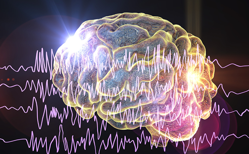
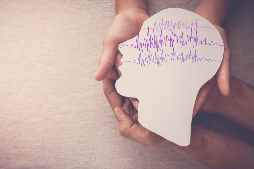
Seizures, paroxysmal neurological symptoms caused by episodic and pathologic neuronal discharging, have myriad associated signs/ symptoms that are dependent upon its anatomical origin and subsequent spread. The underlying causes of “epilepsy” (recurrent seizures) are many and include genetic factors, congenital and/or developmental anomalies, infections, trauma, and tumors. The incidence of epilepsy is approximately 1 %. While antiepileptic drugs (AEDs) can frequently control seizures, 30–40 % of patients fail to achieve control. Various approaches exist for those who fail AED treatment. A surgical approach is important because properly selected patients may be amenable to an excisional operation and possible “cure.” Dietary manipulations, e.g., the ketogenic diet or the modified Atkin’s diet, are often helpful but rarely curative. Brain-stimulation techniques are available for the refractory patient. These techniques include the vagal nerve stimulator (VNS), the responsive neural stimulator (RNS), and transcranial magnetic stimulation (TMS).
Vagal Nerve Stimulator
VNS has the longest history of the three modalities. After more than 10 years in development, the US Food and Drug Administration (FDA) approved it in 1997 for patients over 12 years old with medically refractory partial epilepsy.1 Shortly thereafter the American Academy of Neurology (AAN) issued an advisory stating that patients should undergo evaluations at an epilepsy center to determine the etiology and assess candidacy for excisional surgery. Later, the AAN updated its guidelines to include children with refractory partial or generalized epilepsy who are not surgical candidates.2
The VNS is placed in a subcutaneous surgical pocket under the left clavicle and attached to wire leads that are wrapped around the left vagus nerve. The device is tested in the operating room for functionality and patients return in 1 to 2 weeks to have it activated. Thereafter, they return frequently for adjustments to the stimulating parameters. This is performed through an external wand attached to a computer. While the ultimate stimulating parameters are not clearly defined, they are adjusted as tolerated and according to clinical responses. Stimulations are programmed to occur at regular intervals and the patient can activate the VNS to deliver additional stimulation by swiping a magnet over the device. This is helpful in those patients who have clear auras.
Response rates vary from >30 to 65 % in clinical trials. Early on, it appeared that response rates seemed to increase between the first and third year of use. Responses vary relative to the type of underlying seizure disorder with idiopathic generalized epilepsy being perhaps the most responsive.3 Side effects include hoarseness, which is usually present when the stimulation is on, occasional feelings of shortness of breath, exacerbation of symptoms related to obstructive apnea, and occasional cardiac arrhythmias. However, recent work suggests that the presence of the VNS can reverse pathologic cardiac repolarizations manifest by improvement in patient’s degree of T-wave alternans.4
Responsive Neural Stimulator
In 2013, the FDA approved the RNS for patients over 18 years old with medically refractory partial epilepsy who have one or two well-localized seizure onset foci.5 This programmable device is implanted in the skull with two four-channel contacts, which are implanted as depth electrodes or subdural strips. After implantation, the device records the patient’s electrocardiogram (EEG) activity for a period of time and captures their spontaneous seizures. The system is trained to recognize the earliest neurophysiologic changes associated with their seizures. After training, the programmer directs the device to stimulate once it detects the beginning of a seizure. Hence this system is considered a ‘closed loop’ and ‘responsive’ system doing both detection and stimulation. Like the VNS systems, there is an external programmer capable of altering stimulus parameters according to clinical responses and the patient can activate the RNS with an external magnet.
Early and long-term results show about 55 % patients will have more than a 50 % reduction in their seizure frequency.6 There are isolated reports of patients becoming either free or relatively free of seizures.
There is another programmable deep brain stimulator similar to the deep brain stimulators (DBS) used for movement disorders. Either one or two stimulators can be implanted. The stimulating contacts are directed surgically into the anterior thalamic nucleus. Unlike RNS, the DBS system is not closed loop. It is similar to the DBS used for Parkinson’s disease. The stimulation parameters are manipulated through an external wand and adjusted based upon tolerance and responses. This device has been tested extensively in the US but is not yet FDA approved.
Transcranial Magnetic Stimulation
TMS is an FDA approved form of brain stimulation used in the treatment of drug-resistant depression. In ongoing clinical trials, TMS shows promise in treating certain focal neocortical-based seizures. This technique has been used to assess relative degrees of neocortical excitability.7 These techniques can determine whether changes in excitability are related to changes in the relative levels of excitation or inhibition. In several research protocols, TMS stimulation parameters have been shown to alter the relative degrees of excitation in predictable fashion and subsequently potentially offer antiseizure effects.
TMS works according to Ohm’s law. When an electrical current is generated, a simultaneous magnetic field is created, oriented at a right angle to the direction of the electrical current. The TMS stimulator is a paddle-shaped device placed just above the scalp. It generates a sudden magnetic flux, which induces an electrical current in superficial layers of the underlying cortex (reversal of Ohm’s observation). These electrical currents are manipulated to generate either increased excitability or increased inhibition. One can calculate the strength of the induced electrical current because the skull, skin, etc., that lie between the stimulator and the underlying brain cortex do not alter magnetic fields. The stimulation itself is usually felt as a mild thud and is almost always well-tolerated.
Recent publications focused on potential uses for TMS in epilepsy. In one study,8 TMS was used to probe the relative degree of hyperexcitability and pathologic hyperconnectedness in patients with disorders of cortical migration. Patients with such conditions are significantly more prone to develop medically poorly controlled seizures. In this study, researchers demonstrated that selected areas of neocortex had excessive excitation that was associated with pathologic connections to the deeper structures in the brain where the migration disorders were located. Work is now ongoing to see if TMS stimulation of the abnormal neocortical regions can alter this hyperconnectivity in clinically relevant ways. The second study treated a patient with a well-localized cortical epileptic focus that was extremely poorly responsive to medical manipulation with TMS. On both the short- and long-term basis, there was a dramatic response that allowed the researchers to gradually withdraw some of the patient’s antiseizure medication.9
Conclusion
The goal in treating patients with epilepsy is to control or eliminate their recurrent seizures and to minimize side effects to treatment. AEDs have been the mainstay of treatment for over 100 years. AEDs are effective in 50–60 % of patients. There are short- and long-term side effects to AEDs that include allergic reactions, drug–drug interactions, long-term metabolic and endocrine complications, potential fetal/genetic effects, and cognitive problems. Surgery to remove an epileptic focus can, in selected cases, be curative. When medications fail and surgery is not an option, physicians look for alternative treatments. Dietary manipulations are often helpful but difficult to maintain. Brain-stimulation techniques, as noted above, have been around for the last 15–20 years. These techniques offer an additional method to gain seizure control and with improved control, the ability to reduce the patient’s medication load and reduce their inherent side effects without adding additional adverse treatment effects.


