Q. What was previously known about brainstem network validation in focal epilepsy and sudden unexplained death in epilepsy (SUDEP)?
The first line of evidence for an involvement of the brainstem had come from those rare cases in whom SUDEP was witnessed.2 The symptoms observed in those patients indicated a breakdown of central autonomic control. The brainstem is one of the brain structures that plays a critical role in autonomic control. The second line of evidence came from animal models of SUDEP that also pointed to the brainstem as a critical structure.3 These observations motivated us to investigate brainstem abnormalities in a small group of patients with temporal lobe epilepsy who had undergone magnetic resonance (MR) imaging for another project. Two patients of this cohort had later died of SUDEP. This study showed that temporal lobe epilepsy is associated with brainstem volume loss/network abnormalities. The two patients who later died of SUDEP had more severe and more extensive damage than the other patients.4 This observation led to this new study, which was presented at AES.1
Q. Could you tell us a little about the design and aims of your recent study?
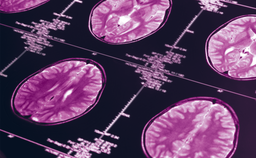
There were two main aims of the study. First, to build on the evidence to date demonstrating an association between brainstem damage in focal epilepsy and autonomic control (assessed by monitoring heart rate variability [HRV]), and second, to replicate the findings of more extensive brainstem damage in a larger SUDEP population. MR imaging was studied in two groups: autonomic population (18 patients with focal epilepsy, 11 controls) and SUDEP population (26 patients with SUDEP epilepsy). Deformation-based morphometry of the brainstem was used to generate profile similarity maps. The resulting Jacobian determinants were further characterized by graph analysis to identify regions with excessive expansion (sigExcROIs) or volume loss.1
Q. What were the major findings of this study?
There were two key study findings. Consistent with existing evidence, volume loss in brainstem regions involved in autonomic control was correlated with reduced HRV in patients with epilepsy. Further, patients who died of SUDEP had widespread brainstem volume loss in their last MR exam before death. More extensive volume loss correlated with a shorter survival time.1
Q. What future studies are planned?
The current project is ongoing. We are still reaching out to academic institutions and families who lost somebody to SUDEP to increase the number of cases. The first step will be to use all the new cases and repeat the current analysis in the brainstem. The next step is to include other regions known to be involved in autonomic control and investigate their contribution. This will be done in collaboration with other researchers who have collected imaging data and data about autonomic/cardiac function in epilepsy patients who did not die of SUDEP. Analyzing their data will give us an opportunity to learn more about how epilepsy affects these regions in patients who do not die of SUDEP and how this damage impacts autonomic parameters. We will then use what we have learned from that study to investigate if these regions are also more damaged in patients who died of SUDEP and how these abnormalities relate to the brainstem damage. The ultimate goal, in the near future, is to use all the information gained by these studies to identify a pattern of brain damage that is only found in epilepsy patients with a high risk for SUDEP, but not in other epilepsy patients. In short, an imaging biomarker for SUDEP.
Q. How are these findings likely to impact on clinical practice?
Although the current findings are promising and a great first step, we are still a long way off from having an imaging biomarker for SUDEP. It also has to be emphasized that there is probably more than one mechanism that can cause SUDEP, and that there are therefore other factors that also need to be considered when assessing an individual patient’s SUDEP risk. That being said, if it is indeed possible to develop this method into a reliable biomarker, it would mean that every time a patient with epilepsy has MR imaging, the imaging would be analyzed using the techniques that were used in this study to investigate to what degree the SUDEP-typical pattern is present or not, and thus to identify and treat those patients with a high risk. Currently, there exists no scientifically approved treatment to prevent SUDEP, but I hope that the work done by my collaborators from the National Institutes of Health-sponsored Center for SUDEP Research will change that.


