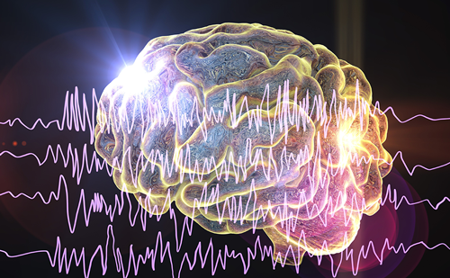
Epilepsy is a term used to describe over 40 different human seizure disorders that vary in clinical and electroencephalographic (EEG) characteristics. It is one of the most common neurological disorders and occurs in about 1% of the population, independent of geography, ethnicity, or gender.1 Seizures occur when the brain is disrupted by abnormal neuronal activity. Having two or more unprovoked seizures is a working definition of epilepsy that is used commonly by physicians. The focus of ‘epileptic activity’ can be centered in any region of the brain, although cerebral cortex, thalamus, and limbic structures are most often involved. In some types of epilepsy, the abnormal activity is confined to a specific brain region, and these are classified as focal, partial, or localization-related epilepsy. In other epilepsy subtypes, the abnormal neuronal activity does not have a clear focus, or may be thalamic or subcortical in nature, and these are classified as generalized epilepsy. Clinical classification of epilepsy subtypes and syndromes is complex and somewhat controversial; however, all agree with a major division between focal and generalized epilepsy.
Causes of Epilepsy
Seizures can be caused by brain tumors, vascular malformation, encephalitis, or as a result of traumatic brain injury. These epilepsies are classified as symptomatic since there is a readily understandable cause for the seizures. However, in most cases the cause of epilepsy is unknown. Epilepsies of unknown cause are divided into two categories: the first is cryptogenic epilepsy, meaning that the cause is suspected to be induced by a pathology that is below the limit of detection of the available diagnostic screens; the second is idiopathic epilepsy, meaning the cause is most likely related to a genetic predisposition. These definitions are used with regularity among neurologists who specialize in treating patients with epilepsy.
Molecules
A significant amount of research on epilepsy is dedicated to understanding the pathophysiology of seizures from a molecular perspective. This research seeks to gain a comprehensive knowledge of how DNA, RNA, and proteins interact to affect the cellular components of the central nervous system (CNS) that regulate neural excitation and inhibition. The molecules and their interactions combine to create cells with specific properties that allow them to form circuit networks and the higher-order electrical characteristics of the brain. Initially, molecular research focused on the main excitatory CNS neurotransmitter, glutamate, and the main inhibitory CNS neurotransmitter, gamma aminobutyric acid (GABA). Thus, glutamate and GABA, their receptors, and the enzymes that regulate their synthesis and degradation have been studied extensively in animal models of epilepsy and in human patients. In addition, many studies of sodium channels have been conducted in epilepsy models as these channel proteins play an important role in the generation and propagation of neuronal action potentials. More recently, unbiased genetic approaches have been used to identify other molecules whose altered expression or function predisposes to seizures in animals and/or epilepsy in humans.
Genetics
Genetic influences contribute to the etiology of many epilepsy phenotypes in humans and animals. The influence of genetics on the etiology of human epilepsy is supported by rigorous epidemiological and genetic linkage data collected using classic family and twin study designs. Several types of human (and animal) epilepsy cluster in families with predictable inheritance patterns. These disorders are caused by mutation in a single gene (monogenic epilepsy), several of which have been identified.2 However, monogenic forms of epilepsy are rare, accounting for only 1–5% of all epilepsy cases. Common forms of human epilepsy have complex inheritance patterns due to variation in multiple genes interacting with environmental factors, which together increase susceptibility to seizures and result in epilepsy. In an effort to identify genetic influences on complex common forms of epilepsy, a number of complementary experimental approaches have been pursued in both animal models and human patient populations. This article highlights some of the successful paradigms employed to identify genetic variation linked to or associated with seizure susceptibility in animals and epilepsy in humans.
Animal Models
For many years, breeders of purebred dogs have known that genetic influences play a major role in predisposition for epilepsy, as certain breeds are at high risk and selective breeding successfully avoids seizure disorders in progeny derived from non-epileptic parents. Molecular genetic studies are under way to identify these gene variations in canine breeds and once identified their homologs in human patients can be studied in detail.3 Other examples of animals with documented spontaneous seizures likely caused by genetic mutation include mice, rats, gerbils, baboons, and even fruit flies. While studies of all of these species have made important contributions, mice are the most widely used animal model for genetic epilepsy research.
The Mouse
Mice are similar to humans with respect to physiology, anatomy, and genetics. The generation and use of inbred mouse strains over the past 100 years has made the mouse an ideal genetic organism and a valuable tool in the search for seizure-causing genes. Computer databases, specialized reagents, and careful phenotyping resources that facilitate genetic studies in the mouse are unrivaled by resources available for genetic studies on any other vertebrate species, including humans. The mouse genome is fully sequenced and the opportunity to generate genetically modified mice through selective breeding, transgenesis, and knock-out/knock-in technology is readily available. Importantly, researchers have identified single gene mutations that lead to spontaneously occurring seizures in mice. Examples include lethargic, slow-wave, stargazer, tottering, and weaver, which are models that exhibit both behavioral and electrographic seizure phenotypes. These monogenic mouse epilepsy models are amenable to genetic dissection techniques, and the mutations underlying their phenotypes have been identified.4–8 In each case, the DNA mutation altered the structure and function of an encoded protein related to ion homeostasis. These findings add support to the popular description of epilepsy as an ‘ion channelopathy’ disease.
In contrast to monogenic mouse epilepsies, studies have also been performed on various inbred mouse strains in which robust differences in seizure susceptibility have been documented. The use of chemoconvulsants, auditory stimuli, and direct electrical stimulation has revealed that there is a large range of quantifiable seizure susceptibilities among the many strains that have been studied. Inheritance patterns in crosses between inbred strains suggest that seizure susceptibility is a complex polygenic trait resulting from variation at multiple genetic loci interacting with the environment. Work from the authors’ laboratories provides an example of how mouse models can be used to identify genetic determinants of seizure susceptibility. The approach is based on two common inbred strains of mice with extremely disparate susceptibility to experimental seizures: C57BL6/J (B6)—relatively resistant—and DBA/2J (D2)—relatively susceptible. Application of a quantitative trait locus (QTL) mapping strategy to these strains led to the nomination9 and identification10 of Kcnj10 genetic variation as a major influence in the model. The Kcnj10 gene encodes an inward rectifier potassium ion channel protein that transports potassium from the extracellular space into glial cells. Potassium is released from neurons during action potentials and extracellular concentrations rise to particularly high levels during repetitive neural activity. High extracellular potassium concentrations make neurons hyper-excitable; however, Kir4.1-mediated potassium buffering prevents this and allows maintenance of neuronal membrane excitability. Translation of the mouse studies into a human genetic association study involving over 400 epilepsy patients and 284 controls revealed a mis-sense coding variation in KCNJ10 homolog (Arg271Cys) in which the Arg271 allele confers significant risk for the development of common forms of epilepsy including idiopathic generalized epilepsy (IGE) and temporal lobe epilepsy (TLE).11 In a confirmatory study involving 550 IGE patients and 660 controls,12 the Arg271 allele was again revealed to increase epilepsy risk significantly. Taken together, these results comprise potentially important translational research—a unique success in translational research involving mouse and human genetic studies of epilepsy that has resulted in the identification of a novel target for antiepilepsy treatment development.
Human Epilepsy
Despite recent advances in the development of antiepileptic medications, clinical management of epilepsy is often complicated and adequate control of symptoms is difficult to maintain.13 Moreover, many of the drugs currently used to treat epilepsy can have deleterious side effects and precipitate serious drug–drug interactions.14 Thus, epilepsy represents a major health problem, and a critical need exists to identify its underlying cause in order to ultimately develop new and more effective treatments. Based on genetic, clinical, and epidemiological studies, epilepsy is recognized as a complex disorder with a highly variable phenotype. The paroxysmal nature of epilepsy involves seizures that may appear to be completely spontaneous or that are triggered by various environmental factors such as drugs, stress, or sensory stimuli. The heterogeneous nature of seizure phenotypes among individuals with epilepsy is also striking, with many behavioral and electrographic patterns distinguishable even among affected members of a single family. Concordance rates determined from twin studies15 as well as studies of multigenerational family pedigrees16 have established that there is a significant genetic component involved in the development of epilepsy.
The heterogeneous nature of epilepsy complicates the choice of exactly which phenotypes or forms of epilepsy to include in genetic studies; this has been a vexing problem for a number of years.17 Thus, to date, genetic studies in human epilepsy have focused on rare phenotypes in isolated families where disease is inherited in a fashion that is predictable based on the laws of Mendel. Such inheritance patterns in a given family suggest involvement of a single genetic locus18 and have led to the discovery of a number of ‘epilepsy-causing’ gene mutations. These discoveries, although most relevant to only a very small fraction of all epilepsy patients, have helped to provide information related to the general biology of seizures. On the other hand, there are fewer studies that have focused on common forms of epilepsy that are multifactorial in nature and result from the effects of many genetic variants working in concert with environmental factors.19 Thus, our current understanding of how most cases of epilepsy develop is still poor and the impact of genetics on the diagnosis and treatment of epilepsy somewhat uncertain.20
Genetic Linkage Studies
In the monogenic forms of epilepsy, genome-wide linkage analysis followed by positional cloning has led to the identification of several causative gene mutations. These include KCNQ2 and KCNQ3 in benign familiar neonatal convulsions,21,22 CHRNA4 and CHRNB2 in autosomal dominant nocturnal frontal lobe epilepsy,23,24 and GABRG2,25 GABRD,26 SCN1A,27 SCN1B,28 and SCN2A29 in the syndrome of generalized epilepsy with febrile seizures plus. Similar success has not been achieved in common forms of epilepsy, however, where linkage approaches are less useful due to polygenic inheritance. It may not be surprising then that a limited number of studies have been reported involving full-genome scans in multiple pedigrees that segregate common forms of epilepsy. Published work includes a recent large study in 126 multiplex families with IGE in which a complex genetic architecture was documented, including susceptibility loci on chr 5q34, 6p12, 11q13, 13q22-q31, and 19q13.30 Another large linkage study on IGE involving 96 families found strong evidence for a locus common to most IGEs on chr 18 and other loci that may influence specific seizure phenotypes for different IGEs, including a locus on chr 6 for juvenile myoclonic epilepsy (JME), a locus on chr 8 for non- JME forms of IGE, and weaker evidence for linkage of absence seizures to loci on chr 5.31 In a genome-wide linkage study of 130 IGE families, results revealed significant evidence for a novel IGE susceptibility locus on chr 3q26 and suggestive evidence for IGE loci on chr 14q23 and 2q36.32 The first linkage study in a common form of epilepsy involved families with JME and is credited with generating initial evidence for an IGE susceptibility locus on chr 6 in or around the major histocompatibility (HLA) region of chr 6.33 This locus has since been the focus of intensive study and some controversy.34,35 Overall, it is evident that there is low correspondence between the results of previously published full genome linkage studies in epilepsy.
More common than full-genome linkage studies are studies that focus linkage analysis specifically on regions of the genome where epilepsy-causing genes have already been identified in order to confirm or refine prior mapping results, or in regions where genes of high biological plausibility are located. In this way, linkage studies have led to the formal identification of underlying genetic causes of epilepsy involving mutations in the genes GABRA1,36 CACNB4,37 CACNA1A,38 CACNA1H,39 and CLCN2.40 It is noteworthy that most of the mutations discovered in the families segregating epilepsy as a single-gene trait are found in genes that encode ion channels or that encode neurotransmitter receptors linked directly to ion channels. As a result of such discoveries, a dominant perspective on the pathogenesis of epilepsy is that it is related in part to ion channel dysfunction.
Although it is clear from studies in Mendelian forms of epilepsy, as well as from animal studies, that ion channels are involved intimately in controlling neuronal excitability and that defects in ion channel genes can cause epilepsy, it is now established that mutations in other types of gene (i.e. non-ion channel genes) can also cause epilepsy. Such genes fall into a number of different classes, including those that encode transcription factors, enzymes for intermediary metabolism, and second messenger molecules. The first non-ion-channel gene discovered as a cause of epilepsy was CSTB (cystatin B). Mutations in this gene were found to underlie a rare form of progressive epilepsy;41 however, the way in which dysfunction of the product of CSTB, a cysteine protease inhibitor, leads to epilepsy is still not clear. Recent evidence that epilepsy can result from mutations in the gene for malic enzyme 2,42 an important component of intermediary metabolism, is of great interest given the antiepileptic effects of dietary treatment strategies such as the ketogenic diet.43 Another non-ion-channel gene linked to epilepsy is EFHC1. This gene encodes a novel axonemal protein and, although mutations in it have been identified as a cause of JME,44–45 there is no clear mechanism established by which such defects result in epilepsy. LGI1 is another gene that has been shown to cause epilepsy when mutated,46,47 as is BRD2.48 This latter gene has been suggested to contribute to linkage signals on chr 6.33 Again, however, the largely unknown function of the product of these genes has hindered attempts to use the discoveries to gain useful insight into the etiology of epilepsy.
Thus, although a number of rare, single-gene forms of epilepsy involve defects in proteins related to ion flux, it is clear that epilepsy is more than simply a disorder of channels (or a series of ion channelopathies) and that genes encoding molecules involved in diverse aspects of cell biology may lead to dysfunction of neurons and/or their connections and cause epilepsy when mutated.
Genetic Association Studies
The majority of genetic studies on common forms of human epilepsy involve association analysis of candidate genes. In general, these studies have examined the association between one or more polymorphisms of a candidate gene and various epilepsy phenotypes. Candidate genes are selected on the basis of a putative role in the pathobiology of epilepsy, map position in relation to previous linkage results, or translational data from animal models. The advantages and disadvantages of association studies of epilepsy are similar to those for other complex trait disorders and although the limitations have been reviewed,49 the criteria for designing uniform studies and defining significance are not fully established. This latter circumstance often confounds comparison of studies from different laboratories, and as a result there are few confirmed (and many unconfirmed) genetic associations in the field of epilepsy research.
Of the numerous case-control studies reported, the largest and most comprehensive is a recent study of 279 prime candidate epilepsy susceptibility genes in 2,700 patients with various types of epilepsy and 1,100 controls recruited from four different geographical regions across the world.50 Although this latter study uncovered several interesting genes for further study, including KCNAB1, KCNMB4, GABRR2, ALDH5A1, and SYN2, there were no strong findings reported, probably because genetic susceptibilities are ethnicity-specific, thus reducing the effective power of the study.
As discussed above, one of the only independently confirmed genetic associations in epilepsy involves KCNJ10.11,12 Given the current state of genetic analysis of the epilepsies,45 whole-genome association studies offer one of the next best methodologies to potentially identify the genetic determinants of complex common forms of human epilepsy.
Heterogeneity
There is substantial literature support for the concept that genetic factors affect risk for common forms of epilepsy. It is generally believed that in any genetic study, the more homogeneous the patient cohort with respect to phenotype, the more power the method will have to detect genes of influence. However, it is also clear that divergent epilepsy subtypes and endophenotypes from various syndromes may have some predisposing genetic factors in common. Animal models that approximate both focal and generalized epilepsy share certain genetic susceptibility loci).9,10
Human epidemiological studies support this notion as well, and evidence suggests that some genetic factors are relevant to divergent epilepsy types.51 For example, about 30% of individuals who suffer from childhood absence epilepsy (CAE) go on to develop myoclonic epilepsy as adults (JME). In another recent study, the type of epilepsy was documented for affected first-degree relatives of an epilepsy patient (proband). In about 30% of the cases, the first-degree relative suffered from a different epilepsy subtype or syndrome compared with the proband.52 Thus, studying cohorts of patients with diverse common forms of epilepsy may identify genetic predisposition to seizure susceptibility in general, whereas studying homogeneous patient cohorts may lead to the discovery of genetic factors that influence a specific subtype or symptom of epilepsy.53
Rare versus Common Variations
An unresolved issue for those studying genetic influences on human epilepsy is whether the causal genetic variations are common or rare. The typical genetic association study is designed to collect data on gene variations that occur with a minor allele frequency of 5% or greater, i.e. common variations that many in the population share.
The rationale for expecting that some common gene variations are linked to or associated with epilepsy is supported by the observation that many patients with the same or similar kinds of seizure disorder exhibit consistent electrographic and behavioral deficits that are diagnostic for specific epilepsy subtypes or syndromes. This is exemplified in patients with CAE as they typically exhibit extremely consistent EEG patterns of 2–5Hz spike and wave activity. It is reasonable to suggest that these similar endophenotypes have a common precipitating factor and that individuals with CAE may share some universal genetic variation that would contribute to the consistently observed EEG abnormalities.
In contrast to the idea that common gene variation underlies susceptibility to common forms of epilepsy, it is also possible that rare gene mutations are responsible. If rare mutations in genes of large effect lead to common forms of epilepsy, linkage and association studies as currently employed will not be useful to identify them. In a practical sense, this would be a major setback for prospects of identifying epilepsy-causing gene mutations. Even though DNA sequencing technology is advancing rapidly and it is now possible to sequence the entire genome of an individual in five working days (at a cost of ~US$70,000), the results of such sequencing would be difficult to utilize. If rare variations in candidate genes for epilepsy were found by intensive sequencing, there would still be no proof that these are causal of the epilepsy phenotype. Indeed, until the entire genome of an individual can be sequenced and analyzed in a short time-frame at a reasonable cost (e.g. one month, $1,000), there is little hope in devising individualized therapy based on a unique genetic profile.
Although it is clear that rare mutations can lead to rare epilepsy phenotypes, the role of rare variants in common forms of epilepsy is more difficult to align with the phenotypic evidence presented by large numbers of patients with similar symptoms and syndromes.
Antieplieptic Drug Response
Based on the large differences observed between individual epilepsy patients’ responses to antiepileptic drug (AED) treatment, it is reasonable to suggest that genetic variation plays a role in AED efficacy and tolerability. The field of pharmacogenetics offers great potential for patients and poses serious challenges for researchers and clinicians. Drug efficacy is complicated to study from a pharmacogenetic perspective since it is difficult to distinguish lack of sensitivity of target molecules from inadequate drug access to targets. Drug levels can be measured in serum or plasma, but it is not known how accurately these measurements reflect the levels to which therapeutically critical brain regions are exposed. Thus, genetic variation in both the targets of drug action and molecules regulating pharmacokinetic parameters must be considered.
As AED tolerability is also critical, there is hope that pharmacogenetic advances will be able to identify patients likely to experience idiosyncratic side effects. Thus, certain AEDs could be avoided in susceptible patients. A very strong association between human leukocyte antigen (HLA) allele B*1502, carbamazepine (CBZ), and Stevens-Johnson syndrome (SJS) in Han Chinese patients54 has limited new use of CBZ in patients of Asian descent. This finding could become a model of the potential for pharmacogenetics to influence AED choice and benefit patients. However, it should be noted that confirming such associations in a more ethnically diverse population may be challenging. The timing and severity of more chronic AED side effects, such as weight gain, weight loss, decreased bone density, cognitive dysfunction, and mood alteration, may also be influenced by genotype. Identification of patients likely to develop side effects could impact specific AED choice. Better understanding of the mechanisms involved in the development of these adverse effects could lead to strategies to circumvent their occurrence if certain AEDs must be prescribed for particular patients.
There is hope that better understanding of AED pharmacogenetics will explain why some patients are refractory to multiple AEDs. Emphasis has been placed on analyzing polymorphisms in genes encoding drug efflux transporters, which transport AEDs and other drugs out of cells. Such work has the potential to identify patients who could be early candidates for non-AED treatments such as brain stimulation or surgery. With improved understanding of AED transport mechanisms, it may also be possible to develop methods of circumventing drug efflux transporters and improve access of AEDs to critical sites of action in the brain.
Although it is not yet possible to make general recommendations for the incorporation of genetic data into the decision-making process for AED therapy, recent studies are beginning to provide a foundation for the future establishment of treatment guidelines.
Conclusion
Epilepsy is a genetically complex group of CNS disorders that affects about 1% of the population globally. Identification of a variety of rare, causative epilepsy mutations, primarily in ion channel genes such as SCN1A, KCNQ2, and CLCN2, but also in genes not directly related to ion channel function such as CSTB, has helped to guide research but has not led to breakthroughs in understanding or treating the disease. Identification of susceptibility genes for the vast majority of epilepsy cases is a matter of ongoing investigation. To date, only a small number of susceptibility genes have been confirmed, including KCNJ10, which was nominated as a candidate from a mouse model of seizure susceptibility. Utilization of translational strategies, such as genetic animal models, in combination with advances in technology such as those facilitating comprehensive genome-wide association, mutation screening, and gene expression studies in large numbers of patients, will help to elucidate the complex genetic determinants underlying the risk for developing epilepsy and identify new targets for AED pharmacotherapy. Furthermore, the establishment and application of well-defined clinical criteria for determining the resistance and responsiveness of patients to AEDs will permit analysis of the genetic determinants that influence successful treatment outcome. Such pharmacogenetic studies can be conducted in parallel with those designed to discover susceptibility genes, thus maximizing the value of the large amount of genetic data that will be generated. Despite current limitations in the practical value of genetic data with regard to diagnosing and treating epilepsy, the future holds much promise for patients and clinicians alike. ■


