Proposed Mechanisms of Vagus Nerve Stimulation
The mechanism by which VNS exerts its effect has not been fully elucidated.However, several studies have shed light on possible mechanisms of VNS action (for recent reviews see references 8 and 9). Vagus nerve afferents are made up of three fiber types:
- large-diameter, myelinated A-fibers;
- intermediate-diameter, myelinated B-fibers; and
- small-diameter, unmyelinated C-fibers.
Destruction of C-fibers does not attenuate the antiseizure response to VNS10 and electrophysiological studies indicate that typical VNS parameters do not activate C-fibers.11 Thus, A and B-fibers likely mediate VNS responses.
Vagus afferent fibers innervate several medullary structures, with the nucleus of the tractus solitarius (NTS) receiving bilateral inputs totaling 80% of all vagal afferents.
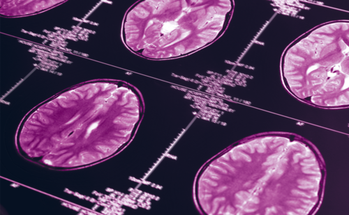
NTS has widespread projections, including direct or multiple synaptic projections to the parabrachial nucleus, vermis, inferior cerebellar hemispheres, raphe nuclei, periaquaductal gray, locus coeruleus, thalamus, hypothalamus, amygdala, nucleus accumbens, anterior insula, infralimbic cortex, and lateral prefrontal cortex, making delineation of the area mediating VNS effects difficult. NTS output has been shown to be important in modulating susceptibility to seizure in rats.12 Blocking function of the locus coeruleus attenuates the antiseizure effect of VNS in rodents.13 Similar brain areas show changes in c-fos activity in rodents receiving VNS, suggesting possible changes in gene regulation may underlie prolonged effects.14 Several neuroimaging studies have attempted to clarify where VNS effects are mediated. Serial positron emission tomography (PET) studies on 10 patients just prior to VNS placement and again within 20 hours of VNS activation were obtained at either subtherapeutic VNS settings (active controls) or therapeutic levels (treatment group) of stimulation. This study demonstrated increased cerebral blood flow in several brain areas including the right postcentral gyrus, bilateral thalami, bilateral hypothalami, medulla, bilateral insular cortices, bilateral inferior cerebellar hemispheres, bilateral amygdala, bilateral hippocampi, and bilateral posterior cingulate gyrus. In the treatment group, stimulation resulted in additional increased blood flow in the bilateral orbitofrontal gyri, the right entorhinal cortex, and right temporal pole.15 These patients were followed through the chronic stimulation phase, with repeat imaging performed three months after chronic intermittent stimulation began. Increased blood flow persisted in subcortical regions, including the right post-central gyrus, bilateral thalami, bilateral hypothalami, bilateral inferior cerebellar hemispheres, and parietal lobules, but was absent from other areas.16,17 Intriguingly, thalamic blood flow response to VNS stimulation in the acute phase predicted the likelihood of clinical response to chronic, intermittent VNS stimulation.16,17 Previous PET studies also had shown increased thalamic blood flow in response to VNS.18A possible role for thalamic involvement was supported by a recent functional magnetic resonance imaging (fMRI) study that also demonstrated increased activity in several areas of the brain, but was most robust in the bilateral thalami (left > right), and the bilateral insular cortices in epilepsy patients whose VNS was activated for the first time immediately prior to scanning.19 A second study demonstrated two patients in whom thalamic changes on fMRI correlated with the degree of seizure control.20 However, other fMRI studies have demonstrated no change in thalamic activity (for a review, see reference 21), and single photon emission computed tomography (SPECT) studies have shown decreased thalamic activity in response to VNS.22 In summary, contradictory observations make it difficult to draw definitive conclusions; however, thalamic involvement remains a likely candidate underlying VNS efficacy.
Vagus Nerve Stimulation Efficacy in Adults and Children
The first large, randomized, blinded study of VNS in humans (EO3) was preceded by two open pilot studies that had shown decreased seizure frequency with VNS.23,24 In this trial,114 patients at least 12 years old with medically intractable partial (simple, complex, secondary generalized) epilepsy were enrolled. Medically intractable epilepsy was defined as greater than six seizures per year. Patients were randomized to receive high stimulation (treatment group) or low stimulation (active control). There was a 12-week baseline period, a two-week recovery phase after implantation, then a 12-week parallel phase study to measure effects on seizure frequency. The high stimulation group had a statistically significant 24.5% reduction in seizure frequency, with 31% of patients achieving at least a 50% reduction in frequency (see Figure 1). The low stimulation group had a decrease in seizure frequency of 6.1%, with 13% of patients achieving greater than 50% reduction in seizures.25 An open extension of the same trial in which patients were maintained or changed over to high stimulation for 12 months included 100 patients and demonstrated a continued reduction in seizure frequency at 9 12 months of 31.9%.26
Subsequently, a similar multicenter, randomized, double blind trial in the US (EO5) demonstrated similar results.27 This trial included patients 12 65 years old with at least six complex partial or secondary generalized seizures per month in a 12 16 week baseline period. Patients were randomized into high-frequency (treatment group) or low-frequency (active control) stimulation. At three months, 196 patients were evaluated, and the highfrequency group had a statistically significant reduction of seizure frequency of 27.9% compared with 15.2% in the low-frequency group (see Figure 1). An open extension study re-evaluated patients at 12 months, and seizure frequency was reduced by 45%.28
In a compassionate-use, multicenter, prospective, open-label trial (EO4) of 24 patients with medically resistant generalized epilepsy aged 4 40 years,29 there was an overall reduction in seizure frequency of 46%, with 45.8% with greater than 50% reduction in seizure frequency, clearly demonstrating a benefit for generalized seizures, similar to other studies.30,31 Several other studies have confirmed the efficacy of VNS.7,32 35 Compiled long-term data from EO1 EO536 demonstrated that 23% of patients had greater than 50% reduction in seizure frequency at three months, and that this increased to 37% at one year, 43% at year two, and then plateaued, staying at 43% at year three, suggesting improved effect over time. This trend was also shown in a registry study in which patients had stable dosing of AED over 12 months, demonstrating that 45% of patients had greater than 50% reduction in seizures at three months, increasing to 58% at 12 months.37 Additionally, VNS may allow some patients to reduce the number or dose of AEDs taken,38,39 though with non-responders, 17.8% of patients had increased numbers of AEDs.39
Although the initial trials for VNS included patients as young as 12 years old, it was not until later that trials separated pediatric data or designed purely pediatric trials. Murphy et al.40 compiled the data on children from the two blinded, randomized trials25,27 (two children from EO3, 17 children from EO5) combined with 41 patients enrolled in a compassionate-use protocol that included patients with partial seizures, primary generalized seizures, or secondary generalized seizures, for a total of 60 patients. The mean age was 13.5 years, with the youngest recipient aged 3.5 years and 16 children under 12 years of age. Pooled data indicated a mean reduction in seizure frequency of 22% at three months, increasing to 42% at 18 months. No significant difference was seen in children less than 12 years old, and no single seizure type appeared more responsive to VNS therapy.40
The largest pediatric study to date was a retrospective, multicenter evaluation of 125 patients with medically intractable epilepsy.41 Follow-up data at three months was available in 95 patients and demonstrated an average reduction of seizure frequency of 36.1% with a median reduction of 51.5%. Furthermore, 28.4% of patients had a greater than 75% reduction in seizure frequency, and two patients became seizure-free. Data were available from 56 patients at six months, with reduction in seizure frequency of 44.7% and median reduction of 51%. A greater than 50% reduction was seen in 57% of patients, and 30% decreased seizure by greater than 75%. No significant difference was found in patients under 12 years of age. Other studies have shown similar results.42
Fifty children with Lennox-Gastaut syndrome (LGS) were evaluated in a multicenter retrospective study. One month following VNS there was a 42% reduction in seizure frequency, including 43% of patients with a greater than 50% reduction in seizure frequency. At six months, seizure reduction was 57.9%, with 58% of patients decreased by more than 50%, and 38% had a greater than 75% reduction in seizure frequency. At six months, drop attacks had decreased by 88%, atypical absence seizures by 81%, and at three months complex partial seizures by 23%.43 Prior studies that included LGS patients also showed clinical efficacy in children3,31,41,44,45 and adults.30 Recently, Majoie et al. evaluated 19 patients with LGS or LGS-like syndromes and found only 21% of patients having greater than 50% reduction in total seizures at 24 months.46 Vagus Nerve Stimulation Safety
VNS has proven to be safe and well tolerated with rare adverse events and generally mild side effects that are usually tolerable without cessation of therapy. Infection may complicate surgery. Most of these cases can be simply treated with antibiotics, though rarely device removal is necessary (reviewed in reference 47). Lead testing in the course of VNS placement has rarely been associated with transient asystole. Rhythm strips from three such patients suggested complete arteriovenous (AV) block occurring during lead testing.48 This is a rare event, estimated to occur in approximately 0.1% of patients.49 Some of these patients were successfully treated with VNS with no subsequent cardiac complications.48,50 Importantly, comparison of sudden unexplained death in epilepsy patients (SUDEP) rates in patients treated with VNS has been found to be no different from seizure patients without VNS.51
The most common side effects reported in clinical trials are hoarseness, voice change, or cough associated with stimulation. The rate of hoarseness or voice alteration in the first two randomized, blinded trials was 37.2 66.3% at three months, and coughing in 7.5 45.3% at three months.25,27 Importantly, there are no significant cognitive side effects. Compiled data from clinical trials EO1 EO5 looked at side effects at 12 36 months (see Figure 2). By 12 months, only 29% experienced hoarseness and 7.8% had cough, indicating possible attenuation of side effects with time.36Ben-Menachem looked at patients as far out as five years, and hoarseness was reported in 11 out of 64 patients.31 Other commonly reported side effects include dyspnea, paresthesias, and pain at surgical site.25,27,29,31,36 Similar side-effects have been found in pediatric populations.3,40 44,46
Conclusions
VNS has been shown to be efficacious and safe for treatment of medically intractable partial or generalized epilepsy in adults and children. Clinical improvement has been demonstrated within three months, and efficacy generally continues to rise until a plateau is reached in the second or third year. The mechanism by which VNS exerts its effect has not yet been clearly defined, but is being actively pursued on many different research fronts. Nearly 10 years after FDA approval, VNS has developed a proven track record for adjunctive therapy in patients with medically intractable epilepsy.


