Dystonia is a symptom that involves involuntary muscle contractions that are frequently sustained, but can be phasic. These contractions or spasms frequently result in abnormal posturing or repetitive movements in various parts of the body. Dystonia is often accompanied by tremor.
Dystonia is a symptom that involves involuntary muscle contractions that are frequently sustained, but can be phasic. These contractions or spasms frequently result in abnormal posturing or repetitive movements in various parts of the body. Dystonia is often accompanied by tremor.
Dystonia has multiple causes but may be categorised by age at onset or by the underlying aetiology as either primary (idiopathic), which may be sporadic or familial, or secondary, the latter often resulting from a neurodegenerative disorder.1 The most commonly inherited form of primary dystonia results from a defect in the DYT-1 gene.2 The specific syndromes of myoclonus-dystonia and dopa-responsive dystonia are classified under the term ‘dystonia plus syndromes’.
Dystonia can also be categorised anatomically according to the part(s) of the body affected by dystonia:
• focal dystonia – involves a single part of the body, for example spasmodic torticollis (cervical dystonia) or blepharospasm or writer’s cramp;
• segmental – involves two or more contiguous regions of the body, for example cervical dystonia or writer’s cramp;
• multifocal – involves two or more non-contiguous regions of the body, for example blepharospasm or dystonia in one foot; and
• generalised – involves both legs and at least one other part of the body.
Levodopa may produce considerable amelioration of symptoms in doparesponsive dystonia. Similarly, alcohol, used judiciously, can be very effective in suppressing myoclonus-dystonia. In general, however, dystonia responds poorly to oral medication, although high doses of anticholinergics or benzodiazepines may have beneficial effects in some cases. Botulinum toxin is the treatment of choice for focal dystonia and is widely used for managing patients with blepharospasm, vocal dystonia and spasmodic torticollis, as well as other focal or segmental dystonias.3,4 However, in some cases tolerance to treatment, mediated through antibodies to botulinum toxin type A or B, may eventually develop.3,4
In the past, cervical denervation procedures were utilised to manage patients with severe medically refractory spasmodic torticollis, but the long-term benefits appear to be modest.5,6 Before the advent of deep brain stimulation (DBS), thalamotomy had been deployed with some success. However, bilateral thalamotomies were associated with a significant adverse event profile, in particular speech disturbances.7 Subsequently, demonstrable efficacy of pallidotomy and globus pallidus internus (GPi) stimulation for dyskinesia, including dystonia, was shown in patients with advanced Parkinson’s disease. GPi stimulation was then introduced for the management of severe, medically refractory dystonia, and is now widely deployed.8–23
Globus Pallidus Stimulation – Patient Selection
The main determinant of whether a patient with dystonia is suitable for GPi stimulation is that the patient, following discussions with his or her multidisciplinary DBS team, accepts the risks involved in undergoing DBS. The main risk involved is a 1–2% rate of a symptomatic intracranial haemorrhage.24 In addition, there are relatively minor complications involved with DBS that include hardware failure or displacement, wound infections, wound pain, unsightly scars, epileptic seizures and reversible stimulation-related adverse effects.25
Minor revision surgery for replacement of the impulse generator (IPG) is typically required every one to three years because of the high battery consumption involved in treating patients with dystonia. Currently, the rechargeable IPG needs replacement at nine-year intervals. In addition, regular attendance at the outpatient department is required – typically two to six times per year – for stimulator checks.
In view of the risks involved with DBS, candidate patients should have severe dystonia that is associated with significant disability and/or social handicap and have an unsatisfactory response to oral medications or botulinum toxin therapy. There is some evidence to suggest that patients with a normal cerebral magnetic resonance scan have a better outcome. Age does not appear to be a factor in determining the response to GPi stimulation. However, the duration of dystonic symptoms may correlate inversely with the response to GPi stimulation.26
The type of dystonia is important in determining the benefit obtained with GPi stimulation. Improvement averages about 70% for primary generalised dystonia (on the Burke-Fahn-Marsden-Dystonia Rating Scale [BFMDRS]) and 50% for cervical dystonia (on the Toronto Western Spasmodic Torticollis Rating Scale [TWSTRS]).26 The average improvement reported in patients with secondary dystonia treated by pallidal stimulation averages just under 50%, although patients with tardive dystonia/dyskinesia or pantothenate-kinase-associated neurodegeneration may do a little better.26 Currently, it is not clear whether stimulation of the thalamus or internal pallidum is more effective for treating patients with writer’s cramp.
In every case, a multidisciplinary assessment of the patient by the DBS team, which typically consists of a neurologist, neurosurgeon, neuropsychologist and therapists, is good practice. It is important that the patient is not cognitively or psychiatrically vulnerable and has realistic expectations of the benefits obtainable with DBS surgery.
Globus Pallidus Stimulation – Surgical Technique
Prior to surgery, patients undergo a 3mm contiguous-slice T1 magnetic resonance imaging (MRI) scan under general anaesthesia. On the morning of surgery, the base ring of the Cosman-Roberts-Wells (CRW) frame is applied to the skull under general anaesthetic, taking care to keep it parallel to the orbito-meatal plane. A computed tomography (CT) scan is then performed with 1mm contiguous slice reconstruction (zero gantry tilt).
Prophylactic intravenous antibiotics (gentamicin and vancomycin) are given. The pre-operative MRI and CT scans are then fused (using Radionics Image Fusion) and the target co-ordinates for the posteroventral part of the GPi are taken. The Schaltenbrand-Wahren brain atlas is available with the Stereoplan software and is also used to facilitate targeting. The entire trajectories of the two DBS leads are planned on the workstation, with particular care being taken to avoid passing through any sulci on the brain surface and also to avoid crossing the ventricles. Furthermore, the trajectory is aimed at traversing the greater part of the posteroventral pallidum with the inferior margin just above the optic tracts.
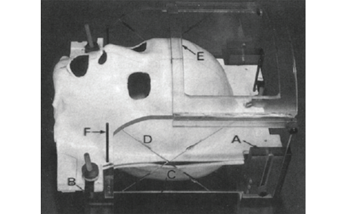
The patient is then positioned supine with the head fixed through the flat adaptor of the CRW frame onto the Mayfield clamp. The scalp is thoroughly prepared with aqueous and alcoholic chlorhexidine. The co-ordinates of one side are transferred to the CRW arc and checked against the phantom for accuracy. The arc is then fixed to the base ring after using the stereotactic frame sterile drape.
After infiltrating local anaesthetic, a curved frontal coronal scalp incision is made, guided by the proposed trajectory. The periosteum is reflected and the proposed site of entry is marked on the skull surface. A mini-plate is screwed in place 1.5cm away from the entry-point, leaving it loose with only one screw in place. A 2.7mm twist-drill craniostomy is then made through the arc with a hand-held drill. The dura is pierced in one motion with the craniostomy. A Radionics TCR electrode (2mm diameter) is then passed to target, measuring the impedance along the tip. Note is made of very low (ventricles) or very high (clot) readings. The Medtronic DBS lead (3387) is then passed to the target.
Although DBS surgery is usually performed for dystonia in an anaesthetised patient, as the dystonic movements preclude, in the main, performance of the operation under local anaesthesia, there is limited on-table feedback. Nevertheless, test stimulation is performed on-table and a clinical assessment is carried out by the neurologist to detect obvious pyramidal effects, such as increased tone. Local field potential monitoring through the DBS lead can also be performed at this stage, along with simultaneous electromyogram (EMG) monitoring to detect capsular responses. The electrode is then fixed in place under the mini-plate, taking care not to crush it against the skull.
The same procedure is repeated on the other side. The right-sided lead is tunnelled to the left and the right scalp wound is closed in two layers. The left side is closed in a single layer. The patient is then taken for another stereotactic CT scan. The position of the DBS leads is checked using image fusion. If satisfactory, the CRW frame is removed and the patient prepared for the second part of the procedure.
Implantation of the Impulse Generator
The patient is positioned supine, with the head turned to the right on the head ring and a jelly roll under the left shoulder. The position for insertion of the IPG is marked on the patient’s skin, having been agreed with the patient earlier. The IPG is generally sited in the left sub-clavicular area for right-handed patients, to allow easy access for self-telemetry using their own programmer.
The scalp, neck and subclavicular areas are prepared and draped. A subcutaneous left sub-clavicular pouch is fashioned. A small incision is made behind the ear along the path of the extension leads. The left scalp incision is then re-opened and the extension leads are passed from the scalp to the pouch. The proximal ends of the extension leads are connected to the DBS electrodes and covered using protective plastic boots. The distal ends are inserted into the IPG (Kinetra, Activa primary cell [PC] or, if rechargeable, the Activa rechargeable cell [RC]. The IPG is placed in the pouch and gentamicin instilled. The wounds are then closed in layers.
Alternative Targets for Deep Brain Stimulation in Dystonia
The use of structures other than the GPi as targets for DBS in patients with dystonic conditions has become infrequent. There are some recent reports on the effectiveness of thalamic (ventralis intermedius and ventralis oralis posterior) DBS in writer’s cramp.27 There are some reports that suggest that the subthalamic nucleus is an effective target for DBS in primary and tardive dystonia.28 It is argued that subthalamic nucleus DBS uses less power and thus prolongs battery life, has immediately beneficial effects (compared with the usual delay of about six months seen after GPi DBS) and is clinically as effective. However, the overwhelming evidence in the published literature on DBS for dystonia is based on the GPi as the target.
Programming Deep Brain Stimulation in Dystonic Patients
Post-operative programming of the implanted pulse generator following electrode implantation in GPi is more difficult in dystonia than other conditions (for example tremor or Parkinson’s disease). This is because the beneficial effects of GPi stimulation take time to appear. Typically, the benefit of GPi stimulation accrues over a period of about six months, but may take up to a year to optimise. In general, some pain relief occurs first, usually within days, and then the phasic components improve before the tonic components of the dystonia.29
As surgery is usually performed under general anaesthesia, limited data will be available from theatre to guide post-operative programming. Currently, the most posteroventral portion of GPi is considered to be the optimum site for stimulation in dystonia and contact closest to this area is usually the deepest.
In the Charing Cross Hospital unit, three days after electrode implantation, each contact is tested with monopolar stimulation set at 135Hz and at a pulse width of 90μs. The voltage is gradually turned up to 4.0V or until adverse effects occur. These may include:
• visual ‘phosphenes’ (flashes);
• capsular effects (pulling or cramp in the contralateral side of the face or limbs);
• a tight feeling in the mouth;
• dysarthria;
• non-specific giddiness; and
• gait disturbances.
The lowest contact on each electrode with which no adverse effects are experienced on stimulation, or the highest threshold to adverse effects is detected, is then chosen for stimulation.
The patients are subsequently monitored regularly as outpatients to assess progress. Increases in the voltage, pulse width or frequency, generally in that order, are considered if a suboptimal therapeutic effect occurs. Typical long-term stimulation parameters in patients with dystonia treated with GPi stimulation at the Charing Cross Hospital unit are shown in Table 1.
Results of Globus Pallidus Stimulation in Primary Generalised Dystonia
Uncontrolled Data
Early reports described improvements of up to 90% in the movement section of the BFMDRS, with the most benefit occurring in children with the DYT-1 deletion.11,30 In subsequent case series, benefit in the order of 40–70% was seen and DYT-1 status did not appear to influence the degree of improvement.10,17,23 Benefit may occur within hours of stimulation but is more often delayed, with progressive improvement seen over months (tending to plateau after about six months).31 The beneficial effects of pallidal DBS in primary generalised dystonia seem to be durable. Several studies include follow-up data at two years, and there are descriptions of small numbers of patients followed post-operatively for over five years.
Prospective, Controlled Data
Recent data from prospective, controlled trials provide more robust evidence for the benefit of pallidal DBS in primary generalised dystonia.19–22 The improvements demonstrated were less than those quoted in several of the uncontrolled studies and there was a significant degree of ‘response variability’, which was largely unexplained. A multicentre French study assessed bilateral pallidal DBS in 22 severely impaired patients with PGD.21,22 Controlled videotaped assessments were performed in a randomised, double-blind manner three months after surgery with the stimulators turned ‘off’ or ‘on’. Uncontrolled assessments followed at six, 12 and 36 months. At three months, there were significant improvements in BFMDRS motor and disablility scale scores compared with baseline, when the stimulators were activated. When ‘on’ and ‘off’ stimulator conditions were compared, BFMDRS motor scores were improved during stimulation but no significant change was detected in the total BFMDRS disability score. At one year, patients had improved by 54.6% in BFMDRS motor and 44% in BFMDRS disability scores compared with baseline. Motor improvement was maintained at three years (mean improvements in the BFMDRS motor and disability scores of 58 and 46%, respectively). Improvement in overall quality of life was noted at one year and maintained at three years. Cognition and mood were not adversely affected.32 There were five adverse events but no permanent neurological sequelae.
In a more recent study, 40 patients (24 with primary generalised dystonia, 16 with segmental dystonia) were randomised to GPi stimulation or sham treatment (electrodes implanted but stimulated at 0 volts) for three months.19 Thirty-six patients had blinded assessments after six months of active stimulation. After three months, BFMDRS motor scores were significantly improved by 39.3% (4.9% improvement in the sham-treated group). BFMDRS disability scores also significantly improved (37.5 versus 8.3%). Following six months of neurostimulation, patients had improved by 44.5% in BFMDRS motor and 41% in BFMDRS disability scores compared with baseline. No adverse cognitive or mood effects were detected. Improvements in quality of life were also demonstrated.20 There were no intracranial haemorrhages, but 22 adverse events occurred in 19 patients (18% were hardware-related complications and the remainder were generally mild, reversible stimulation-related complications).
Results of Globus Pallidus Stimulation in Primary Cervical Dystonia
GPi stimulation for cervical dystonia was first reported in 1999.16 Case reports and small case series, sometimes embedded in descriptions of heterogeneous groups of patients,14,23,32 indicated improvements of 50–65% in the severity of cervical dystonia. In a prospective, single-blind study of bilateral pallidal stimulation in 10 patients with severe, medication-resistant cervical dystonia,15 the TWSTRS severity score had improved by 43% at one year post-operatively. Disability and pain scores were also significantly improved and the total TWSTRS score improved by 59%. Complications were mild and reversible in four patients.
Results of Globus Pallidus Stimulation in the Secondary Dystonias, Dystonia-plus and Heredodegenerative Syndromes
Uncontrolled reports imply that, in general, secondary dystonias are less responsive to pallidal DBS than primary generalised dystonias (average improvement of approximately 49%).26 However, it is difficult to generalise in view of the striking diversity of causes in this group; dramatic improvements may be achieved in selected cases, particularly if cranial imaging is normal. Encouraging results have been reported in patients with panthotenate-kinase-associated neurodegeneration, with short-term improvement in severity of over 60%.26,33 Tardive dystonia may improve by 50–86% following pallidal DBS.26,34,35 In a few small case series of myoclonus dystonia, treatment with pallidal DBS resulted in average improvements in the order of 50%.26,36 Less benefit has been reported in miscellaneous cases of anoxic brain injury, Lesch-Nyhan syndrome, neuroacanthocytosis and GM1-type 3 gangliosidosis.
Adverse Effects
The operative risks are described earlier in this article. Bilateral GPi stimulation in dystonia is preferable to lesion-based surgery in terms of the associated reversibility, adaptability and reduced morbidity. However, hardware-related complications, including infections and electrode lead displacement (see Figure 1) or fracture, may occur more often in DBS for dystonia, where prominent axial movements cause added mechanical stress.37 The most common stimulation-related side effects include dysarthria, hypophonia and dysaesthesia; these are usually reversible.19,21 There are reports of a ‘rebound’ effect with the development of status dystonicus upon stopping stimulation.23 Although most studies indicate that bilateral GPi stimulation in dystonia does not have significant adverse effects on cognition or mood,32 careful monitoring is warranted.
Conclusion
Bilateral GPi is now an accepted treatment for carefully selected patients with severe forms of primary and secondary dystonia in whom the anticipated benefits are considered to be worth the risks associated with DBS surgery. There are some recent data to suggest that further exploratory work needs to be carried out to assess the relative merits of the subthalamic nucleus target in comparison with GPi. However, there may still be a role for thalamic DBS in some cases of focal hand dystonia. ■


