Neuroimaging opens the possibility of non-invasive investigation of structural and dynamic processes in the human brain in vivo. It is a helpful tool to explore the impact of structural and functional changes at the cortical and subcortical level in different neurological and psychiatric pathologies. Magnetic resonance imaging (MRI) with its various applications is one of the leading technologies in this field. MR neuroimaging may be used on the one hand for diagnostic procedures and on the other hand for analysis of structural and functional cortical networks. Conventional cortical (and cervical) MRI techniques are complemented by volumetric and morphometric analysis of 3D MRI data (e.g. voxel-based morphometry [VBM]), proton MR spectroscopy (MRS), diffusion tensor imaging (DTI) and functional imaging methods such as functional MRI (fMRI).
There have been extensive advances in computer-based and conventional neuroimaging in motor neuron diseases (MNDs) such as ALS; however, standard protocols for clinical diagnosis and biomarkers are still lacking and work is still in progress. Neuroimaging can make a substantial contribution to clinical evaluation and diagnostic findings. Structural and functional neuroimaging may provide an objective marker for monitoring disease progression and the modifying effect of potential treatments.
For the last decade, advanced acquisition and post-processing techniques have led to a considerable increase in the number of structural and functional studies in severely restricted patients such as those with ALS. This article will evaluate the use of neuroimaging in the clinical setting for ALS/MND.
Amyotrophic Lateral Sclerosis
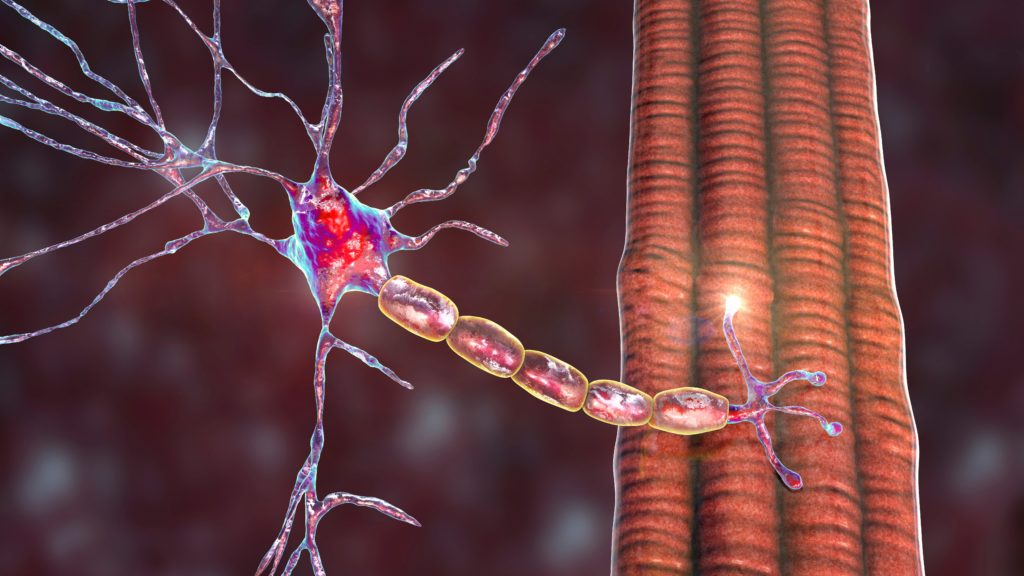
ALS is the most common adult-onset MND. It is characterised by a relentless progressive waste of motor neuron function leading to loss of mobility and verbal communication. The disease terminates in a so-called ‘locked-in’ state where cognitive functions are mostly preserved but afferent and efferent information flow is reduced to a minimum. Autonomous and sensory nervous system function is usually only mildly affected, and sphincter and ocular function is preserved for the the longest. Upper and lower motor neurons are affected in the course of the disease; however, onset of symptoms is usually restricted to either upper or lower motor neurons, i.e. the disease starts either with weakness of the arms or legs (‘spinal’ onset, mostly lateralised to one side) or with speaking and swallowing difficulties (‘bulbar’ onset). Patients die within three to five years, usually from breathing difficulties, unless appropriate measures such as mechanical ventilation and parenteral nutrition are taken.
About 10% of patients present a genetic background, and 10–20% of these genetically determined cases present a mutation in the SOD-1 enzyme. Over the last few years, evidence has emerged supporting the involvement of other genes that seem to be causally linked to ALS, such as dynactin, senataxin, TDP-43 and FUS. In most patients, the aetiology of the disease is unknown.
Structural Imaging in Amyotrophic Lateral Sclerosis
According to the revised El-Escorial criteria, evidence of upper and lower motor neuron development is needed for the diagnosis of ALS. However, there is no in vivo marker available for involvement of the upper motor neurons, and involvement of the lower motor neurons often masks upper motor neuron involvement. There is a pronounced delay between the onset of symptoms and diagnosis. Neuroimaging in ALS is mostly used to exclude other pathologies to increase the probability for the diagnosis of ALS rather than to provide definite evidence. Positive evidence may only be subtle and unspecific, although there are some signs in neuroimaging data with positive predictive value.1 Criteria based on these findings are used for clinical diagnosis.
One of the most widely used structural imaging techniques in the clinical diagnosis of ALS is MRI, which provides high-contrast images of anatomical structures.2 Voxel- and tensor-based morphometry MRI (VBM and TBM) enables quantitative measures of grey- and white-matter volume loss. Diffusion-weighted MRI (DTI) is mostly used for the detection of white-matter pathology. MRS complements MRI by providing information about the histological properties of tissue. Arterial spin labelling (ASL) MRI measures regional brain perfusion and complements the field of connectivity and therefore structural conditions in the brain. Other structural imaging methods such as X-ray radiography and computed tomography (CT), which are based on the fact that different tissues absorb different amount of X-rays depending on their density, have lost their importance in ALS diagnostics and are mostly used in emergency diagnostics.
MRI is based on the fact that atoms with uneven nucleus numbers have a magnetic moment. In living organisms this fact can best be exploited using hydrogen. A patient is placed inside a constant magnetic field and the nuclear spins of hydrogen atoms (for example) are aligned. Radiofrequency pulses (gradients) are used to alter the energetic states of the hydrogen atoms. When the gradients are turned off, the atoms revert to their original energetic state and the spare energy is radiated. Different types of tissue have different ratios of hydrogen and, accordingly, radiate different amounts of energy. T1- and T2-weighted MRI can be best used to assess anatomical structures of brain structure and the cervical spine. Artefacts of bone structures are not a problem, unlike in CT. MRI of the cervical spine is one of the most prominent and significant techniques to rule out, for example, lesions of the myelon in cervical myelopathy or polyradicular lesions. Vascular lesions such as those seen in cerebral microangiopathy in aged people can present signs that resemble ALS. However, great experience is needed to distinguish between artefacts and positive results. Furthermore, MRI has no predictive value for histological properties of tissue. Some further limitations concern the implementation of MRI. No metal is allowed inside the MRI scanner and therefore pacemakers and surgical clips are a contraindication. Claustrophobic patients have difficulties spending half an hour inside an MRI scanner. Open systems have been developed but they have lower field strength and lower resolution, leading to longer scanning times and an increase in the time needed to take measurements.
TBM and VBM can be used to determine volumes of tissues in MR images. Differences in the volume of grey and white matter in the brain can be analysed between groups (e.g. between patients and controls). Evidence of changes in various cortical and subcortical regions in patients with ALS has been provided using this technique.3–5
DTI and DTI-based fibre tracking (FT) are unique ways to learn more about human neuroanatomy, especially white matter, in vivo.6 In ALS, DTI has the potential to non-invasively visualise the involvement of the upper motor neurons. It has the greatest diagnostic potential in ALS and the number of DTI studies has increased markedly over the last few years. DTI is based on the concept of diffusion of molecules according to Brownian motion. Diffusion-weighted MRI (DWI) can be used to determine diffusion within tissue in the human body. Membranes, tracts and other constraints in human tissue reduce free (isotropic) diffusion of molecules. Molecular diffusion is reduced perpendicularly to those barriers and, accordingly, aligns along those barriers. This anisotropy can be measured with DTI. Fractional anisotropy (FA) is a parameter of the disruption of diffusion within tracts of the human body.7 DTI is highly sensitive to the loss of directed fibre tracts such as those in the pyramidal tract. Accordingly, it allows quantitative measurement of white matter loss of cortical and cervical motor neurons in ALS. The first evidence of reduced FA in ALS patients compared with healthy controls emerged as early as the 1990s.8 In regions-of-interest (ROI) analysis, there was evidence that FA reduction correlated with disease severity and clinical affection of the upper motor neuron. This was most prominent for ALS patients with bulbar onset. Further evidence for FA reduction was also given for other parts of the pyramidal tract. However, these ROI-based analyses were highly dependent on intra- and inter-rater variance of ROI placement. Semi-automated techniques have since been developed.9 Recent advances in DTI analysis have led to evidence of affection of the upper motor neurons in ALS before clinical onset of the disease, and therefore this technique might serve as an early clinical marker for the disease in motor and extramotor areas such as the frontal, temporal and occipital areas.10–12 Currently, there are high constraints in using DTI for detecting FA changes in the cervical myelon. The limited spatial resolution and vulnerability for movement artefacts of echo planar imaging (EPI) sequences, which are usually used for DTI measurements, necessitate thorough implementation of acquisition, planning and study design as well as high patient compliance.
There is evidence supporting MRS as a helpful technique for monitoring disease progression.13,14 Based on measurements of the nuclear properties of a substrate, metabolites can be detected in the brain; for example, cortical N-acetyl-aspartate (NAA) can be measured non-invasively using MRS. Reduced NAA levels in the cortical motor areas of ALS patients have been shown to correlate with the severity of the disease. Furthermore, cortical NAA has been associated with reduced survival time and might therefore have potential as a biomarker for ALS.15 Proton MRS has been suggested as a useful tool for reflecting the characteristic changes of metabolites in ALS.14 MRS has been applied clinically in medical treatment trials.16 ASL perfusion has similarly been suggested to be a useful tool for monitoring disease progression in ALS.17
Functional Imaging in Neuroscience
Neuroscience aims to explore the functional state of the brain as well as the capacity of the adult brain to functionally compensate for a progressive loss of neurons. Non-invasive functional neuroimaging techniques that can effectively map brain functions have now been available for almost 80 years. Since different techniques have different shortcomings, the development and implementation of new functional imaging techniques have been complementary over these years. Electroencephalography (EEG) is a technique for directly measuring the electrical activity of cortical neurons on the surface of the head. Measurements of the magnetic equivalents of EEG recordings (the electrical activity of cortical neurons induces magnetic fields) ushered in the magnetoencephalography (MEG) era. Since the mid-1970s, methods for measuring brain metabolism and therefore energy expenditure following neuronal activity have been established, such as positron-emission tomography (PET) and single-photon-emission CT (SPECT). These techniques record the dynamic distribution in the human brain of isotopes administered to a subject when assigned to a specific task. Since radioactive substances have to be administered to the patient, these techniques have limited application for clinical and scientific research. fMRI can be used to measure physiological changes not only in metabolism but also in, for example, blood flow following oxygen expenditure in the brain.18 fMRI has to a large degree displaced other functional neuroimaging procedures, such as PET and SPECT. A new approach to the assessment of cortical function in diseased brains is resting state (RS) MRI, which investigates synchronised fluctuation in functional networks. RS analysis can be used to assess disease-related neurofunctional alterations in widespread brain networks in ALS.19 With the help of functional neuroimaging, our understanding of the pathophysiology and plasticity of the human brain has increased substantially. Cortical and subcortical plasticity in the adult human brain compensates for structural and functional deficits following neurodegeneration, atrophy and trauma.20 In ALS, functional imaging has contributed substantially to our understanding of functional changes in the motor and extramotor systems during progressive motor neuron loss.21,22 In the future, fMRI may have the potential to complement the diagnostic procedures and help to determine the stage of functional loss in ALS patients.
The clinical potential of a non-invasive probe of brain function with the option of repeated measures over time (owing to the lack of radiation charge), in addition to the high feasibility of MRI scanners in many hospitals and research centres, has extended the application of fMRI in clinical science and contributed to an exponential increase in scientific publications on fMRI over the last decade.23
Functional Imaging in Clinical Settings
Study designs in clinical neuroimaging require that all paradigms are easily accomplishable by physically restricted patients, e.g. with ALS. Frequency and applied strength in motor tasks and required reaction time have to be adjusted accordingly. In an active paradigm (e.g. fMRI), a task is performed by a subject inside the MRI scanner (e.g. hand movement or viewing images). In a passive paradigm (e.g. resting state MRI) no special task is given and subjects are asked to lie still. Using that approach, baseline cortical activity is measured. The duration of a task has to take into account that subjects must lie supine inside the scanner for half an hour on average with minimum head movement (<1.5 mm within 5 minutes). The performance of a group of patients is usually compared with the performance of a healthy control group measured by the identical acquisition protocol. Sequences that are best suited for functional neuroimaging should be both fast and sensitive to the signals of interest such as echo-planar imaging (EPI) sequence in fMRI. Other sequences (e.g. in MRI fast low angle shot [FLASH]) might facilitate access to better imaging quality, including higher spatial resolution, but cause higher scanning times,24 which is not always favourable in clinical settings.
However, as functional neuroimaging signals e.g. in fMRI, derive from the interaction of multiple parameters (e.g. perfusion, metabolic turnover of neurons, density of venous vasculature of tissue, medication etc.), have mostly small amplitudes (e.g. 1–5% in BOLD) and may vary between brain areas and individuals as well as experimental and clinical settings, quantitative analysis in absolute terms is precluded; identification of functionally specialised cortical areas is challenging.25,26
Functional Imaging in Amyotrophic Lateral Sclerosis
Functional neuroimaging supports the notion of altered cortical network functioning in motor and extramotor areas in ALS patients. In fMRI studies, ALS patients present higher volumes of activated brain areas in motor tasks compared with healthy controls, thus providing evidence for functional reorganisation and cortical plasticity in fMRI studies.19,21,27–30 It has been proposed that these changes may represent cortical plasticity, as new synapses and pathways are developed to compensate for the selective loss of pyramidal cells in the motor cortex.31 Whether the changed pattern of activity in other motor functional areas and higher cognitive areas during motor tasks represents the recruitment of redundant parallel motor system pathways or whether they map functional compensation or reorganisation is a matter of speculation. The additional recruitment of different cortical areas might be a (futile) way to compensate for ALS-associated progressive functional loss. More research needs to be performed on how well ALS patients with advanced stages of the disease retain the potential for compensatory activity and how training of, for example, movement imagery might influence the ‘compensatory’ process. fMRI seems to be an appropriate way to gain more knowledge on this issue.
Functional Imaging of Extramotor Paradigms in Motor Neuron Disease
The multisystemic character of ALS has been supported by findings of functional imaging studies for cognitive, socioemotional and sensory pathways. About 2–5% of ALS patients present with an ALS/dementia complex, but there is increasing evidence that patients with classic ALS without obvious clinical evidence of cognitive deficits may have subtle changes in frontal cortical function as well.32,33 Cognitive impairment has been reported as more pronounced in ALS patients with a bulbar onset compared with patients with spinal onset.34–37
Longitudinal investigation of ALS patients revealed that cognitive dysfunction in ALS occurred early in the disease course and that the cognitive deficits may not progress in synchrony with motor decline, but may occur distinctly more slowly.37 Functional neuroimaging has supported the clinical findings of frontal cortical involvement not only in patients with an ALS/dementia complex but also in patients with ALS and subclinical cognitive impairment: hypoperfusion in the frontal cortex in ALS with or without cognitive deficits measured with PET32 and fMRI38 and association of reduced frontal executive function and reduced activity in fronto-parietal areas measured with PET.33,39 There is no evidence of additional recruitment in other areas to compensate for the functional loss in the frontal cortex. Cognitive functions of frontal areas do not exhibit redundancy (in contrast to motor pathways, for example); therefore, compensation might not be possible. Further longitudinal fMRI studies of different cognitive functions in ALS might improve our understanding of subclinical cognitive deficits in ALS.
Further differences in cortical pattern activation in fMRI have been observed in ALS patients during processing of socio-emotional and sensory stimuli.40,41 Differences in functional processing of sensory stimuli supported earlier findings of EEG studies42 and were supported by findings of structural changes measured by DTI.41 Future studies will highlight other changes in non-motor pathways in ALS with no obvious clinical equivalent. Neuroimaging can provide keys to the understanding of the overall pathological changes in the course of ALS.
Current Status
Neuroimaging is a clinical tool with a great deal of potential, although its clinical application of functional neuroimaging with respect to ALS remains speculative. To summarise, non-invasive structural and functional brain imaging substantially adds to our understanding of the pathophysiology of ALS. Structural neuroimaging is currently able to support the clinical diagnosis of ALS in routine clinical practice. However, work is in progress to improve the feasibility and quality and therefore the predictive value of this technique for ALS diagnosis and prognosis. There is increasing evidence that functional changes beyond the primary motor network exist in ALS, i.e. in frontal areas. The functional involvement of visual, auditory and somatosensory cortical areas seems to be associated with ALS, although anatomical and clinical evidence of dysfunction of sensory processing is limited. Whether these functional changes will be of diagnostic relevance in the future or whether they are instead an epiphenomenon evident in certain patients, in which case occurrence may depend on selection bias in studies, is unknown. Further research, for example using functional imaging, is necessary.
The clinical implications of structural neuroimaging are undoubted, but in the case of functional studies the implications remain controversial. The lack of sufficient standardisation hampers the comparability of studies. General guidelines for neuroimaging studies in the clinical setting would be helpful. Functional studies may define the heterogeneity of cognitive/extramotor changes in ALS and related conditions. However, the diagnostic relevance of imaging results may be limited by individual anatomical and functional differences, and this in turn may limit the use of functional imaging as a diagnostic marker. A further limitation to the clinical application of fMRI is the cost of the procedure to the patient and to the overall health service budget. A thorough cost–benefit analysis is a prerequisite for any clinical application.
Future Perspectives
The pharmaceutical industry and clinical researchers are still looking for an objective biomarker of disease progression or functional improvement to determine the efficacy of disease-modifying agents.23 Structural and spectroscopic non-invasive imaging techniques have been discussed as promising candidates.43,44 In the future, functional imaging technigues such as fMRI might add to this field as potential in vivo progress markers.45 In the future, it can be expected that more studies will combine structural and functional neuroimaging in the same patient cohort. The two techniques complement one another. Degradation of, for example, ascending and descending pathways of white-matter fibres (measured by DTI) may cause disruption of cerebral function (measured by fMRI). Cortical volume loss (measured by VBM) may cause changes in cortical network functioning (again measured by fMRI). Combined non-invasive neuroimaging techniques may be useful tools to assess prognosis and study rehabilitation strategies for ALS patients. However, the combination of structural and functional imaging techniques requires careful interpretation of findings. Co-existence of changes on a structural and functional level does not automatically imply causality. Lack of evidence for functional or structural disruption does not imply that findings on the other level are not trustworthy and should be regarded as false-positives. Acquisition and analysis methods strongly influence results and outcomes. Overall, neuroimaging may in the future extend its use as a tool in clinical diagnosis and as a biomarker of disease progression and efficacy of therapeutic treatment. The matter of preservation of cognitive functions in the course of ALS is a key issue in the discussion of life-sustaining treatments in ALS patients. An improved understanding of structural and functional changes in ALS is a prerequisite for providing adequate treatment to ALS patients at any stage of the disease, and neuroimaging may help us to achieve this objective. ■