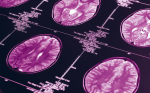
The gloomy prognosis of high-grade gliomas is well known, especially for glioblastoma multiforme (GBM). Until recently, surgery followed by irradiation was the standard treatment, with a median survival of around 11 months. Since the results of the European Organization for Research and Treatment of Cancer (EORTC) trial with adjuvant and concomitant temozolomide, which showed that median survival can be improved a little – from 12.1 to 14.6 months, with a two-year survival percentage of >25%1 – this chemotherapeutic drug has been added to the standard regime for patients who are generally in good condition. Although this improvement is significant, it represents only a small step forwards in the treatment of patients with GBM. Therefore, a lot of research is still needed.
Many trials have been published or are under way concerning a variety of new approaches: the selection of patients for chemotherapy based on methyl guanine methyl transferase (MGMT) status, convection-enhanced delivery of ‘old’ and new drugs, macrotoxins, antiangiogenic drugs, pathway inhibitors and immunotherapy, etc. In most studies, little attention has been paid to the role of surgical resection. Even when a sub-analysis of the role of biopsy versus resection is performed, or when upfront stratification is carried out concerning the radicality of surgical removal, the definition of that radicality is poorly defined, seldom quantified and often based on, and consequently biased by, the surgeon’s impression.
However, there are a few studies in which the size of the resection has been measured carefully, and in which an analysis has been made of the effect of resection (or residual tumour) on overall survival (OS), progression-free survival (PFS) and quality of life (QOL). The results all point towards a positive – and in some studies significant – effect on these three factors.2,3 Furthermore, after sub-analyses on some of the recent drug studies (especially the Stupp study with temozolomide), the conclusion may be made that even when ‘radical resection’ per se does not add too much to survival, the effects of adjuvant therapy, e.g. the chemo-radiation regime, seem to be positively influenced by resection.4 This forms the rationale for attempts to improve radicality in neurosurgical tumour resection.
Radical Resection and Fluorescent Guidance
Surgical approaches aiming at ‘maximal’ or ‘radical’ resection include neuronavigation (with definite limitations due to shifts) and intra-operative magnetic resonance imaging (MRI), computed tomography (CT) or ultrasound, in addition to the experience of surgeons with direct visualisation and tissue ‘feeling’ techniques. Such procedures, though effective, are expensive and also have limitations regarding interpretation.
A promising but simpler method to reach more radical resections seems to be the use of a vital staining of tumour tissue, which can be used in the operating theatre. A group of German neurosurgeons, under the guidance of Stummer and Tonn, have proved over the last few years that a prodrug to protoporphyrin IX, 5-amino levulinic acid (5-ALA), works in this way.5 It is a naturally occurring compound in the human body, but when given in a large amount it is accumulated preferentially in the skin and in certain malignant tumours, especially high-grade gliomas. When cells stained by synthesised protoporphyrin IX are illuminated with a special light (blue, wavelength 375–440nm), they show fluorescence with a red/pinkish light at a wavelength of 635nm, which makes them easily recognisable. The precursor 5-ALA is given to patients by the oral route a few hours before surgery.
In a multicentre, randomised clinical trial comparing patients operated on with and without 5-ALA, Stummer et al.5 were able to find a positive effect of the use of 5-ALA on the radicality of resection: 66 versus 36% in the non-ALA cases showed an absence of contrast-enhancing tumour following post-operative MRI. In a historical series, so-called radicality is reported at between 13 and 20%. PFS at six months was also significantly higher in the 5-ALA group (41 versus 21%). However, OS was not significantly different between both groups.
Recently, the same authors published a further analysis that demonstrated that their positive results were realistic, and that after correction for bias due to certain inclusion criteria the significance of the 5-ALA effect was still present.6 The most recent analysis of the German data shows that 5-ALA-based surgery did add to morbidity, but in their experience this morbidity was only temporary without permanent inadvertent sequelae (Stummer, personal communication).
Our Experience
From June 2006 to January 2008, we operated on 40 patients with a pre-operatively presumed high-grade glioma with the use of 5-ALA. These patients were also selected based on a pre-operatively presumed chance of complete resection of the contrast-enhancing parts of the tumour.
A histopathological investigation revealed 38 high-grade gliomas, one nocardia-abcess and one (non-fluorescent) metastasis. Interestingly, one of these was a combined grade II and grade III glioma, in which only the grade III part was fluorescent. In 10 patients new neurological morbidity occurred: aphasia, paresis, apraxia, drowsiness and worsening of epilepsy. This was temporary in nine, but permanent in one. There was one post-operative epidural haematoma, and deep venous thrombosis in one case. There was no procedure-related mortality.
Currently, a total of 24 patients are available for analysis on six-month PFS and on radicality of resection. PFS at six months yielded six cases (25%), which is considerably lower than in the German series (41%); however, due to the small number of cases this is not significant.
Radicality of resection may be a question of definition. In the first 18 cases we reached 12 resections >95% (66%), which was a cause for optimism because these figures compared favourably with the German data (66%). However, we became increasingly aware of difficulties in the interpretation of the post-operative scans.
After thorough inter-observer studies with four experienced surgeons and radiologists, we reviewed many of the scans and could definitively decide on 21 of the 24 patients with longer follow-up than six months. The resection percentage now ranged from 74.3 to 99.2%. An analysis of the resection percentage versus PFS was performed, with two cut-off points: 90 and 95% resection, respectively (see Table 1).
The findings show there was no relation in this (small) series of radicality and PFS at six months. Moreover, in our patients no relation was found between the percentage of resection and the post-operative Karnofski Performance Score or complication rate. Further analysis with more patients reaching six months and over is needed. Thus far, the use of 5-ALA has alerted us to the possibility of more radicality in our resections, and apparently without adding more morbidity. Whether this will result in significantly better outcomes has yet to be proved.
During the German study, it became clear that the use of such a fluorescent drug may harbour pitfalls and dangers. Therefore, a series of training courses was organised for colleagues from interested centres in other countries in Europe, and it is planned that such training courses will be continued and include more countries.
Basic problems to acknowledge, discuss and experience during such a course are:
• the pitfalls of pre-operative interpretation of MRI scans;
• logistics in timing of drug delivery and actual surgical tumour removal;
• availability of the right light source (originally featured only on the Zeiss NC4 microscopes [no longer available] and the Pentero™, which is now available);
• microscope settings;
• handling the combination of light sources;
• interpretation of normal versus abnormal and evident tumour tissue;
• awareness of changing anatomical relationships with the progression of tumour removal and subsequent adaptation of surgical techniques; and
• the risk of doing too much, adding to morbidity instead of improving the patient.
Conclusion
Following the introduction of 5-ALA into the clinic, and after the first randomised, controlled trial in Germany, it has become clear that this fluorescent drug – now on the market as Gliolan® – can be safely used in the hands of experienced surgeons. In turn, ‘radical’ resection – which will never be ‘total’ or ‘totally safe’ – seems to add to better survival, and may make the difference for the possible success of adjuvant treatment. In a practical clinical situation, the lessons we learned from our own experience with the drug are:
• more radicality of surgery can be reached;
• training in the effective and sensible use of 5-ALA is necessary and helpful;
• interpretation of post-operative images is still difficult and debatable;
• eloquent areas should still be respected, even when showing fluorescence; and
• not every presumed GBM on the scan is a GBM.
In due time, the word ‘drug’ may be avoided and become replaced by ‘pharmacological tool’, since it is an adjuvant substance based on naturally occurring molecules that helps the surgeon in visualising where and how far to go. In the future, it may still be considered a drug when used in the setting of photodynamic therapy (a combination of 5-ALA-derived protoporphyrin and laser-light irradiation). Studies are under way to explore the possibility of its use for deep-seated high-grade brain tumours.7 ■