In spite of their lack of propensity for hematogenous dissemination, the prognosis for adults afflicted with gliomas has not substantially improved, even with advances in neurosurgery, neuro-oncology, and radiation oncology.1,2 Although microsurgical resection remains the initial treatment of choice for most gliomas, these tumors may grow in eloquent areas, making gross total resection impossible. They are on occasion capable of local or distant recurrence, providing a challenging dilemma for the treatment team.3 First conceived by Lars Leksell in 1951, stereotactic radiosurgery (SRS) is a neurosurgical modality that combines stereotactic technique with highly focused high-energy radiation treatments, making it possible to deliver large doses of radiation to an extremely small target.4,5 By keeping the individual radiation highly focused, the normal brain parenchyma is protected while still allowing a large dose of radiation to be delivered to the desired target. In some cases, glioma tumors are well-suited for radiosurgery, as recurrent or residual tumors may be small and well circumscribed, thereby permitting easy targeting for radiosurgery. This article reviews the effectiveness of radiosurgery in the treatment of gliomas.
Radiobiology of Gliomas and Utility in Stereotactic Radiosurgery
Malignant tumors usually contain a proportion of hypoxic cells, which are often resistant to damage by X-rays or gamma-rays.6 In assessing survival for these cells, there appears to be a point where the accumulation of sublethal insults at low doses has a cumulative effect. This may be the result of DNA being a target for cellular damage by ionizing radiation, and the resultant double-stranded breaks causing cell-cycle arrest and cell death.
Typically, mammalian tumor cells have a characteristic shape after a single dose of irradiation (see Figure 1). During this period, there is a change in cell viability (often as high as 2.5–3 times) as aerated cells are non-viable and the response to radiation is dominated by hypoxic cells.
Radiosurgical treatment for malignant gliomas takes advantage of the natural difference in susceptibility of pathological and normal tissue. The relative radioresistance of normal brain tissue relates to its low mitotic activity and ability for cellular repair. The gamma knife curtails injury to normal brain tissue by producing a rapid radiation fall-off and minimizing radiation to adjacent normal structures. The rate at which a total dose is applied is also important. A higher dose rate (the same total dose applied over a shorter period of time or a larger dose in an equivalent amount of time) increases the lethality of a dose due to greater interference with intrinsic cellular repair mechanisms during irradiation. The significance of this effect is seen most clearly at a threshold dose rate of 1Gy/min.7 The normal tissue surrounding a target not only receives a markedly lower dose but also receives it in a lower dose rate.
Radiosurgery for Low-grade Gliomas
Low-grade gliomas consist primarily of the World Health Organization grade I–II astrocytomas and oligodendrogliomas, as well as mixed tumors, which have histological properties of both. Each individual subtype of tumor represents a distinct histological subtype of glial neoplasm with differences in prognosis.8,9 For low-grade glial neoplasms, adverse location, tumor recurrence, or progression despite surgery require the use of other treatment modalities.10
The optimal treatment for low-grade gliomas remains controversial in the absence of prospective randomized trials. Most patients with fibrillary low-grade astrocytomas eventually die and only approximately 15% of patients have long-term survival.9 Post-operative external-beam radiotherapy appears to prolong survival.11 In a randomized study of 203 consecutive adult patients with non-anaplastic, non-pilocytic astrocytomas, 83 patients (41%) had died at a follow-up six years after external-beam irradiation compared with 55% of non-treated patients.8 Other studies of radiotherapy for low-grade gliomas also show an improved survival.9–11 Post-radiotherapy recurrence and progression, however, are common.8
The role of SRS for low-grade gliomas has remained incompletely defined. Unlike implanted seeds or wafers,13 SRS allows for non-invasive administration of focal radiation. The majority of studies on radiosurgery for low-grade gliomas are restricted by small patient numbers and limited follow-up. Indications of early results, however, are comparable to those following external-beam radiotherapy or 125I seed implantation. In a study of 10 patients with low-grade gliomas who received radiosurgery, one patient died 66 months after treatment and nine were alive 22–82 months after treatment.14 Barcia et al. studied 16 patients with low-grade gliomas, six of whom received conventional external fractionated radiotherapy, six of whom received fractionated stereotactic radiotherapy, and four of whom received no prior radiation.15 In that study, tumors disappeared in eight cases and significantly decreased in size or ceased growth in five cases. Unfortunately, survival was not reported in this study. Heppner et al. conducted a retrospective review of 49 patients with low-grade gliomas, with 63-month median follow-up, and noted a median clinical progression-free survival time of 44 months and median radiological progression-free survival of 37 months. Complete radiological remission was seen in 14 patients (29%), and mortality due to tumor progression occurred in seven patients (14%) (see Figure 2).16
These studies, along with others (see Table 1),17,18 support the role of SRS as an effective adjuvant to surgery in the treatment of low-grade gliomas. Some have postulated that in situations where tumors are found in eloquent brain areas, where the strength of evidence is not enough to risk major neurological complications during surgery, a maximal, safe resection, with adjuvant gamma knife surgery to treat the residual tumor, could provide satisfactory disease control in patients with low-grade gliomas. Following the results of the European Organization for Research and Treatment of Cancer (EORTC) study evaluating the effects of radiotherapy in low-grade glioma patients, standard fractionated radiotherapy can additionally be considered to delay time to progression in high-risk patients.19
Radiosurgery for High-grade Gliomas and Glioblastoma Multiforme
High-grade Gliomas
The role of radiosurgery in the initial treatment of malignant gliomas continues to be a subject of debate, as the use of a highly focal treatment for a diffuse disease does not seem intuitively pleasing. Nevertheless, given the shortcomings of all known treatments for malignant gliomas, the goal of radiosurgical treatment in high-grade gliomas involves striking a balance between increasing the length of survival and maintaining an acceptable quality of life for the patient.
Surgical resection followed by external-beam radiation therapy (XRT) is considered to be the standard management for glioblastoma multiforme (GBM).20,21 Radiosurgery has been evaluated as an adjuvant treatment modality, and there has been considerable debate regarding its efficacy. Gamma knife surgery has not been evaluated as a sole treatment for GBM; all studies address the role of gamma knife surgery as an adjuvant therapy after initial resection or at the time of tumor recurrence.
The addition of radiosurgery to conventional treatment (surgery and external-beam radiotherapy) in the initial management of malignant gliomas appears to result in only a modest improvement in survival compared to that in historical reports. For 41 patients with malignant gliomas enrolled in a prospective study, radiosurgical doses of 1,200cGy after conventional radiotherapy resulted in a 76% survival with a median follow-up of 19 months.1 Gannett et al. described a planned SRS boost as part of the primary management of patients with malignant gliomas.22 In that study the one- and two-year diseasespecific survival rates measured from the date of diagnosis were 57 and 25%, respectively.22 Other studies show similar results. For 11 patients receiving planned external-beam radiotherapy and linear accelerator (linac) radiosurgery, Buatti et al. found the median survival to be 17 months, with all patients having progression of disease within one year of radiosurgery.23
Radiosurgery has also been explored for the treatment of recurrent malignant gliomas. In one study of 35 patients treated for recurrence of malignant gliomas, survival after radiosurgery was eight months (21 months after initial surgery).24 For 22 patients with recurrent malignant gliomas receiving fractionated stereotactic radiotherapy (30–50Gy in six to 10 fractions), the median survival after radiotherapy was similar, at 9.8 months.25 Finally, in a study of 86 patients having radiosurgery for recurrent malignant gliomas, the median survival was 10.2 months, and the survival after 12 and 24 months was 45 and 19%, respectively.26
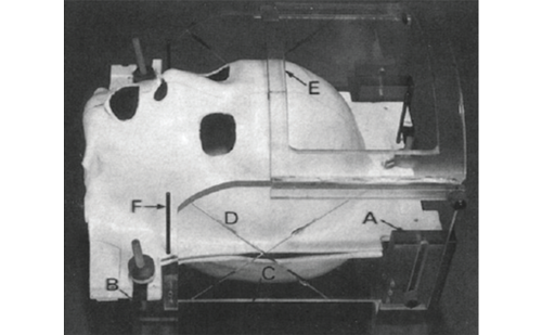
Radiosurgical modalities other than the gamma knife (see Figure 3) may also be potentially useful and beneficial for the treatment of highgrade gliomas (see Table 1). The utility of linac-based radiosurgery has also been reported.27–29 Souhami and colleagues did not distinguish between patients treated with linac-based radiosurgery and those treated with gamma knife surgery, whereas Shireve and colleagues exclusively used linac radiosurgery. They report that linac radiosurgery may provide a survival benefit to selected patients with GBM, identifying two important prognostic factors: age and Radiation Therapy Oncology Group (RTOG) class. While patients below 40 years of age had a median survival of 49 months, patients over 40 years of age had a median survival of 18.2 months (p<0.001). Moreover, patients with RTOG class III GBM had a significantly longer median survival than those with RTOG class IV and V: 29.5 versus 19.2 and 18.2 months, respectively.27 Other than the patients included in Souhami and colleagues’ report, further significant studies of linac-based radiosurgery have not been reported.
Hypofractionated radiotherapy using linac technology (intensity-modulated radiotherapy [IMRT]), however, has been investigated. IMRT is designed to deliver a relatively high dose of radiation to targeted lesions while sparing adjacent normal tissue, but in a fractionated manner.30–32 Voynov and colleagues found a median survival of 10 months when IMRT was used at the time of recurrence in patients with high-grade gliomas.32
For high-grade glioma patients, IMRT has also been investigated as an alternative to upfront standard radiation therapy, but with limited success. Floyd and colleagues reported no improvement in time to progression or survival when IMRT was used in place of standard radiation therapy. Instead, they found an increased rate of need for surgery due to necrosis.30 Narayana and colleagues reported similar findings in a series of patients who were treated with IMRT in place of XRT.31 The authors suggest that although IMRT does not improve overall survival, it may be advantageous in some scenarios due to the decreased time required for administration of radiation.30,31
Gliomblastoma Multiforme
In spite of advances in medical and surgical treatment of gliomas, the prognosis for most patients with GBM has hardly changed in the last decade. The vast majority of patients succumb to disease within one year of diagnosis. Studies involving the role of gamma knife surgery in the setting of GBM have yielded mixed results (see Table 2).20,28,33–35
Souhami and colleagues, reporting on behalf of the RTOG, were the first to conduct a multicenter randomized trial looking at the addition of SRS, including both gamma knife surgery and linac-based radiosurgical techniques, to standard XRT for the treatment of GBM. SRS was used as part of the initial management of GBM in these patients, as opposed to being used as a salvage therapy. They randomly assigned 203 patients with supratentorial GBM to receive either post-operative SRS (prescription dose 15–24Gy) followed by XRT and carmustine (BiCNU®), or XRT and carmustine without the preceding SRS. Among the patients who were eligible to enroll in the trial, they observed no difference between the two groups with regard to the primary end-point of survival: the SRS group had a mean survival time of 13.5 months whereas the control group had a mean survival of 13.6 months.28 Although Souhami and colleagues did not find a significant effect of adding SRS to the initial management of GBM, a role for gamma knife surgery and other stereotatic radiosurgical techniques in the treatment of GBM should not yet be excluded, especially in light of a number of individual institution reviews suggesting encouraging results.1,20,33,34 Different treatment regimens, e.g. post-XRT boost or at the time of recurrence, and SRS for patients with gross total resection (who were excluded from Souhami and colleagues’ study) may prove beneficial. The rationale for such investigations is provided by numerous institutional reviews suggesting a life-prolonging effect of gamma knife surgery in the management of GBM.
Nwokedi and colleagues performed a retrospective analysis of patients treated at the University of Maryland over six years. Due to a shift in their institutional treatment philosophy, the authors changed their practice of administering gamma knife surgery at the time of tumor recurrence to giving a scheduled gamma knife surgery boost within four weeks of the completion of XRT. This change in practice allowed the authors to compare the two treatment paradigms using overall survival as a measurement of efficacy. They observed a nearly two-fold increase in mean survival time (25 versus 13 months) in the group that received gamma knife as a planned boost within four weeks of XRT.20 This treatment paradigm differs from that of Souhami and colleagues, who administered SRS one week prior to the initiation of XRT.
Other studies have further supported the role of radiosurgery as adjuvant therapy in GBM. Kondziolka and colleagues compared their series of GBM patients treated with gamma knife surgery to historical controls. Again, in contrast to the study by Souhami and colleagues, in this series patients generally received gamma knife surgery five to eight months after initial diagnosis (after other therapies had been completed, including XRT and chemotherapy) or at the time of recurrence. Patients who received gamma knife surgery (mean prescription dose of 15.2Gy) as part of the initial treatment regimen had an increased median survival of 20 months compared with 11.2 months in the control group. They also reported a median survival of 30 months after gamma knife surgery for those patients who received this treatment at the time of tumor progression.34
Loeffler and colleagues treated patients with radiosurgery (prescription dose ranging from 10 to 20Gy) two to four weeks after XRT, similar to that reported by Nwokedi and colleagues, and likewise observed a median overall survival of 26 months for those patients.1 Reports suggest that SRS particularly benefits high-grade glioma patients with a Karnovsky performance status of at least 90 and those patients who received adjuvant chemotherapy.33
At the University of Virginia, our unpublished experience of 56 patients appears to support the use of gamma knife surgery in GBM, providing a small survival advantage. Although the results of these institutional reviews present encouraging data, the patients receiving gamma knife surgery in each series usually represent a highly selective group; in none of the series were all of the patients at an institution treated with gamma knife surgery. Therefore, one must take caution in interpreting each institution’s results and realize that such results cannot necessarily be generalized.
From a theoretical standpoint, it is difficult to understand how a highly invasive tumor such as a GBM can be treated effectively with such a focused treatment as gamma knife surgery. However, it can be used to treat the largest concentration of the residual tumor or the resection cavity based upon the neuroimaging studies. It seems clear that no single treatment modality in the neuro-oncology armory is a magic bullet for such tumors and, as such, this multimodality approach (i.e. SRS, XRT, cytoreductive surgery, and chemotherapy) to GBMs may be prudent.
Conclusions
For patients with low- and high-grade gliomas, SRS offers a precise, local administration of radiation that may result in both tumor control and prolongation of survival. Further assessment of SRS for the treatment of gliomas requires prospective, randomized clinical evaluation. ■


