Chronic inflammatory demyelinating polyneuropathy (CIDP) denotes a spectrum of acquired, chronically progressive or recurrent, immunemediated disorders of the peripheral nervous system with variable pathology and pathogenesis.1 The estimated prevalence may be up to nine per 100,000 population.2,3 A universally accepted definition of disease does not exist. A variety of clinical and investigational criteria have been proposed and applied in attempts to include the different presentations of CIDP.4,5 Perhaps the future development of biological markers will help reliably identify patients with CIDP.4
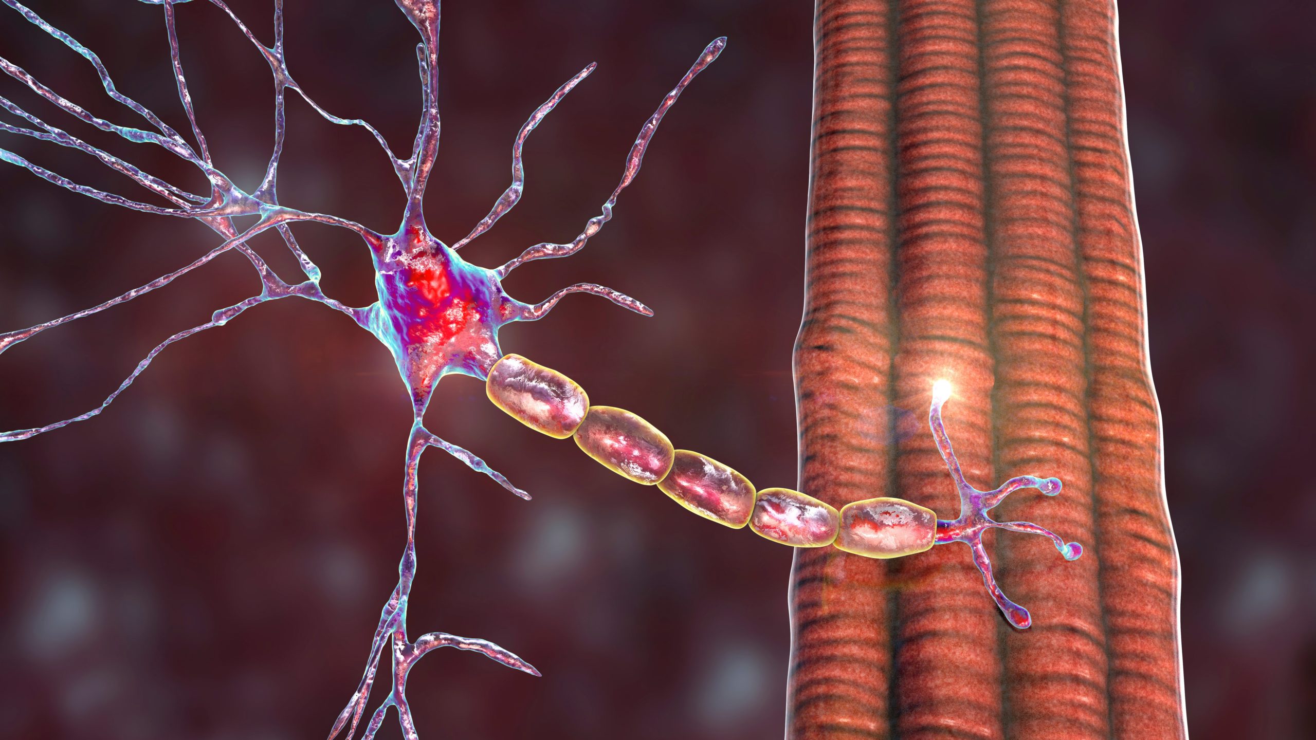
The pathogenesis of CIDP is not fully understood.1,6,7 Cell-mediated and/or humoral immune mechanisms are involved in an attack against unidentified target antigen(s) of the myelin sheath and/or Schwann cells, at times leading also to secondary axonal injury.
Randomised controlled trials (class I evidence) showed that most (60–80 %) patients respond at least to a degree to treatment with intravenous immunoglobulin (IVIG),8–11 plasmapheresis,12,13 or corticosteroids.14 Yet, there exists insufficient (class IV) evidence (from open label case series, randomised and quasi-randomised trials) to assess the benefits of conventional immunosuppressive agents (azathioprine, cyclosporine, methotrexate, cyclophosphamide or mycophenolate mofetil) in patients with CIDP.15,16 Most patients require long-term maintenance treatment.17,18 Due to the unpredictable efficacy, financial burden and/or toxicity of ‘broad-spectrum’ immunomodulatory/-suppressive drugs, a need exists to develop effective and safe therapeutic strategies.19,20 Research is hindered by the current lack of consensus on appropriate outcome measures and definition of treatment failure, as well as the tendency to study potentially effective therapy exclusively on refractive CIDP patients.21,22 In order to realise the full potential of any new drug/agent, it may be advisable to identify and exclude from future drug trials any patients with predictably unresponsive (chronically stable and inactive) disease.20 The future development and application of biomarkers could assist in the selection of effective therapy at initiation and long-term.23 This review offers an updated summary and analysis of the genetically engineered, biological therapeutic agents considered potentially useful in patients with CIDP. Specific recommendations on management strategies are not proposed, as reviews on this subject have been published.24–28 Biological therapeutics may rarely induce dysimmune inflammatory neuropathies (discussed elsewhere).29
T-lymphocytes
Activated T-lymphocytes invade peripheral nerve and partake in the pathogenesis of CIDP.6,7,30
Natalizumab
Natalizumab (Tysabri®) is a monoclonal antibody (mAb) targeted at the α4 subunit of α4β1 (VLA4) and α4β7 integrins that are expressed on the surface of activated lymphocytes and thus blocks the α4-mediated adhesion of lymphocytes to specific receptors (e.g., VCAM) on the luminal surface of activated vascular endothelium. Moreover, natalizumab perhaps acts in vivo by inhibiting the interaction of α4-expressing leukocytes with corresponding ligand(s) in the extracellular matrix and on parenchymal cells, thus preventing additional recruitment and activity of stimulated immune cells.31 In a rat model of EAN, α4-integrin-blockade led to apoptosis of peripheral nerve infiltrating T-lymphocytes and improvement of clinical disease.32 In the single literature report, a single 300 mg intravenous dose of natalizumab was ineffective in a patient with refractive CIDP,33 despite demonstration of perivascular T cell activation (α4 integrin-expression) on sural nerve biopsy and mAb attachment to target antigen on circulating T cells. Natalizumab is apparently planned for a CIDP trial,20 but no study is registered (www.clinicaltrials.gov). Optimum patient selection for T-cell-targeted therapy could potentially be based on the development and demonstration of indicators of T-cell activation e.g., (a) increased DR antigen expression in circulating T cells; (b) increased serum concentrations of soluble IL-2 and IL-2R; (c) increased concentrations of IL- 6, IL-8 and IL-17, and percentage of IFNγ+ IL-4 T cells in CSF, and (d) expression of α4 integrin on perivascular T cells.
B-lymphocytes
The concept of an aberrant B cell response is generally accepted as a key element in the pathogenesis of peripheral nerve immune-mediated demyelination.6,30,34
Rituximab
Rituximab (Rituxan®) is a chimaeric mAb directed against the CD20 surface antigen of B cells. Rituximab (RTX) is an IgG1κ antibody created by attaching complementarity-determined regions of mouse anti- CD20 antibody (2B8) to human IgG1κ heavy-chain constant region sequences.35 In case reports, RTX (usually 4-weekly 375 mgm2) was offered to patients with CIDP that responded progressively less well to IVIG or conventional immunosuppressants; two childhood-onset cases were described.36,37 CIDP variants occurred in patients with Morvan syndrome and myasthenia gravis,38 sodium-losing nephropathy,39 lymphomas (EBV-associated,40 small lymphocytic B cell41 and marginal zone B cell42), idiopathic thrombocytopaenic purpura,43 diabetes,44 Evans syndrome,45 or elevated anti-SGPG IgM antibody;46 1 patient developed an IgM band (anti-disialosyl antibodies), cryoglobulins and cold agglutinins, and fulfilled criteria for the CIDP subset, chronic ataxic neuropathy with ophthalmoplegia, M-protein, agglutinin, and anti-disialosyl antibodies (CANOMAD).47 Probable reporting bias of case histories selected for patients responsive to treatment. Some showed no benefit,36,48 or improved after transient worsening that coincided with a serum IgM flare-up.42 In one patient, the effect of RTX was difficult to assess because concurrent immunosuppressants were used.40 Explanations for the inconsistent treatment response may include: (1) possible differences in immunopathogenesis of CIDP patients with IVIG-dependence versus IVIG- resistance, e.g. CD20+ lymphocytes may play a variable role in different patients; (2) existence of concurrent diseases that may have confounded treatment response; (3) lack of ‘standard’ CIDP regimen so that some patients may require higher RTX doses; and (4) an undetermined element of secondary axonal damage unresponsive to treatment.
A retrospective email survey of all 105 members of the Inflammatory Neuropathy Consortium reported on the experience of 11 physicians with RTX on 20 more-or-less refractory CIDP patients.49 Treatment was considered beneficial (various outcome measures) in 12 of 20 patients, though two patients relapsed (one patient was re-treated). Of these 12 responsive patients, 11 suffered concomitant autoimmune or haematological diseases. In a nationwide Italian retrospective analysis of various immunosuppressive/-modulatory drugs in 110 refractory CIDP patients,50 six of 18 patients responded to RTX treatment (defined as improvement by one point on the Rankin Scale). The percentage of patients responsive to RTX was similar to the other empirically chosen immune altering drugs, so that RTX was not proven a superior treatment modality. Generally, only the presence of a serum monoclonal bandpredicted a less favourable therapeutic response.
A recent retrospective, observational multicentre study reported onRTX therapy (four doses of weekly 375 mg/m2) in 13 refractory CIDP patients.51 Nine patients (seven patients with blood disorders) responded to treatment (i.e., improved >2 points on ‘standard’ clinical scales, or maintained improvement during ongoing IVIG/plasma exchange). RTX benefit manifested after a mean of two months and lasted a mean of one year. From the above mentioned information, it seems reasonable to consider RTX when treatment-resistant, active CIDP occurs in the context of other B cell-mediated diseases that might also respond to anti-B cell therapy. Treatment earlier in the course of disease seems to result in a better response. An unstated consensus implies that the lack of controlled trials and risk for adverse events presently preclude the generalised use of RTX in CIDP.
Conceivably, markers could provide useful information on any RTX-induced changes of B cell homeostatic regulation in patients with CIDP. For instance, measurements of serum BAFF (B cell-activating factor) proved a potential useful predictor of responsiveness after the administration of RTX to patients with anti-MAG polyneuropathy.52 Moreover, in treatment-naïve CIDP patients, naïve B cells showed impaired expression of Fcγ-receptor IIB and failed to up-regulate as cells progressed to the memory compartment;53 this under-expression was partially restored by clinically effective IVIG treatment. Perhaps the application of FcγRIIB expression could serve as a candidate prognostic marker for therapy directed at autoantibodies i.e., B cells).
B- and T-lymphocytes
Alemtuzumab
Alemtuzumab (Campath 1H®) is a recombinant humanised mAb (IgG1-κ isotype) directed at CD52 antigen that is present on the surface of most B- and T-lymphocytes, macrophages and monocytes. After binding, alemtuzumab facilitates complement-dependent cytolysis, antibody-dependent cellular cytotoxicity and apoptosis.54 A single intravenous infusion results in rapid and marked lymphopaenia. B-cell numbers return to/above normal in about 27 months, CD8+ T cells in 30 months and CD4+ T cells in 60 months.55 It has been suggested that the efficacy of this mAb in autoimmune diseases rests on a rearrangement of the lymphocyte repertoire and not solely on T cell depletion. That said, CD52 expression on mononuclear cells in CIDP patients has not been specifically researched.
In a case report, alemtuzumab administration (30 mg/daily for 5 days) to a patient with IVIG-dependent, relapsing CIDP induced a delayed (8 weeks), relatively long (16 months) clinical remission.56 It could not be established whether the clinical efficacy of alemtuzumab correlated with suppression of any particular lymphocyte subsets, because post-treatment counts were too low. However, a return to normal lymphocyte levels correlated with return of neuropathy clinical activity.
In a small, multicentre, uncontrolled, retrospective study, alemtuzumab was offered to seven patients with refractory, IVIG-dependent CIDP.57 Patients received 1–2 infusions of alemtuzumab (12–30 mg/day; maximum 180 mg/course). Following treatment, the mean monthly IVIG requirement decreased by 26 %, and IVIG administration interval increased from a mean of 22 to 136 days. Response to treatment (or re-treatment) was inconsistent: prolonged remission was obtained in two patients; partial response was obtained in two patients and three patients showed no demonstrable benefit. Responding patients had a younger age at disease onset (teens), acute-onset illness (Guillain-Barré syndrome-like) and a shorter duration of disease (i.e., possibly less axonal injury). Secondary autoimmunity developed in three patients and may have been triggered by elevated serum IL-21 levels.58 In order to help improve the prediction of alemtuzumab treatment-response, any future CIDP trials should include a close study on depletion/reconstitution of lymphocyte subsets relative to clinical response. To help improve the benefit-risk ratio, it may be necessary to identify and exclude patients predisposed to autoimmunity e.g., genetically determined high levels of IL-21 secretion.59
Interferon Type 1
Interferons (IFNs) are extracellular protein molecules with antiviral, anti-proliferative and immunomodulatory properties that are deemed important for maintaining homeostasis and in-host defense responses.60,61 If a molecule is capable of changing a pro-inflammatory cellular immune response (during active inflammation) into an anti-inflammatory response (during recovery),62 it should prove useful to treat autoimmune neuropathies; this was the logic behind the experimental use of IFNs type 1 in patients with CIDP.
Interferon-alpha
The method by which IFNα may improve CIDP theoretically rests on the complicated immunomodulating effect exerted via reduction of pro-inflammatory cytokines (e.g., IFNγ and TNFα) that take part in the process of inflammatory demyelination.
Case histories report on the benefit of IFNα therapy for refractory CIDP,63–65 including a 3-year-old child66 and a pregnant woman;67 in an HCV-infected patient, CIDP responded well to an IFNα protocol aimed against the virus.68 The IFNα doses and schedules varied. Patients reported onset of improvement as soon as 2 days after the first infusion. Clinical recovery was documented as soon as 15 days after treatment onset. Nerve studies showed improved motor nerve conduction velocities and distal M-response amplitudes, and reduction of conduction blocks. Improvement persisted as long as 25 months after the last infusion. After exacerbation of CIDP, patients responded to IFNα dose increase or reintroduction, which suggested that improvement of neuropathy during IFNα therapy was not merely coincidental.
As a result of the positive experience in a single patient,69 an open-label, prospective, pilot study was launched to treat 16 patients with refractory CIDP with IFNα-2a (3 mU 3x/week) for 6 weeks.70 Nine patients improved with gains in the mean MRC and leg sensory scores, though without change of mean grip strength and Rankin disability score. Clinical improvement generally began after several weeks; in five patients benefit was sustained during follow-up periods as long as 15 months.
Electrophysiological measures did not improve. IFNα therapy elevated baseline increased serum TNFα levels; therefore, it could not be proved that IFNα exerts its immunomodulatory effects on inflammatory demyelination by down-regulating pro-inflammatory cytokines (i.e., TNFα). The limitations of this study included the unblinded, uncontrolled treatment of a small number of patients for only a relatively brief period.
Another open-label study hinted at the usefulness of IFNα therapy in 12 patients with refractory CIDP.71 Follow-up periods ranged from 4 months to 8 years. Six patients showed marked and sustained improvement (Rankin scale). The onset of improvement was noted as soon as 2 to 4 days after initiating treatment. No improvement was measured in 6 other patients and further 2 patients with respiratory failure died. This small, uncontrolled study demonstrated a meaningful improvement in half the patients and suggested that IFNα therapy could be considered a potentially effective therapeutic option for otherwise intractable CIDP. In the abovementioned nationwide Italian retrospective analysis of various immunotherapy agents in 110 patients with refractory CIDP,50 4 of 11 patients responded to IFNα therapy. The presence of axonal injury, patient age at disease onset and disease duration proved not to be predictors of poor therapeutic response. Based on existing information, it seems reasonable to carefully consider IFNα therapy as a possible strategy in patients with CIDP who are resistant to, or intolerant of, current therapeutic strategies. IFNα may newly induce or exacerbate pre-existing autoimmunity, but indicators of such a risk have not been determined;72 any future study must consider autoimmunity in a risk-benefit analysis.
Interferon-beta
IFN has shown success in the management of patients with multiple sclerosis. The interest in the use of IFNβ in CIDP patients was partly based on the apparent immune response similarities shared by this inflammatory neuropathy and multiple sclerosis,31 though the exact method of action in CIDP has not been established.30 Case histories report on the benefit of IFNβ-1a therapy for refractory, relapsingremitting73– 75 and progressive pure motor76 CIDP. Improvement or stabilisation of various measures was noted as early as 2 weeks after treatment onset. Patients suffered no relapses during long-term therapy (up to 40 months), regained or maintained functional independence and showed electrophysiological improvement. Longer durations of treatment and follow-up periods were possible reasons for the observed efficacyof IFNβ therapy.
However, in the abovementioned Italian nationwide retrospective analysis of the effects of various immunosuppressive drugs in 110 patients with refractory CIDP,50 none of three patients treated with IFNβ responded to therapy (defined as improvement by one point on the Rankin Scale). Open-label and randomised controlled studies also report on a less-than-impressive effect of IFNβ treatment in patients with refractory CIDP. In a 6-month prospective open study, four patients with refractory CIDP were treated with subcutaneous IFNβ-1a (6 mU 3x/week for 1 week, then 12 mU 3x/week for 23 weeks).77 Overall, patients demonstrated no statistically significant benefit in the chosen primary outcome measures (Neurologic Disability Scale, a functional disability scale, timed 10-metre walk test and the Hammersmith Motor Disability Test); electrophysiological studies showed no improvement in summed motor responses. Yet, any potential therapeutic benefit was marred by poor prognostic indicators i.e., long- standing disease that proved refractory to other treatment and that axonal loss on electrophysiological studies. Yet, combination treatment with IVIG (mean interval 8 weeks) resulted in significant improvement in patients who relapsed during, or did not respond to, IFNβ-1a therapy and hinted at a possible synergistic treatment effect. In addition, a long-term (up to 29 months) follow-up report on the same patients seemed to underscore the impression that combination therapy of IFNβ/IVIG resulted in a better outcome in patients who had not respond to either treatment alone.78 A 6-month, multicentre, prospective, open-label study evaluated the safety and efficacy of intramuscular IFNβ-1a (30 μg/week) in 20 patients with IVIG-resistant CIDP.79 There were no serious adverse events. Clinical disease improved in seven patients, stabilised in 10 patients and continued to worsen in three patients. The primary efficacy endpoints (quantitative Neurological Disability Score and clinical grading scale) showed significant improvement according to the protocol analysis; however, there was no change in grip strength post-treatment. Electrophysiological studies showed improved mean motor nerve potential areas. Improvement was independent of patient age or gender, duration of disease and clinical form of CIDP. These encouraging results led to two placebo-controlled randomised trials. In a 3-month, randomised, double-blind, crossover study, 10 patients with refractory CIDP received subcutaneous placebo or IFNβ-1a (11 μg 3x/week for 2 weeks, then 22 μg 3x/week for 10 weeks), followed by a 4-week wash-out period and then opposite treatment for 12 weeks.80 Results demonstrated no significant treatment differences on any of the chosen clinical or neurophysiological outcome measures. It was concluded that IFNβ was safe but ineffective in refractory CIDP. However, any potential benefit of this cytokine may have been negatively skewed by the resistant nature of the neuropathy (e.g., irreversible axonal injury) and the relatively short duration of this low-dose treatment protocol. The second study was a multicentre, randomised, double-blind, placebo-controlled trial on 67 patients with IVIG-dependent CIDP treated with intramuscular IFNβ-1a (30 μg or 60 μg) or placebo once or twice weekly.81 The primary outcome measure was total IVIG dose required from week 16 to 32. After 16 weeks, IVIG was discontinued, but re-initiated at relapse (>2 point worsening of MRC sum score and 1 grade on ODSS) or termination of study at 32 weeks. On comparing treated versus placebo groups, there was no difference in the total IVIG dose required, or in the relapse rate after discontinuing IVIG. Thus, IFNβ-1a provided no significant dose reduction in IVIG-dependent CIDP. The evidence from the observational studies provides inconclusive support for the use of IFNβ-1a. Randomised trials showed significant benefit and differences in design and outcome measures prevented meta-analysis.16 With further trials, it may be worth pursuing the use of IFNβ-1a in the CIDP patient subgroup requiring high-dose IVIG maintenance therapy.
Tumor Necrosis Factor-alpha
Tumor necrosis factor alpha (TNFα) is a cytokine with pro-inflammatory and immune regulatory properties and thus plays significant roles in various aspects of immune system development, immune-response regulation, and T-cell-mediated tissue injury.82-84 The pathogenic role of TNFα in the inflammatory demyelinating neuropathies was recently reviewed.85
Tumor Necrosis Factor-alpha Antagonists
No individual case reports have been published on the treatment of CIDP with TNFα antagonists. Limited available data have not proved that TNFα is directly involved in the pathogenesis of CIDP,30 so that this cytokine may merely be marker of an activated immune system. A single retrospective, uncontrolled study reported on the possible efficacy of etanercept therapy in 10 patients with refractive CIDP.86,87 Patients received ‘standard’ dose (25 mg subcutaneously twice weekly) etanercept. Significant improvement was determined in three patients and possible improvement in three patients, respectively, judged by manual muscle strength testing, sensory thresholds and functional disabilities 4–6 months after starting treatment. Any benefit of treatment with etanercept was probably mediated by inhibition of the pro-inflammatory role of TNF-α in the pathogenesis of inflammatory demyelination neuropathies.88,89 This open-label, retrospective study has its inherent limitations and the full potential of anti-TNF-α treatment of immune-mediated neuropathies warrants further study preferably not confined only to treatment-resistant patients; however, no trials are registered. Theoretically, alteration of cytokine activity by TNF-α antagonists has a potential as an antigen-non-specific treatment approach to inflammatory demyelination of the peripheral nervous systems. Potential benefits of TNF-α blockade must be measured against drug side effects.90–92
Complement
Data implicate complement activation in the pathogenesis of immunemediated myelin damage.30,92 Moreover, in vitro and in vivo murine models of anti-GQ1b antibody-mediated demyelinating neuropathy showed significant neuroprotective effects by inhibiting the formation of the C5b-9 membrane attack complex.94–96
Eculizumab
Eculizumab or h5G1.1-mAb (Soliris®) is a recombinant humanised IgG2/4κ mAb that prevents cleavage of human complement component C5 and thus blocks the formation of C5b-9 and subsequent generation of pro-inflammatory molecules.97,98
There exist no reports on the use of this mAb in patients with CIDP. Of potential interest is a 14-week phase 1 study of eculizumab (three doses 600 mg weekly; five doses 900 mg twice-weekly) in 13 patients (10/13 with concomitant IVIG) with multifocal motor neuropathy. The mAb was deemed safe; there was no difference in the secondary outcome measure (IVIG dosing interval).99 A small treatment effect was noted in patientrated subjective scores and selected clinical and electrophysiological measurements. To fully explore the potential of eculizumab, longer-term, placebo-controlled studies are necessary. Complement inhibition with eculizumab seems a theoretically attractive agent for therapeutic trials also in patients with CIDP,20 because one proposed mechanism of effective IVIG treatment of CIDP includes complement binding.100 In an 8-month open-label study of eculizumab in patients with CD59 deficiency, primary outcome measures will assess (amongst other outcome measures)whether this mAb can improve the baseline neurological deficits or reduce the relapse rate in patients with relapsing CIDP and alter the cumulative steroid and IVIG dosage before compared to after treatment [ClinicalTrials.gov identifier: NCT01579838]. The study completion date is scheduled for March 2013; provisional data are unavailable.
Conclusion
‘Standard’ randomised controlled trials are difficult to apply to CIDP because: (a) there is difficulty enrolling large patient numbers in a relatively rare disease; (b) there exists no universal agreement on disease definition; (c) CIDP may not be a single disease; (d) the natural disease course varies; (e) there exists no universal agreement on treatment outcome measures; and (f) there exists no universal agreement on the definition of therapeutic failure. Moreover, the full potential of investigational drugs will be unlikely realised if studies are restricted only to refractive patients (i.e., with indeterminate component of axonal injury), thus inadvertently creating a negative selection bias. The success rate of any future randomised controlled studies with biological agents could be potentially improved by: (a) enrollingsufficient patient numbers from multiple trial centres; (b) applying clear definitions of patient selection that are both disease specific and sensitive; (c) using validated outcome measures to establish therapeutic response; (d) ensuring study periods that reflect the chronic nature of disease; (e) enrolling patients with earlier, perhaps more responsive phases of disease; and (f) developing and applying molecular biologicaltechniques to discover/ascertain biological markers that help specify appropriate targeted immunotherapy for specific CIDP patient subgroups. Finally, the viability of biological therapeutics in the modern health care environment will have to consider and apply risk-benefit and cost-benefit analyses.