Hunter syndrome (mucopolysaccharidosis II, OMIM 309900), is a rare progressive X-linked lysosomal storage disease caused by deleterious mutations in the iduronate-2-sulfatase (I2S) gene, leading to a deficiency of the enzyme.1,2 I2S is required for the catabolism of the glycosaminoglycans (GAGs) dermatan sulphate and heparan sulphate; in the absence of I2S, these GAGs accumulate in tissues and organs.1 Hunter syndrome occurs with a reported incidence of 0.3–0.71 per 100,000 live births1,3,4 and almost exclusively affects males, though rare cases of females having the disease are known. Patients suspected of having Hunter syndrome are often first screened by assessing urinary GAGs (quantitative and qualitative tests are available). However, a definitive diagnosis requires enzyme assay in leukocytes, fibroblasts, dried blood spots or plasma, using substrates specific for I2S. Molecular testing can confirm the diagnosis and may be used to screen family members when the type of MPS and the family mutation is known.5,6
Charles Hunter first described the condition in 1917 in two brothers presenting with developmental delay.7 Hunter syndrome is associated with multiple somatic symptoms affecting nearly every organ, including the cardiovascular, respiratory, gastrointestinal and endocrine systems.8 Patients also develop characteristic dysmorphic facial features and stunted growth (see Figure 1).8
Although the clinical phenotype of Hunter syndrome covers a wide continuous spectrum from mild to severe disease, patients are usually considered to be in one of two groups: those having the severe (neuronopathic) form of the disease and those having the attenuated (non-neuronopathic) form.5,9 Approximately two-thirds of patients have neuronopathic Hunter syndrome and develop both the somatic symptoms and cognitive impairment.8 This form of the condition is characterised by developmental delay that generally becomes apparent at 2–4 years of age and cognitive decline that occurs from approximately 4 years of age onwards (see Figure 2).8,9 Neuronopathic Hunter syndrome is life-limiting, and the natural course of the disorder results in death in the second decade of life.8,10 Enzyme replacement therapy (ERT) has been available for almost 10 years in Hunter syndrome and positively affects specific somatic symptoms.11–13
Patients having the non-neuronopathic form of Hunter syndrome are more likely to have normal cognitive abilities but may experience some other neurological disorders related to accumulation of GAGs (e.g., seizures, carpal tunnel syndrome, myelopathy) in addition to somatic symptoms.5,9 However, somatic symptoms are themselves life-limiting, nd such patients usually die in early adulthood.5,9 This article reviews the neurological manifestations of Hunter syndrome and provides information on the diagnosis and management of these manifestations.
Cognitive Decline and Primary Neuronal Parenchymal Involvement
In the absence of a known family history, general developmental delay is often the neurological presenting symptom that leads to diagnosis of Hunter syndrome.8 In neuronopathic Hunter syndrome, children’s cognitive, language and motor skills plateau and subsequently decline from the age of 4 years onwards.8,9 Behavioural problems often precede cognitive decline and are often the first symptoms parents report.14–16 Behavioural problems include tantrums, obstinacy and hyperactivity and can be misdiagnosed as attention deficit hyperactivity disorder or other neurodevelopmental disorders.14–16 Children having both forms of Hunter syndrome tend to have hearing loss and limited mobility; this, combined with declining cognitive function in neuronopathic disease, can cause patients to become frustrated and may exacerbate behavioural difficulties.9,15 Hyperactivity may appear to lesson with age, but this is usually because patients becoming increasingly physically disabled as the disease progresses.8,9 Patients may ultimately enter a vegetative state.8,9 Delays in speech development may occur in conjunction with cognitive decline in severely affected children, and many patients never learn to speak in full sentences.9,14 Patients having non-neuronopathic disease may show a delay in acquiring language skills despite normal cognitive development, owing to hearing loss. In some patients, mild residual cognitive disability may occur secondary to early sensorial isolation.9 Thus identifying neuronopathic disease can be complicated in some individuals. Sleep disturbances caused by either obstructive apnoea or central apnoea or both are three times more common in patients who have neuronopathic disease than in non-neuronopathic patients,15–17 indicating a significant role for neuronal dysfunction. Reported sleep disturbances include difficulty initiating or maintaining sleep, decreased rapid eye movement sleep and sleep onset insomnia.15,17 Seizures and seizure-like behaviours also occur early in the disease. Although more prevalent in those having neuronopathic Hunter syndrome, seizures have been reported in patients having either form of the disease. Tonic–clonic seizures are the most common type of seizure in Hunter syndrome, but absent and myoclonic seizures have also been reported.15 It is possible that absence seizures (petit mal) are under-reported; neurologists should be aware of this and seek to determine whether they occur in a patient. Some patients experience more than one type of seizure; if Hunter syndrome has not been suspected, a primary diagnosis of epilepsy may be made.15,18,19
Secondary Neurological Disorders
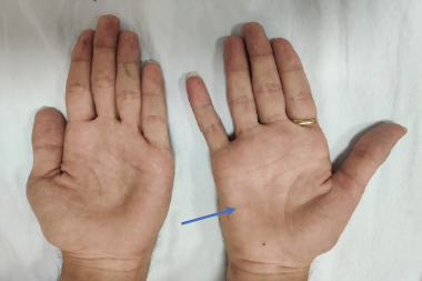
Secondary neurological disorders affecting the central or peripheral nervous systems can be seen in both neuronopathic and nonneuronopathic Hunter syndrome. Some severe problems, such as hydrocephalus, are more common in neuronopathic disease, whereas others disorders (such as carpal tunnel syndrome and spinal cord compression) are equally likely in both forms of the condition.15 Chronic communicating hydrocephalus, which has been hypothesised to be due to impaired resorption of cerebrospinal fluid, occurs in 80–100 % of patients who have Hunter syndrome.17,20 It can be difficult to differentiate between communicating hydrocephalus, which requires prompt surgical intervention, and progressive brain atrophy, for which there is no treatment.20 This may lead to delayed or inappropriate surgery.20 Optic nerve head swelling and optic atrophy have been reported in 20 % and 11 % of patients, respectively.21,22 It has been suggested that GAG accumulation in ganglion cells or thickening of the sclera leads to compression of the optic nerve, which in turn causes optic atrophy.17 Although poor peripheral and night vision are common symptoms of Hunter syndrome, these are caused by retinal degeneration rather than by optic nerve defects.23 As already noted, nearly all patients experience hearing loss, but this is predominantly caused by conductive defects.9 However, patients do suffer some sensorineural hearing loss thanks to compression of the cochlear nerve, reduced numbers of spiral ganglion cells and loss of hair cells.9,24 Carpal tunnel syndrome caused by entrapment of the median or ulnar nerves has been reported to affect 96 % of children who have Hunter syndrome but may be underdiagnosed, for patients’ cognitive impairment often prevents them from accurately describing their symptoms.17,25 Manual clumsiness, avoidance of manual activity and abnormal patterns of grasping may indicate that a child has carpal tunnel syndrome. Hunter and other mucopolysaccharidoses syndromes are the most common cause of carpal tunnel syndrome in children, indicating that these disorders should be considered in children presenting with this condition.26 Spinal cord compression is a common complication and can cause cervical myelopathy (as well as myelopathy on other levels); this can have devastating consequences such as progressive and irreversible loss of motor function and sensation in all four limbs, spasticity or neurogenic bladder.17,20 Thus it is important that spinal cord compression be detected as early as possible.
Morphological Changes in the Central Nervous System
All patients who have Hunter syndrome display neurological abnormalities on magnetic resonance imaging (MRI) or at autopsy, even in the absence of clinical neurological symptoms.20,27,28 Neurodegenerative changes in the periventricular and subcortical white matter, the corpus callosum and the basal ganglia that give a sieve-like or honeycomb appearance are common (see Figure 3A).20,27,28 A review of multiple radiology studies found that the three most consistently reported neurological findings in Hunter syndrome patients were enlargement of the periventricular spaces, ventriculomegaly and brain atrophy (see Figure 3B).27 Although these changes are all seen in other conditions, they are rarely seen in combination with sieve-like changes – hence the co-occurrence of these signs is highly suggestive for Hunter syndrome.27 Closed cephaloceles, Chiari malformation, giant cisternae and J-shaped sellae are also common radiological findings in Hunter syndrome.20,29,30 The extent of sieve-like changes has been reported to be inversely correlated to ventricular enlargement and atrophy, suggesting that sieve-like lesions develop first and lead to more extensive neural damage in white matter, followed by atrophy.27 The observed correlation between disease duration and white matter lesions and atrophy in a cohort of 31 Hunter syndrome patients supports this theory.31 Although some studies have shown that symptom severity corresponds with the extent of changes found on MRI scans, other studies have found no such correlation.27,29 Indeed, patients having the non-neuronopathic phenotype have been reported to have extensive neural atrophy and ventriculomegaly but no cognitive retardation.27 However, a recent study has reported an association between reduced volumes of white matter and corpus callosum and attention deficits in patients with nonneuronopathic disease who show no IQ or memory deficits.32 Further research is required to define the relationship between morphological changes in the brain and disease progression. Cervical spine MRI abnormalities include dens hypoplasia, periodontoid thickening and disc abnormalities in almost all patients having Hunter syndrome, regardless of phenotype.20,33 Spinal stenosis is present in almost 50 % of cases.20,33
Management of the Neurological Aspects of Hunter Syndrome Because of the complexity of sign and symptoms, patients with Hunter syndrome require regular follow-up and the provision of care by a multi-disciplinary team.34 Because of the extensive neurological manifestations of Hunter syndrome, particularly in its neuronopathic form, paediatric neurologists should play a major role in this team. ERT is available for the somatic symptoms of Hunter syndrome, but symptomatic treatment is currently the only option for neurological symptoms13 (see Table 1). Antipsychotic agents (both typical and atypical agents) and attention stimulants (such as methylphenidate) might improve behavioural concerns associated with Hunter syndrome. However, published studies of such agents in patients having this disorder are lacking. When prescribing, physicians must be aware of the expectations, limitations and potential risks associated with these agents. Thus psychoactive agents should be prescribed only by a neurologist or psychiatrist experienced in these matters. Stimulating environments, special schooling or speech therapy to promote maximal learning during the early stages of the disease, are also recommended to aid behavioural and cognitive development.5 Anticonvulsant therapy can reduce the frequency of seizures and might also improve sleep, and cognitive and behavioural symptoms.19 Polysomnography can be used to assess patients’ sleep disturbances; considering the occurrence of seizures, it is best that a full electroencephalograph be recorded.
Communicating hydrocephalus can be effectively treated by insertion of a ventriculoperitoneal shunt, which can improve motor development in early stages of the disease.17 It is suggested that nerve conduction studies be performed in patients every 1 to 2 years from age 4–5 onwards to monitor peripheral nerve function and the development of carpal tunnel syndrome.34 If detected early, carpal tunnel syndrome can be treated successfully by decompression surgery.5 However, without such an intervention, carpal tunnel syndrome might result in loss of hand function. MRI of the spine should be performed if patients develop any symptoms of spinal cord compression, such as back pain, hyper-reflexia, sudden loss in muscle strength or loss of bladder control. If spinal cord compression is found, decompression surgery must be undertaken promptly, preferably by an experienced team, because these patients are at risk of serious surgical complications, such as pulmonary or cardiac obstruction.35 Spinal cord compression can have a devastating effect on patients, and prompt treatment can avoid development of these symptoms.5 Thus, if a patient is undergoing MRI for reasons other than suspected spinal cord compression, MRI of the spine should be performed. Somatosensory evoked potential (SSEP) can also be used to assess the extent of any myelopathy present.36 It is widely accepted that both MRI and SSEP are useful tools for monitoring the pathologic progression of neurological disease,33,36 but there is no evidence for or consensus on ideal frequency of these assessments before patients become symptomatic. Parents and carers should be made aware of the signs and symptoms of spinal cord compression and should be informed that medical advice should be sought immediately so that investigations and treatment can begin promptly. ERT with idursulfase (Elaprase®, Shire, Lexington, MA, US) has been reported to reduce some of the somatic manifestations in up to 92 % of patients who have severe Hunter syndrome.13 Idursulfase does not cross the blood–brain barrier and so has no effect on neurological manifestations.13,18,20 However, an investigational formulation of idursulfase that can be delivered via an intrathecal device directly into cerebrospinal fluid and target cognitive impairment is being investigated.37,38,39
Conclusions
Neurologists have an important role in the diagnosis and management of Hunter syndrome because of the extensive neurological involvement in this disease. Patients affected with neuronopathic Hunter syndrome display developmental delay and cognitive impairment, which becomes evident in early childhood and progressively worsens, usually leading to premature death in the second decade of life. Neurological signs and symptoms that may be evident in patients having either the neuronopathic or non-neuronopathic phenotypes include seizures, optic nerve compression, hearing impairment, sleep apnoea, hydrocephalus, carpal tunnel syndrome, spinal cord compression and cervical myelopathy. Neurologists should also be aware of the somatic signs and symptoms of the disease so that they can recognise the condition in undiagnosed patients. All patients who have Hunter syndrome show neurological abnormalities on MRI scans. Though the extent of neurological change does seem to correlate with age, it does not always correlate with clinical severity. Because the manifestations of Hunter syndrome can vary greatly from patient to patient, a highly tailored approach to clinical care is required involving physicians with expertise in the disorder. With such an approach, current management options may alleviate some of the neurological symptoms of Hunter syndrome, but treatments that target accumulation of GAGs in the central nervous system are required if we hope to further improve the prognosis of patients who have neuronopathic Hunter syndrome.


