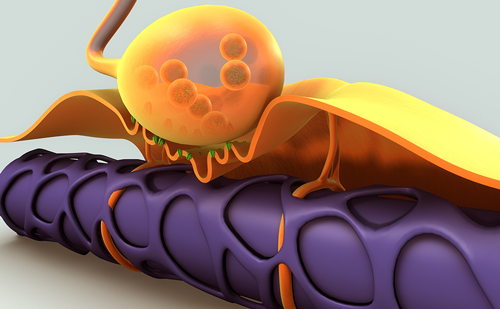
Amyotrophic lateral sclerosis (ALS) is a neurodegenerative disorder with selective and progressive loss of upper and lower motor neurons. Although this disease was identified more than 140 years ago by Charcot, the pathogenesis has yet remained unknown. Recent advancements in neuroimaging, neurophysiology and neuropathology have elucidated subclinical or clinical involvements of the non-motor systems, in addition to the upper and lower motor neurons. Accumulating evidence shows that autonomic nervous abnormalities are found in both early and advanced stages of ALS.1 The autonomic dysfunction may be clinically evident and devastating, especially in advanced stage when ventilators are required. Here, the autonomic dysfunctions in ALS are reviewed.
Clinical and Haemodynamic Studies
The autonomic nervous system is divided into sympathetic and parasympathetic divisions, each with a central and peripheral component. The central component is known as the central autonomic network and regulates the balance between the sympathetic and parasympathetic regulations of the visceral organs. The term ‘autonomic failure’ is mainly used to describe impairment of the sympathetic vasomotor efferent systems, including the intermediolateral nucleus (IML) of the spinal cord and sympathetic ganglion. Autonomic failure represents orthostatic or postprandial hypotension or syncope attacks caused by reduced sympathetic tone, which are often observed in multiple system atrophy, but patients with ALS rarely develop such symptoms. Contrary, cardinal autonomic dysfunctions in ALS are sympathetic hyperactivity and sympathovagal imbalance, the clinical manifestations of which often resemble the symptoms of autonomic overactivity or baroreflex failure.2,3 Differential diagnosis between autonomic failure and baroreflex failure should be carefully performed, since the therapeutic approaches for these two are quite different.3 In early stage of ALS, the sympathetic hyperactivity may be subclinical. Patients with ALS do not usually show overt hypertension needing treatment. However, some patients, especially, the ones with bulbar type of ALS experience palpitation, facial flushness and hot sensation around the face. These symptoms might be caused by increased sympathetic tone. Haemodynamic studies have shown that compared to healthy subjects, patients in early stage of ALS have higher resting heart rate and blood pressure.4–7 Plasma norepinephrine level was also reported to be elevated and not correlated with the extent of motor disability.8–12 Since plasma norepinephrine level reflects the amount of norepinephrine released from the peripheral sympathetic nerve terminals, its elevation is an indirect indicator of increased sympathetic tone. Although intravenous norepinephrine infusion test has shown preserved adrenoceptor function in the peripheral blood vessels, some patients on ventilators exhibit blunted response to infused norepinephrine, indicating down-regulated hyposensitivity of peripheral adrenoceptors induced by constantly high level of plasma norepinephrine.13
Muscle Sympathetic Nerve Activity Direct measurement of sympathetic activity is possible as muscle sympathetic nerve activity (MSNA) by recording postganglionic nervous impulses in peripheral nerves, using the microneurographic recording technique. MSNA is considered to reflect sympathetic activity related to cardiovascular control of vascular resistance. Many investigators have reported that MSNA was increased in patients in early stage of ALS.5,14–17 MSNA was elevated in ALS when quantified as the number of sympathetic burst (bursts/min or bursts/100 beats).14,16 Response of MSNA following head-up tilt, however, is less in patients with ALS than in control subjects.17 Similarly, various stimulation techniques such as Valsalva manoeuvre, apnoeic stimulation, painful stimulation, cold water immersion, glucose ingestion and lower body negative pressure, showed blunted responses of MSNA in patients having ALS.5,14 These reduced responses were attributed to the ‘ceiling effect’ caused by the constantly elevated sympathetic tone at resting state.14 The level of MSNA in ALS did not correlate with motor disability, respiratory function, or disease duration, suggesting a primary phenomenon as well as motor neuron degeneration. Although Shindo et al. reported that MSNA level gradually decreased with disease progression in individual patients,15 chronological variation of MSNA remains in argument.14 In addition, the chronological improvement of MSNA is inconsistent with the markedly increased sympathetic tone and autonomic storm in patients at the advanced stage of ALS when ventilators are required.10
Variability of Heart Rate and Blood Pressure
Another effective technique to measure the sympathetic and parasympathetic function is the time and frequency domain analysis of heart rate and blood pressure, especially the power spectrum analysis. Heart rate variability (HRV) in the low-frequency (LF) band (0.04–0.15 Hz) is mediated by both sympathetic and parasympathetic influences, whereas oscillation in the high-frequency (HF) band (0.15–0.40 Hz) is derived from cardiovagal modulation.18 A combination of HRV and blood pressure variability analysis can indicate not only baroreflex sensitivity (for the LF band), but also functioning of cardiorespiratory transfer (for the HF band).4
Previous reports on HRV or power spectrum analysis have reported that the abnormalities in ALS include increased sympathetic tone or impaired cardiovagal function, both of which lead to the sympathovagal imbalance. Pisano et al. first reported the imbalance of sympathovagal function due to an increase in LF/HF ratio.6 This alteration was not related to the clinical features or disease duration. Similar observations were made by other researchers.4,19–21 For example, Linden et al. observed reduced baroreflex sensitivity and diminished cardiorespiratory transfer during normal breathing in ALS due to decreased LF and HF bands, respectively.4 These findings were similar to those reported for essential hypertension sharing a common central autonomic derangement. By using sinusoidal neck suction method to stimulate carotid baroreceptors, Hilz et al. showed impaired cardiovagal response with preserved sympathetic vasomotor control.19 In addition, in early stage of ALS, patients with bulbar involvements show more predominant cardiovagal dysfunction than patients with non-bulbar involvement.20
Other Observations
A limited number of reports have shown decreased sympathetic outflow in patients with ALS. The corrected QT interval (QTc) on electrocardiography (ECg), a measure of sympathetic influence on the heart, was reported to be prolonged in length and increased in dispersion in patients with ALS, indicating reduced sympathetic activity.22 It should be noted that this report did not evaluate the extent of sympathetic tone and the co-existence of constantly increased sympathetic tone and QT interval prolongation at the terminal stage of ALS does not seem contradictory. Another evidence for impaired cardiac sympathetic function was provided by a 123I-metaiodobenzylguanidine (MIBg) uptake study by Druschky et al., which evaluated the function of the postganglionic sympathetic terminals in the heart. These authors studied MIBg-single photon emission computed tomography in early stage of ALS and found decreased heart/mediastinum ratio (mean 1.82 ± SD 0.27) in patients with ALS compared to normal controls (2.16 ± 0.26).23 They concluded the presence of cardiac postganglionic sympathetic denervation. However, there are no similar reports to date and their conclusion needs further validation.24
Observations in Advanced Stage of Amyotrophic Lateral Sclerosis
Although the abovementioned autonomic dysfunctions are usually subclinical, patients in most advanced stage of ALS, when ventilators are used, often show critical autonomic manifestations. Marked fluctuation of blood pressure and heart rate, namely ‘autonomic storm’, may occur during clinically stable stage on ventilator. The autonomic storm presents paroxysmal hypertensive crisis: more than 250 mmHg of systolic blood pressure and tachycardia without counter-regulation of heart rate (see Figure 1).10,13,25 During the hypertensive crisis, plasma norepinephrine level is usually markedly high, indicating sympathetic hyperactivity. The presence of tachycardia strongly suggests central resetting of baroreflex sensitivity. Patients often exhibit facial flushing, jaw clonus-like involuntary movements, pseudobulbar affect, and disinhibition of emotional expression during the crisis. These observations may suggest a limbic origin for the autonomic storm, although the precise pathophysiology remains to be elucidated.
The hypertensive stage is often followed by successive blood pressure fall (circulatory collapse) without counter-regulation of heart rate, which may lead to sudden death.10 The circulatory collapse is likely to occur during sleep at night and hyposensitivity of peripheral adrenoceptors might enhance the pressure decrease by a sleep-associated decrement of sympathetic tone.13 The adrenoceptor hyposensitivity is probably attributed to its down-regulation due to the constant high-level secretion of norepinephrine from sympathetic nerve terminals.
All these features resemble symptoms of baroreflex failure, whose cardinal manifestations include hypertensive crisis, volatile or labile hypertension, orthostatic tachycardia and malignant vagotonia.3 Similar symptoms are also found in the acute stage of guillain-Barré syndrome, which involves vagal afferent and sympathetic preganglionic efferent nerves.26,27 Differential diagnosis of baroreflex failure includes central nervous disorders, psychological or metabolic diseases such as pheochromocytoma and disorders of peripheral baroreflex deafferentation. A recent study on patients having ALS with circulatory collapse showed a quantitative preservation of vagal visceral branches, excluding the possibility of peripheral deafferentation of baroreflex arc.28 Although there have been reports of pathological involvement of peripheral sensory and small fibers in ALS,29,30 the autonomic storm cannot be attributed to such peripheral nervous lesions.
Disease Specificity of Autonomic Storm
The question to be resolved is whether the autonomic storm is primary or secondary to ALS. The factors that should be considered are the long-term bedridden state, severe muscle atrophy, long-term ventilator support and psychological stress.10 At present, there is no evidence that certainly supports or rules out these potential effects. However, autonomic storm in ALS is a very peculiar phenomenon that seldom occurs in other neuromuscular disorders. given that not all patients who have ALS with a long-term use of ventilator and severe muscle wasting develop autonomic storm and that ventilator-dependent patients with Duchenne muscular dystrophy having similar muscle atrophy do not usually show any circulatory fluctuation, the autonomic storm may be primary to ALS.10
Sudomotor Function
Patients with ALS often complain of increased or reduced sweating in their hands or feet, altered skin temperature, or skin discolouration. Studies of sudomotor function have reported that denervation of sympathetic postganglionic cholinergic nerves could occur in ALS. The sudomotor axon reflex test showed reduced sweating rate in ALS, indicating mild postganglionic sudomotor dysfunction.31,32 On the other hand, Beck et al. documented that compared to healthy subjects, patients in early stage of ALS had higher sweating rate in the palms, potentially supporting the idea of increased central sympathetic tone in ALS.33 However, they also reported a reduced sweating rate in advanced stage of ALS, suggesting progressive peripheral sudomotor dysfunction and resultant atrophy of sweat glands.
In addition, studies have electrophysiologically evaluated the sweat function by using sympathetic skin response (SSR) and skin sympathetic nerve activity (SSNA).34–38 SSR was reported to be abolished in 40 % of the patients in early stage of ALS and its latency was prolonged, suggesting subclinical involvement of sudomotor fibres in ALS.35 Interestingly, in contrast to these reports of SSR, SSNA assessment by microneurography showed high resting frequency and reduced response to mental stress, accompanied by slight prolongation of SSNA reflex latencies.34,38 These findings support basic sympathetic hyperactivity of central origin. The reduced response may reflect the ‘ceiling effect’ as in the case of MSNA.14 Both the vasomotor and sudomotor systems might be hyperactive, even though some patients show peripheral sudomotor hypofunction.
Neuropathology
Neuropathological studies on the autonomic dysfunction in ALS have been limited. IML of the thoracic spinal cord, one of the most efferent neurons of the baroreflex arc, was reported to be moderately decreased in number in patients with ALS.39,40 This reduction has been often considered as a potential cause of the sympathetic dysfunction, although the reduction of IML neurons itself does not explain the increase in sympathetic tone. It may explain the reduced size or delayed latency of SSR in ALS and it is not contradictory to the idea of sympathetic hyperactivity shown in ALS. The reported reduction of IML neurons in ALS is not severe and the remaining neurons are considered hyperactive. Consistent with this, IML, dorsal vagus nucleus and solitary tract nucleus are well preserved in most advanced stage of ALS,41,43 when a considerable number of patients who are on ventilator develop ophthalmoplegia or a totally locked-in state.41,42 There are a few reports on the neuropathology for sympathetic hyperactivity, especially in the brainstem and the limbic system. Shimizu et al. and Kato et al. reported that some patients with blood pressure fluctuation in the advanced stage of ALS showed lesions in the lateral hypothalamus, the central and basolateral nuclei of the amygdala and the brainstem reticular formation.10,43 It should be noted that these pathological findings were variable and infrequent across patients. The primary autonomic centres in the medulla oblongata, however, were intact. Although the brainstem reticular formation was previously suggested to be primarily involved in ALS,44 Kato et al. observed preservation of catecholaminergic neurons in the medullary reticular formation in patients with ALS and blood pressure fluctuation.45
It will be difficult to neuropathologically elucidate the precise central lesions corresponding to the clinical autonomic manifestations. generally, the central autonomic neural control is very complex and subtle imbalance between excitatory and inhibitory pathways could activate various autonomic nuclei and induce autonomic symptoms.46,47 If destruction of nervous tissue is severe, the lesions could produce constant autonomic symptoms like orthostatic hypotension in multiple system atrophy, but the fluctuations of symptoms may be attributed to mild alterations that cannot be detected by classic neuropathological methodology. Recent advancement in TDP-43 proteinopathy for sporadic ALS revealed a wide distribution of the protein aggregates in the central nervous system,48 including the limbic system, also called the ‘central autonomic network’ (see Figure 2).46 In addition, the limbic motor and bulbo-respiratory motor systems are anatomically and physiologically linked to each other.49 Furthermore, ALS is often accompanied by involvement of the frontotemporal lobes, which overlaps the limbic system. However, there have been no reports on a correlation between cognitive dysfunction and sympathetic tone, and further studies are needed.
Prognostic Significance
For patients in early stage of ALS, sympathetic hyperactivity rarely influences survival. A major cause of death is usually respiratory muscle paralysis, not circulatory collapse. Pinto et al., however, reported that decreased variability of heart rate might predict sudden death (cardiac arrest) in ALS.50 Four patients in their report showed very low values of the heart rate coefficient variation (<0.20), and three of them died suddenly within the following two months, despite normal nocturnal oxygenation. The prolongation of QT interval on ECg may also be an indicator of poor prognosis.22 The autonomic storm or circulatory collapse in advanced stage of ALS with ventilator use predicts poor survival prognosis. Patients with hypertensive crisis may die within several months, if not treated appropriately.10 Ventilator-dependent patients showing blood pressure fluctuation should be continuously monitored for vital signs, especially at night, because sudden pressure fall is more likely to occur during sleep.
Familial Amytrophic Lateral Sclerosis
Recent development of gene analysis in familial ALS has disclosed many types of gene abnormalities. Although precise distributions of disease-specific central nervous lesions have not been elucidated in each type of familial ALS, superoxide dismutase 1 (SOD1)-related ALS may show variable symptoms and lesions across families or locations of mutation. Some patients with SOD1-related ALS show severe degenerative lesions in the autonomic nuclei, including IML, dorsal vagus nucleus and solitary tract nucleus. Autonomic failure, such as orthostatic or postprandial hypotension and atonic bladder, was reported to develop in a sporadic case of SOD1-related ALS (V118L).51 Since cases of classical sporadic ALS never showed autonomic failure, sporadic ALS and SOD1-related ALS must have pathogenic and phenotypic differences. Like classic sporadic ALS, SOD1-related ALS may also show sympathetic hyperactivity. A ventilator-using patient with g93S mutation showed paroxysmal hypertension, prominent sensory disturbances and pressure fall during sleep at night.52 The norepinephrine level was very high and the bladder function showed an overactive detrusor type. In addition, Kandinov et al. reported a higher heart rate at rest and following stress in transgenic mice carrying a SOD1-g93A mutation than in wild-type mice.53 Blood pressure elevated even before the appearance of motor dysfunction and gradually decreased with the progress of the disease.54 Thus, SOD1-related ALS may show variable phenotypes of autonomic dysfunction across different genotypes.
Treatments
Therapeutic approaches for the sympathetic hyperactivity in ALS have not yet been established. Since most patients with ALS die from respiratory failure, it has not been clarified whether treatment of the sympathetic hyperactivity would bring a better effect on disease progression or survival. If the sympathetic tone is one of the prognostic indicators, it should be treated by medicine such as beta-adrenergic antagonists. No evidence, however, has been obtained so far. Avoiding hypoxic episodes may reduce reflex sympathetic activation.55 Hypertensive crisis or circulatory collapse in advanced stage of ALS should be immediately treated, given the severe and poor prognosis it indicates. Direct vasodilators, such as calcium antagonists, however, should be avoided since these may induce critical over-reduction of the blood pressure. Ohno et al. reported that tamsulosin hydrochloride, by acting as central alpha-adrenergic blocker, might modulate blood pressure and reduce plasma norepinephrine level.9Benzodiazepines might also prove effective for centrally increased sympathetic tones by potentially mediating central gABAergic functions.3,25 To establish an appropriate therapy, further studies are needed.
Conclusion
The cardinal features of the autonomic nervous dysfunction in ALS are the sympathetic hyperactivity and sympathovagal imbalance. Their clinical significance is obscured in early stage, but critical in advanced stage of the disease, when ventilators are required. Details of the involved central nervous lesions and appropriate therapeutic approaches remain to be determined. No evidence has been found to date of the pathophysiological correlation between the neurodegenerative process of ALS and the autonomic abnormalities. Further studies may be needed to establish the pathognomonic significance of autonomic dysfunction in ALS.


