Clinical Features of Amyotrophic Lateral Sclerosis
Clinical Features of Amyotrophic Lateral Sclerosis
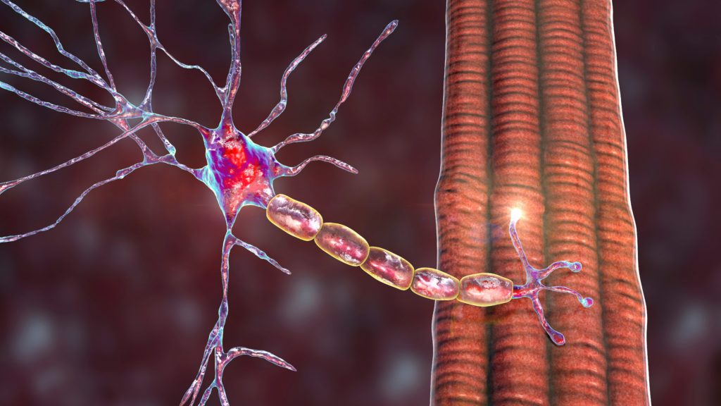
Amyotrophic lateral sclerosis (ALS), or Lou Gehrig’s disease, is a neurological disorder characterized by the selective loss of upper and lower motor neurons in the brain and spinal cord. ALS is characterized by progressive muscle weakness, atrophy, and spasticity. The typical age at onset for most ALS patients is between 45 and 60 years, with an average survival of less than three years. The final fatal event is often the loss of the motor neurons that innervate the respiratory muscles and diaphragm. The worldwide incidence of ALS is one to two new cases per 100,000/year, whereas its prevalence is four to six affected individuals per 100,000, with a lifetime risk of ALS of one in 1,000. Most cases—approximately 90%—of ALS are sporadic (SALS), thus lacking an overt genetic cause. The remaining 10% of cases are mostly inherited in a dominant manner and are thus referred to as familial ALS (FALS). Sporadic and familial patients are largely clinically indistinguishable.1 ALS is traditionally viewed as lacking in cognitive impairment; however, a diagnosis of frontotemporal dementia (FTD), a neurological disorder characterized by neurodegeneration of the frontal lobe of the brain, has been reported in 3–15% of ALS cases.2 The human impact of ALS is enormous as it significantly affects a patient’s quality of life, with patients losing their ability to eat, speak, and live independently. At present, while significant advances have been made in palliative therapies, there is no cure or means to significantly slow disease progression. Indeed, currently just one Canadian Agency for Drugs and Technologies in Health (CADTH)/US Food and Drug Administration (FDA)-approved therapy exists (Riluzole), and it offers only a modest slowing of disease progression.
Molecular Mechanisms of Disease in Amyotrophic Lateral Sclerosis
A major breakthrough in ALS research occurred in 1993 with the discovery that mutations in the Cu–Zn superoxide dismutase gene (SOD1) lead to ALS.3 Altogether, over 120 different mutations spread throughout the gene have been identified for SOD1 (alsod.iop.kcl.ac.uk/ Als/index.aspx); however, they account for only 15–20% of all FALS cases (or 1–2% of all cases).4 Based on various mutant (mut) SOD1 mouse/rat models expressing a range of SOD1 proteins with divergent biochemical properties, it is well accepted that disease-linked SOD1 mutant proteins provoke a selective loss of motor neurons through acquisition of one or more unidentified toxic properties, not through the loss of its endogenous dismutase activity but through an as yet unknown toxic gain of function. This is further emphasized by the complete lack of a motor neuron phenotype in mice that have their SOD1 gene deleted.5,6 SOD1 rodent models develop hind-limb paralysis and motor neuronal degeneration,7 and are arguably the single most robust recapitulation of any human neurodegenerative disease. Studies using these mice have identified several molecular mechanisms that may be crucial in ALS pathogenesis and motor neuronal degeneration. These include glutamatergic excitotoxicity, axonal damage, failure of the protein quality control machinery, mitochondrial dysfunction, and oxidative damage.8 Interestingly, motor neuron loss results from damage incurred both within the motor neurons themselves and in the surrounding glial cells. Thus, ALS is considered to be non-cell-autonomous.9 The mut SOD1 models have also been valuable to ALS researchers for the evaluation of several therapeutic agents, many of which were found to be efficacious in delaying the onset of symptoms and increasing lifespan. Unfortunately, none of these pharmaceutical compounds has proved to modify disease progression or improve the quality of life in ALS patients. Thus, the role of SOD1 in human patients may only be important to those with mutations in the SOD1 gene, while other genes and molecular mechanisms may be responsible for the majority of ALS cases. Furthermore, the absence of efficacious pharmaceutical treatments for ALS patients highlights the importance of the discovery of new genes and, subsequently, development of new animal models that can be used to explore additional mechanisms in ALS and aid in the development of effective patient therapies.
Pathology of Amyotrophic Lateral Sclerosis and TDP-43 Identification
A common feature of many neurodegenerative diseases is the formation of protein aggregates/inclusions. Whether these aggregates are toxic, harmless, or even protective in the disease process remains a topic of considerable debate. In the context of human ALS, ubiquitin is often part of these aggregates; however, the exact composition of these aggregates remains largely unknown. Inclusions have been found with distinct features and shapes in ALS patients, many of which are positive for Tau, a gene known to be mutated in patients with FTD.10 Other inclusions are Tau-negative but ubiquitin-positive. In an effort to resolve the composition of these aggregates, Trojanowski and Lee generated antibodies against isolated ubiquitin-positive inclusions and subsequently used them to screen a panel of ALS and FTD patient spinal cord samples.11 It was found that some cytosolic aggregates were preferentially labeled with these antibodies. Mass spectroscopy analysis of these extracted cytosolic aggregates identified an amino-terminal truncation product of the TAR DNA binding protein (TDP-43).11,12 This finding was supported by the observation that a lower-molecular-weight isoform of ~25kDa was indeed present exclusively in patients with ALS and FTD, but not in Alzheimer’s patients or non-affected controls.11 A large portion of TDP-43 present in these aggregates was found to be hyperphosporylated.13 These novel findings led to an explosion of subsequent reports evaluating the presence of TDP-43 in aggregates in ALS tissues and examining its potential role in disease.14–18 Interestingly, it has also been reported that TDP-43 inclusion bodies are not present in FALS patients with SOD1 mutations nor in mut SOD1 transgenic mice, leading some to propose that motor neuron disease marked by TDP-43 aggregates may be distinct from mut SOD1-mediated pathology,15 although this is highly controversial at present.
Identification of a Genetic Link Between TDP-43 and Amyotrophic Lateral Sclerosis
In light of the presence of TDP-43 in these aggregates and the distinct possibility that TDP-43 may play an as yet undetermined role in ALS pathogenesis, several groups screened the coding region of the TARDBP gene, which encodes the TDP-43 protein in ALS and FTD patients. Our group identified eight distinct heterozygous missense mutations in nine patients (six SALS, three FALS) including one segregating A315T mutation after sequencing 200 cases (see Table 1).19 Western blot analysis of lymphoblast cell lines indicated that a lower-molecular-weight TDP-43 protein product of approximately 28kDa was present exclusively in samples from patients with mutations.19,20 Concurrently, another group identified three different mutations (two familial mutations and one sporadic), also in the glycine-rich C-terminal region of TDP-43.21 Of note, the M337V mutation segregated in a pedigree that was large enough to retrospectively perform a genome scan and confirm that linkage in the family was exclusive to the TARDBP locus (ALS10) on chromosome 1p36. Injection of mutant, but not wild-type, TDP-43 RNA in chick embryos resulted in abnormal limb and tail development.21 Another independent group detected two missense mutations in TARDBP in FALS cases, including one that was positive for TDP-43 inclusions upon immunohistochemical analysis of autopsied tissue.22 A Q343R FALS mutation was also identified,23 and the A315T mutation was also reported to segregate in another family where individuals are described as having lower motor neuron disease without upper or bulbar motor neuronal involvement.24 Two recent reports also validated several of the already published mutations and identified three novel mutations in TARDBP.20,25 Finally, our group recently screened a cohort of French SALS patients and identified three novel mutations, including the first frameshift mutation, which creates a premature stop codon, Y374X26 (see Table 1). This mutation will consequently lead to the expression of a truncated protein, thus removing the last 41 amino acids of the TDP-43 protein.
Altogether, the rate of TARDBP mutations in certain populations can be quite variable, from 0.5 to 4.5%, and two studies did not identify any variants in the TARDBP gene in a Belgian and American cohort (see Table 2).27,28 For the most part, in these studies none of the patients with TDP-43 mutations had a personal or family history of FTD. An important control step was also undertaken to ensure that the TARDBP gene did not have a high degree of variance in the general population. When the gene was fully screened in 190 control individuals, only one missense change, A90V, was found,19 and this was detected in other control screens as well.21 The last exon of TARDBP has been sequenced in over 2,000 control DNA samples and no missense variants were reported (see Table 2). Thus, the C-terminal missense and truncating variants appear exclusive to ALS patients, and overall TARDBP is very highly conserved with little sequence variation.
Some insight can be gained regarding the position of the mutations in the TDP-43 protein and their potential function. Altogether, 19 of these 20 mutations were identified in the C-terminus of TDP-43, where a number of phosphorylation sites are predicted to exist. Most of the mutations identified in our screen are predicted to increase TDP-43 phosphorylation, as predicted by bioinformatics phosporylation programs and the fact that six of the mutations create a threonine or serine residue, thus indicating that phosphorylation of TDP-43 may be an important step in mutant-TDP-43-caused toxicity.19 The G348C mutation may increase the aggregation propensity of TDP-43 because the cysteine residue that is introduced may lead to abnormal disulfide bridges in the protein. The D169G mutation is noteworthy because it is the only one not in the C-terminal region of the protein; however, it is present in the first RNA recognition motif (RRM) of TDP-43 and is predicted to affect RNA binding. Finally, the Y374X truncating mutation can be expected to alter or abrogate the interaction of the TDP-43 C-terminal region with heterogeneous nuclear ribonucleoproteins (hnRNPs) A/B and A1a29 (see below).
TDP-43 Function
TDP-43 is highly conserved from humans to Caenorhabditis elegans and is widely expressed in a variety of human tissues, including brain and spinal cord.14 The protein structure of TDP-43 is predicted to contain two RNA-recognition motifs (RRM1 and RRM2), one of which partially overlaps with a putative nuclear export sequence.30,31 The C-terminal region contains multiple Phe–Gly and Trp–Gly motifs, features usually found in nuclear pore proteins. Finally, the C-terminal region mediates an interaction between TDP-43 and three members of the hnRNP family: A1, A2/B1, and A3.29 The TDP-43 protein was originally identified as capable of binding the TAR DNA sequence motifs of HIV and thus named the TAR DNA-binding protein (TDPDBP).32 Subsequent work demonstrated that TDP-43 binds a distinct RNA repeat consisting of six or more UG repeats via its RNA recognition motif (RRM1) domain33 and functions in vitro as a transcriptional repressor.34 Specifically, the interaction between TDP-43 and hnRNPs results in exclusion of the exon adjacent to the UG repeats that have been recognized by RRM1 of TDP-43.29 Therefore, based on structure and this reported in vitro function, TDP-43 is predicted to participate in mRNA splicing and/or nucleocytoplasmic transport. Mutations in TDP-43 could thus interfere with the protein’s normal interactions or its speculated transport through the nuclear pore complex, contributing to the progressive accumulation of TDP-43 aggregates reported in ALS patients.11,12,15
The transport of mRNA to synapses followed by local protein synthesis is now a well-recognized means to maintain and contribute to synaptic activity/function, and is especially relevant to the very large motor neuron. In light of the substantial amount of data that exists implicating mRNA transport and processing in other neurodegenerative diseases, such as spinal muscular atrophies, establishing the functional significance of disease-causing TDP-43 mutations in ALS pathogenesis is essential and timely. Furthermore, relevant to motor neurons and ALS, TDP-43 has previously been shown to interact with the survival motor neuron (SMN) protein35 and contribute to the alternative splicing of SMN2 exon seven.36 This has interesting correlations since copy number variants of the SMN1 and SMN2 genes have been suggested as susceptibility factors for the development of ALS.37 Finally, TDP-43 has been demonstrated to be localized in the neuronal RNA granules and to play a role in dendrite maintenance in motor neurons.38 Also, TDP-43 has been shown to have a nuclear localization signal and to be mainly localized in the nucleus.39 It is possible that TDP-43 mutations may disrupt the nuclear signal and its transport to the cytosol, where TDP-43 may be more likely to be aggregated. TDP-43 has also been shown to be cleaved by caspases and thus could represent a marker of disease pathology and apoptotic processes.40 However, the function that phosphorylated TDP-43 and its export from the nucleus plays, and whether it has a role in motor neuron demise, are not well understood.
Finally, in a mechanism that would draw parallels with SOD1, TDP-43 mutations could yield an as-yet unknown toxic gain of function that is selectively vulnerable to motor neurons, thus leading to their demise in ALS patients. Using in vivo and in vitro models, our group and collaborators in the ALS field are currently exploring these possibilities in order to understand the functional significance of TDP- 43 mutations in ALS pathogenesis. These models will be a great tool to further our knowledge of ALS pathophysiology. Most importantly, pharmacological treatments that prolong disease duration and onset in mut SOD1 transgenic mice can be tested also using these mut TDP- 43 animal models prior to entering the very labor-intensive and expensive road of human clinical trials. ■