touchNEUROLOGY touchNEUROLOGY
Neuromuscular Diseases
Read Time: 5 mins

The Role of Neuromuscular Ultrasound when Diagnosing Chronic Inflammatory Demyelinating Polyneuropathy

Copy Link
Published Online: Nov 3rd 2008 US Neurology, 2013;9(1):52–4 DOI: http://doi.org/10.17925/USN.2013.09.01.52
Authors: Antonios Kerasnoudis
Quick Links:
Abstract
Article
Article Information
Abstract:
Overview

Chronic inflammatory demyelinating polyneuropathy (CIDP) is the most common acquired immune-mediated inflammatory disorder of the peripheral nervous system. The diagnosis is based in classic cases, on the distribution pattern of the neurologic semiology, and pathologic changes of nerve conduction studies (NCS). However, in cases with subtle clinical presentation, an extended diagnostic workup may be needed (cerebrospinal fluid examination, laboratory tests, nerve biopsy). NCS remain fundamental for the diagnosis, follow up, and measurement of response to immune-treatment in CIDP. However, new challenges arose on how best to acquire a static and dynamic imaging of the peripheral nerves, with the aim of providing a holistic approach to the nerve impairment. According to the literature, neuromuscular ultrasound is able to detect in cases of CIDP thickened or swollen roots, peripheral nerves, or brachial plexus, findings that are consistent with ongoing inflammation. This review provides a timely update on the nerve ultrasound findings of CIDP and future possibilities of neuromuscular ultrasound are also discussed.

Keywords

Chronic inflammatory demyelinating polyneuropathy, nerve ultrasound, nerve hypertrophy, conduction block, immune-mediated neuropathies, brachial plexus

Article:

Chronic inflammatory demyelinating polyneuropathy (CIDP) is an acquired immune-mediated inflammatory disorder of the peripheral nervous system with an estimated prevalence of about 0.5 per 100,000 children and 1 to 2 per 100,000 adults.1,2 Typical CIDP arises between the ages of 30 and 60 years and is characterised from a progressive, symmetric proximal and distal muscle weakness, paresthesias, sensory dysfunction, impaired balance and reduced or diminished tendon reflexes which evolve slowly over at least 8 weeks. The course can be either monophasic with stepwise progression or relapsing with spontaneous remissions.3 The diagnosis is based, in typical cases, on the time course, distribution pattern of nerve impairment and results of the nerve conduction studies (NCS), that are frequently consistent with a motor and sensory demyelinating polyradiculoneuropathy, with evidence of conduction block and temporal dispersion.4,5 These findings may be further supported from evidence of cytoalbuminologic dissociation in cerebrospinal fluid analysis, but this is not obligatory for the diagnosis.6 Different criteria, such as Inflammatory Neuropathy Cause and Treatment (INCAT), American Academy of Neurology (AAN) and Saperstein, can be used to define the disease.7,8

While the NCS remain nowadays fundamental to confirm the presence, pattern and severity of this type of polyradiculoneuropathy,6 new challenges arose in the last few years, how to acquire the best static and dynamic imaging of the relevant nerve structures in CIDP, aiming to provide a complementary and holistic approach to nerve impairment. Using magnetic resonance imaging (MRI), various hypertrophic changes have been demonstrated in peripheral nerves, roots or brachial plexus in studies on CIDP patients.9–14 Although MRI is a very accurate diagnostic method in imaging soft tissues, it has the disadvantages ofbeing expensive, time-consuming, affected from artefacts (for example metal) and not practical, especially when a number of nerves need to be examined over a long course in patients with polyneuropathy. The role of neuromuscular ultrasound in the diagnostic workup of CIDP and polyneuropathies in general, remains less well defined and parallels the beginnings of research of entrapment neuropathies. Only a few studies in the literature have used ultrasound to examine the pathological changes in immune-mediated neuropathies, highlighting mainly pathological changes of the cross sectional area of peripheral nerves and their correlation with clinical and electrophysiological findings.15–21 This review provides a timely update on the diagnostic role of neuromuscular ultrasound in the diagnostic of CIDP, while possible future possibilities of neuromuscular ultrasound are also discussed.

Quantification of Ultrasound Findings in Immune-mediated Neuropathies
Cross sectional area (CSA) reference values for peripheral nerves and brachial plexus have been reported in various studies in the literature.22–27 The difficulty, however, to differentiate a normal from a pathological heterogeneity of cross sectional area changes in peripheral nerves, especially in CIDP cases, remains an important limitation of neuromuscular ultrasound in clinical practice. Two novel ultrasound measures, aiming to quantify pathological ultrasound changes of peripheral nerves in immune-mediated polyneuropathies, have been recently introduced in the literature: (1) the intranerve cross sectional area variability (for each nerve), defined as maximal cross sectional area / minimal cross sectional area28 and (2) the internerve cross sectional area variability (for each patient), defined as nerve with maximal intranerve cross sectional area variability/nerve with minimal intranerve cross sectional area variability,28 (3) the side to side difference ratio of the intranerve cross sectional area variability (SSDIVA) (for each nerve), defined as side with maximal intranerve cross-sectional area variability / side with minimal intranerve cross-sectional area variability and29 (4) the intraplexus cross sectional area variability defined as: maximal cross sectional area of the brachial plexus / minimal cross sectional area of the brachial plexus (see Table 1).29

Using the intranerve cross-sectional area variability the sonographer may differentiate in immune-mediated neuropathies focal (higher values) from diffuse (lower values) nerve enlargement, while the internerve cross sectional area variability may reveal possible distribution patterns of peripheral nerve impairment.28,29 On the other hand the side to side difference ratio of the intranerve cross sectional area variability may be useful in detecting any lateralisation of pathological changes and the intraplexus cross-sectional area variability in differentiating focal (higher values) from diffuse (lower values) brachial plexus enlargement.29

Ultrasound Findings
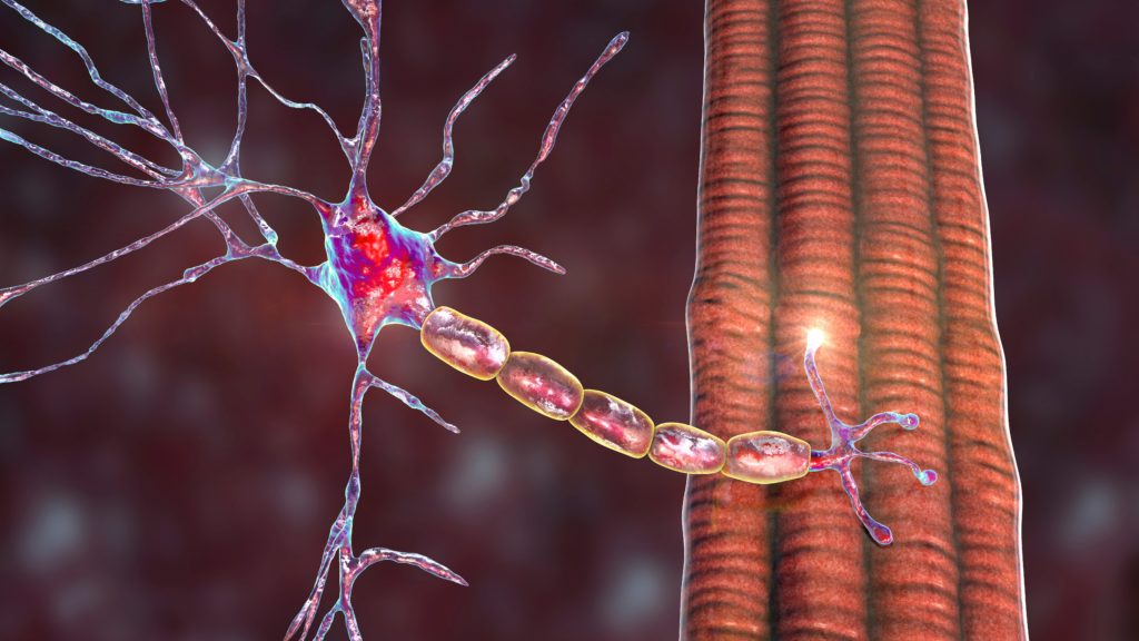
Currently ten studies (evaluating a total of 74 cases) on nerve sonography in CIDP patients have been published (see Table 2). The first description of the sonographic findings in CIDP was published from Taniguchi et al.30 In this report, the authors documented a brachial plexus hypertrophy on both sides and peripheral nerve hypertrophy at several sites of the median, sciatic and femoral nerve. Similar findings had only been eported in MRI studies until then.9–14 A possible explanation of the documented hypertrophy (see Figure 1) could derive from the classical ‘onion-bulb’ histological appearance of the nerves in CIDP, as a result of recurrent episodes of demyelination and remyelination.31

After this initial publication, several years passed until the first systematic ultrasound study of CIDP patients was published. In 2004, Matsuoka et al. reported the ultrasound findings of the cervical roots in 13 patients with CIDP and 35 healthy individuals.32 The authors demonstrated a hypertrophy of the cervical roots in nine out of 13 patients with CIDP, a finding that seemed to correlate with the elevated levels of protein in cerebrospinal fluid. Another ultrasound study of 36 CIDP patients from Zaidman et al. confirmed the presence of diffuse nerve enlargements in peripheral nerves in this type of immune-mediated nerve injury.25 Similar findings have been reported in several case reports in the following years.33–36 These findings showed a correlation with the disease duration and nerve conduction findings in a small group of CIDP patients.37 On the other side, Rajabally et al. compared the distal median nerve cross sectional area of 14 CIDP patients to 14 patients with sensory axonal neuropathy of various aetiologies (including alcoholism, vitamin deficiency, impaired glucose tolerance, vasculitis, idiopathic). The authors concluded, that the cross-sectional area of the median nerve was greater in CIDP, when compared to other polyneuropathies (sensitivity of 57 % and specificity of 93 %).38

Another important aspect in the field of sonography in CIDP, is the possible use of this method for identifying nerve conduction blocks. The localisation of the nerve conduction block is often difficult to be made in the nerve conductions studies, especially when dealing with proximal parts of the nerves. By overlooking this typical electrophysiological finding of this disease, a delay in the diagnosis and therefore treatment can occur. In three CIDP cases in the literature, a correlation between the site of hypertrophy detected with ultrasound and the site of conduction block detected with nerve conduction studies was demonstrated.33,39–40 Although this seemed to be a promising development, it is worth noting that Zaidman et al. failed to confirm these findings in a later study.15 Systematic studies on the sensitivity and specificity of this finding failed in the literature. A novel approach to the quantification of the pathological findings in CIDP was recently published in the literature.28,29 Using two new measures, the intranerve- and internerve cross-sectional area variability, in a small group of immune-mediated neuropathies, Padua et al. and Kerasnoudis et al. were able to demonstrate that the CIDP shows preferably a diffuse pattern of nerve enlargement (lower values of intranerve cross-sectional area variability), when compared to other immune-neuropathies, such as the multifocal motor neuropathy (MMN) (higher values of intranerve cross-sectional area variability).

Two CIDP cases reported tried to highlight the possible value of neuromuscular ultrasound as a screening tool of immune therapy. In both cases, following intravenous immunoglobulin or prednisolone therapy the patients bought a remarkable clinical improvement, but the sonographic follow-up did not show any improvement of the pathological findings.33,35 The same observation was done in three cases of multifocal acquired demyelinating sensory and motor neuropathy (MADSAM).20,21 Systematic data on the sensitivity and specificity of sonography as a screening method of immune therapy do not exist.

Conclusions
As the main uncertainties regarding the diagnostic criteria of CIDP are steadily resolved, new challenges continuously arise on how to acquire the best static and dynamic imaging of the relevant nerve structures in this type of immune-mediated disease, aiming to provide a complementary and holistic approach to nerve impairment. Although the first nerve ultrasound studies on CIDP are rewarding to both clinicians and patients, the challenge remains to quantify ultrasound changes and to highlight a possible unique distribution pattern of pathological findings. The newly proposed measurements in the literature28,29 may help to achieve this goal, but multicentre prospective validation and clinical correlations are needed.

Article Information:
Disclosure

The authors have no conflicts of interest to declare.

Correspondence

Antonios Kerasnoudis, MD, Department of Neurology, St Josef-Hospital, Ruhr University, Gudrunstr 56, 44791 Bochum, Germany. E: antonis.kerasnoudis@gmail.com
An erratum to this article can be found below.

Received

2013-02-09T00:00:00

References

  1. Mcleod JG, Pollard Jd, Macaskill P, et al., Prevalence of chronic inflammatory demyelinating polyneuropathy in New South Wales, australia, Ann Neurol, 1999;46:910–3.
  2. lunn MP, Manji h, choudhary PP, et al., chronic inflammatory demyelinating polyradiculoneuropathy: a prevalence study in south east England, J Neurol Neurosurg Psychiatry, 1999;66:677–80.
  3. dalakas Mc, advances in the diagnosis, pathogenesis and treatment of cidP, Nat Rev Neurol, 2011;7:507–17.
  4. lewis Ra, chronic inflammatory demyelinating polyneuropathy, Neurol Clin, 2007;25:71–87.
  5. Koller h, Kieseier Bc, Jander S, hartung hP, chronic inflammatory demyelinating polyneuropathy, N Engl J Med, 2005;352:1343–56.
  6. Rajabally Ya, Nicolas G, Pieret F, et al., Validity of diagnostic criteria for chronic inflammatory demyelinating polyneuropathy: a multicentre European study, J Neurol Neurosurg Psychiatry, 2009;80:1364–8.
  7. Saperstein dS, chronic acquired demyelinating polyneuropathies, Semin Neurol, 2008;28:168–84
  8. cornblath dR, Feasby TE, hahn aF, et al., Research criteria for diagnosis of chronic inflammatory demyelinating polyneuropathy (cidP), Neurology, 1991;41:617–8.
  9. Midroni G, de Tilly lN, Gray B, Vajsar J, MRi of the cauda equina in cidP: clinical correlations, J Neurol Sci, 1999;170:36–44.
  10. duggins aJ, Mcleod JG, Pollard Jd, et al., Spinal root and plexus hypertrophy in chronic inflammatory demyelinating polyneuropathy, Brain, 1999;122:1383–90.
  11. Eurelings M, Notermans Nc, Franssen h, et al., MRi of the brachial plexus in polyneuropathy associated with monoclonal gammopathy, Muscle Nerve, 2001;24:1312–8.
  12. Sinclair cd, Morrow JM, Miranda Ma, et al., Skeletal muscle MRi magnetisation transfer ratio reflects clinical severity in peripheral neuropathies, J Neurol Neurosurg Psychiatry, 2012;83:29–32.
  13. Mizuno K, Nagamatsu M, hattori N, et al., chronic inflammatory demyelinating polyradiculoneuropathy with diffuse and massive peripheral nerve hypertrophy: distinctive clinical and magnetic resonance imaging features, Muscle Nerve, 1998;21:805–8.
  14. Schady W, Goulding PJ, lecky BR, et al., Massive nerve root enlargement in chronic inflammatory demyelinating polyneuropathy, J Neurol Neurosurg Psychiatry, 1996;61:636–40.
  15. Zaidman cM, al-lozi M, Pestronk a, Peripheral nerve size in normal and patients with polyneuropathy: an ultrasound study, Muscle Nerve, 2009;40:960–6.
  16. Smith Ec, hobson-Webb ld, Massey E, Nerve ultrasound in motor conduction block: pre- and posttreatment findings, Muscle Nerve, 2008;38:1369.
  17. Granata G, Pazzaglia c, calandro P, et al., ultrasound visualization of nerve morphological alteration at the site of conduction block, Muscle Nerve, 2009;40:1068–70.
  18. Beekman R, van den Berg lh, Franssen h, et al., ultrasonography shows extensive nerve enlargements in multifocal motor neuropathy, Neurology, 2005;65:305–7.
  19. Kerasnoudis a, correlation of sonographic and electrophysiological findings in a patient with multifocal motor neuropathy, J Neuroimaging, 2012 dec 17. doi: 10.1111/j.1552- 6569.2012.00757.x.
  20. Scheidl E, Böhm J, Simó M, et al., ultrasonography of MadSaM neuropathy: focal nerve enlargements at sites of existing and resolved conduction blocks, Neuromuscul Disord, 2012;22:627–31.
  21. Kerasnoudis a.ultrasonography of MadSaM neuropathy: focal nerve enlargements at sites of existing and resolved conduction blocks, Neuromuscul Disord, 2012 Nov;22(11):1032.
  22. cartwright MS, Passmore lV, Yoon JS, et al., cross-sectional area reference values for nerve ultrasonography, Muscle Nerve, 2008;37:566–71
  23. cartwright MS, Shin hW, Passmore lV, Walker Fo, ultrasonographic reference values for assessing the normal median nerve in adults, J Neuroimaging, 2009;19:47–51
  24. cartwright MS, Shin hW, Passmore lV, Walker Fo, ultrasonographic findings of the normal ulnar nerve in adults, Arch Phys Med Rehabil, 2007;88:394–6
  25. Yalcin E, onder B, akyuz M, ulnar nerve measurements in healthy individuals to obtain reference values, Rheumatol Int, 2012; Epub ahead of print.
  26. . Won SJ, Kim BJ, Park KS, et al., Measurement of cross-sectional area of cervical roots and brachial plexus trunks, Muscle Nerve, 2012;46:711–6.
  27. haun dW, cho Jc, Kettner NW, Normative cross-sectional area of the c5-c8 nerve roots using ultrasonography, Ultrasound Med Biol, 2010;36:1422–30.
  28. Padua l, Martinoli c, Pazzaglia c, et al., intra- and internerve cross-sectional area variability: new ultrasound measures, Muscle Nerve, 2012;45:730–3.
  29. Kerasnoudis a, Klasing a, Behrendt V, et al., intra- and internerve cross-sectional area variability: new ultrasound measures’’, Muscle Nerve, 2013;47:146–7.
  30. Taniguchi N, itoh K, Wang Y, et al., Sonographic detection of diffuse peripheral nerve hypertrophy in chronic inflammatory demyelinating polyneuropathy, J Clin Ultrasound, 2000;28:488–91
  31. Matsuda M, ikeda S, Sakurai S, et al., hypertrophic neuritis due to chronic inflammatory demyelinating polyradiculoneuropathy (cidP): a postmortem pathological study, Muscle Nerve, 1996;19:163–9.
  32. Matsuoka N, Kohriyama T, ochi K et al., detection of cervical nerve root hypertrophy by ultrasonography in chronic inflammatory demyelinating polyradiculoneuropathy, J Neurol Sci, 2004;219:15–21
  33. imamura K, Tajiri Y, Kowa h, Nakashima K, Peripheral nerve hypertrophy in chronic inflammatory demyelinating polyradiculoneuropathy detected by ultrasonography, Intern Med, 2009;48:581–2.
  34. Jang Jh, Yoo JK, Kim BJ, ultrasonographic findings in chronic inflammatory demyelinating polyneuropathy, Am J Phys Med Rehabil, 2012;91:546–7
  35. Kerasnoudis a. Nerve ultrasound in a case of chronic inflammatory demyelinating neuropathy, Muscle Nerve, 2013 Mar;47(3):443-6.
  36. Kerasnoudis a, re: ultrasonic findings in chronic inflammatory demyelinating polyneuropathy, Am J Phys Med Rehabil, 2013 Jan 30. [Epub ahead of print]
  37. Kerasnoudis a, Gold R, Klasing a, et al., correlation of morphological changes in cidP with the disease duration. dGN congress. 85. Kongress der deutschen Geselschaft für Neurologie (dGN), 27.09.12, hamburg Germany. P555.
  38. Rajabally Ya, Morlese J, Kathuria d, Khan a,Median nerve ultrasonography in distinguishing neuropathy sub-types: a pilot study, Acta Neurologica Scandinavica, 2012;125:254–9.
  39. Smith Ec, hobson-Webb ld, Massey E, Nerve ultrasound in motor conduction block: pre- and posttreatment findings, Muscle Nerve, 2008;38:1369.
  40. Granata G, Pazzaglia c, calandro P, et al., ultrasound visualization of nerve morphological alteration at the site of conduction block, Muscle Nerve, 2009;40:1068–70.

Further Resources

Share this Article
Related Content In Neuromuscular Diseases
  • Copied to clipboard!
    accredited arrow-down-editablearrow-downarrow_leftarrow-right-bluearrow-right-dark-bluearrow-right-greenarrow-right-greyarrow-right-orangearrow-right-whitearrow-right-bluearrow-up-orangeavatarcalendarchevron-down consultant-pathologist-nurseconsultant-pathologistcrosscrossdownloademailexclaimationfeedbackfiltergraph-arrowinterviewslinkmdt_iconmenumore_dots nurse-consultantpadlock patient-advocate-pathologistpatient-consultantpatientperson pharmacist-nurseplay_buttonplay-colour-tmcplay-colourAsset 1podcastprinter scenerysearch share single-doctor social_facebooksocial_googleplussocial_instagramsocial_linkedin_altsocial_linkedin_altsocial_pinterestlogo-twitter-glyph-32social_youtubeshape-star (1)tick-bluetick-orangetick-red tick-whiteticktimetranscriptup-arrowwebinar Sponsored Department Location NEW TMM Corporate Services Icons-07NEW TMM Corporate Services Icons-08NEW TMM Corporate Services Icons-09NEW TMM Corporate Services Icons-10NEW TMM Corporate Services Icons-11NEW TMM Corporate Services Icons-12Salary £ TMM-Corp-Site-Icons-01TMM-Corp-Site-Icons-02TMM-Corp-Site-Icons-03TMM-Corp-Site-Icons-04TMM-Corp-Site-Icons-05TMM-Corp-Site-Icons-06TMM-Corp-Site-Icons-07TMM-Corp-Site-Icons-08TMM-Corp-Site-Icons-09TMM-Corp-Site-Icons-10TMM-Corp-Site-Icons-11TMM-Corp-Site-Icons-12TMM-Corp-Site-Icons-13TMM-Corp-Site-Icons-14TMM-Corp-Site-Icons-15TMM-Corp-Site-Icons-16TMM-Corp-Site-Icons-17TMM-Corp-Site-Icons-18TMM-Corp-Site-Icons-19TMM-Corp-Site-Icons-20TMM-Corp-Site-Icons-21TMM-Corp-Site-Icons-22TMM-Corp-Site-Icons-23TMM-Corp-Site-Icons-24TMM-Corp-Site-Icons-25TMM-Corp-Site-Icons-26TMM-Corp-Site-Icons-27TMM-Corp-Site-Icons-28TMM-Corp-Site-Icons-29TMM-Corp-Site-Icons-30TMM-Corp-Site-Icons-31TMM-Corp-Site-Icons-32TMM-Corp-Site-Icons-33TMM-Corp-Site-Icons-34TMM-Corp-Site-Icons-35TMM-Corp-Site-Icons-36TMM-Corp-Site-Icons-37TMM-Corp-Site-Icons-38TMM-Corp-Site-Icons-39TMM-Corp-Site-Icons-40TMM-Corp-Site-Icons-41TMM-Corp-Site-Icons-42TMM-Corp-Site-Icons-43TMM-Corp-Site-Icons-44TMM-Corp-Site-Icons-45TMM-Corp-Site-Icons-46TMM-Corp-Site-Icons-47TMM-Corp-Site-Icons-48TMM-Corp-Site-Icons-49TMM-Corp-Site-Icons-50TMM-Corp-Site-Icons-51TMM-Corp-Site-Icons-52TMM-Corp-Site-Icons-53TMM-Corp-Site-Icons-54TMM-Corp-Site-Icons-55TMM-Corp-Site-Icons-56TMM-Corp-Site-Icons-57TMM-Corp-Site-Icons-58TMM-Corp-Site-Icons-59TMM-Corp-Site-Icons-60TMM-Corp-Site-Icons-61TMM-Corp-Site-Icons-62TMM-Corp-Site-Icons-63TMM-Corp-Site-Icons-64TMM-Corp-Site-Icons-65TMM-Corp-Site-Icons-66TMM-Corp-Site-Icons-67TMM-Corp-Site-Icons-68TMM-Corp-Site-Icons-69TMM-Corp-Site-Icons-70TMM-Corp-Site-Icons-71TMM-Corp-Site-Icons-72