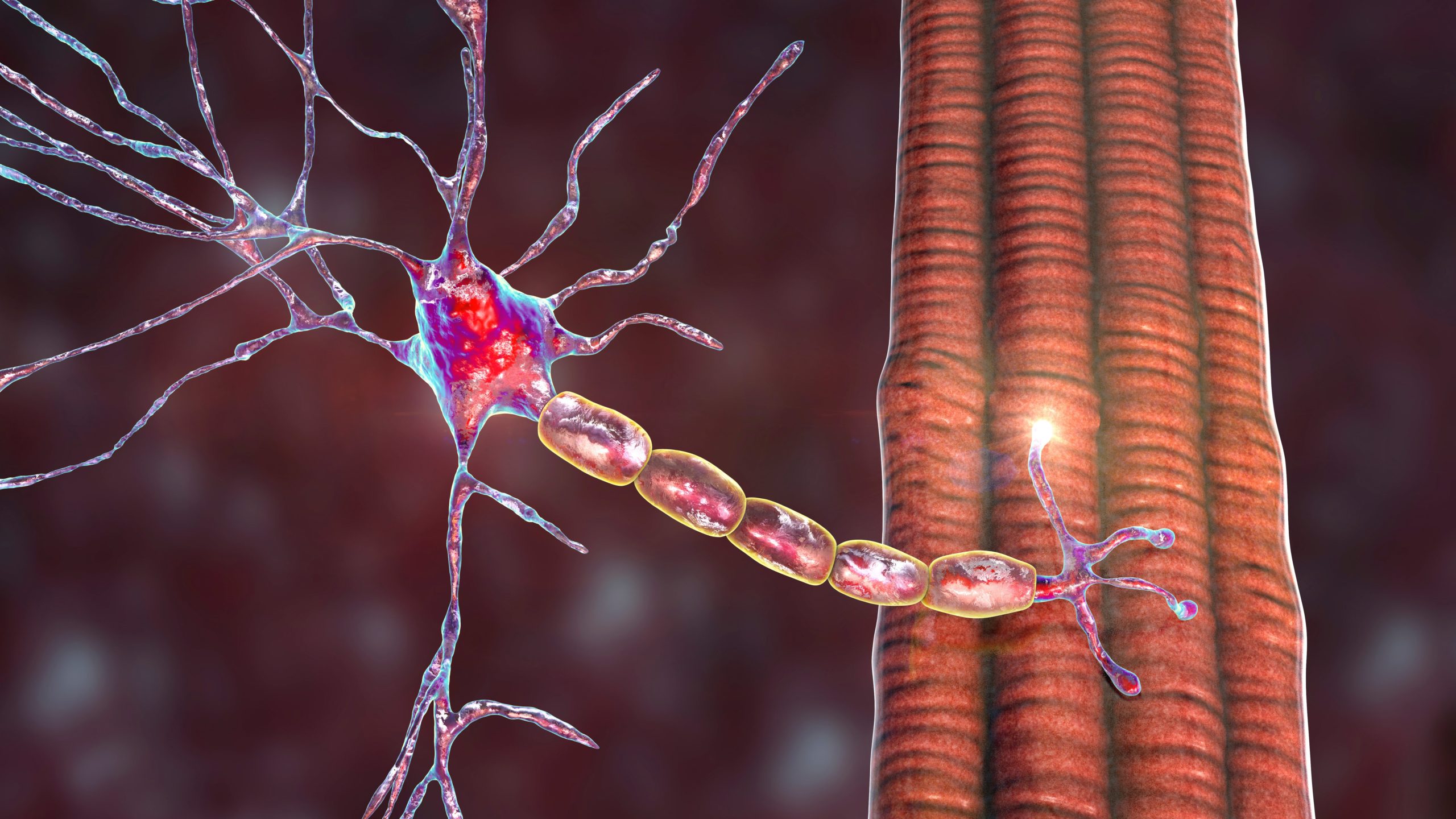
Motor neuron disorders (MNDs) are a group of neurological disorders in which motor neurons are destroyed. Motor neurons are large cells that control voluntary muscle activity, including speaking, walking, breathing, swallowing and general movement of the body. MNDs are generally progressive, but there are exceptions. In upper MNDs, cells in the motor cortex of the brain are affected, as well as their projections in the corticospinal tract. Signs of upper motor neuron damage include muscular weakness, spasticity, brisk reflexes and the Babinski sign. In addition, some subjects exhibit cognitive changes, and a small proportion exhibit frontotemporal dementia.1 In contrast, degeneration of neurons located in the ventral horns of the spinal cord (lower motor neurons) with projections through the ventral roots to the muscle cells lead to weakness and muscle atrophy. Examples of MNDs are given in Table 1.
Apart from the specific upper MNDs in Africa mentioned below, there are also disorders with both upper and lower motor neuron symptoms, such as amyotrophic lateral scleros. Below, a pure lower MND, poliomyelitis, is mentioned, since polio immunisation campaigns are highly topical in Africa. As a differential diagnosis to MND, tropical ataxic neuropathy (TAN) is mentioned. The main aim of this review is to describe clinical and neurophysiological findings in konzo, a non-progressive upper MND reported in several remote rural areas of the Central African Republic, Mozambique, Tanzania and the Democratic Republic of Congo (DRC).2–9
Upper Motor Neuron Disorders in Africa
Konzo was first described in 1938 in the Bandundu province of the DRC.2 It is characterised by a sudden onset of a spastic paraparesis or quadriparesis in severely affected subjects. In 1996, the World Health Organization (WHO) suggested the following diagnostic criteria for konzo: “a visible symmetric spastic abnormality when walking and/or running, a history of abrupt onset (<1 week), a non-progressive course; onset in a formerly healthy person, showing bilaterally exaggerated knee and/or ankle jerks without signs of disease of the spine.”10 Criteria for the degree of severity of konzo according to the WHO are: mild form – subject is able to walk without support; moderate form – subject uses one or two sticks to walk; and severe form – subject is unable to walk.10
In addition to motor symptoms, some affected subjects may present with blurred vision, nystagmus and speech difficulties of pseudobulbar type.11 The disease mainly affects children and women of childbearing age.10,12 Konzo is named after a local designation among the Yaka ethnic group of the Bandundu province in the DRC and means ‘tied legs’, referring to the scissoring gait of affected subjects.13 Epidemiological studies have shown that the disease is associated with a high exposure to cyanogenic compounds in a diet dominated by insufficiently processed bitter cassava.14–18 Persons consuming poorly processed cassava in large quantities are susceptible to neuropathologies caused by cyanide.19,20 Affected subjects also have a low intake of sulphur amino acids, which are needed to detoxify cyanide in the human body.14–20
Neurolathyrism is another toxic nutritional disorder known in Africa. It is an irreversible spastic paraparesis caused by excessive consumption of grass peas (Lathyrus sativus). Neurolathyrism is known in Ethiopia, where neurolathyrism epidemics appear in connection with food shortages and famines.21–25 Neurolathyrism has been around for a very long time, and epidemics have occurred in areas such as India, the Middle East, Russia and southern Europe.26,27 Neurolathyrism affects more men than women, and, as in konzo, young age is associated with an increased risk of paralysis.25 At onset there are often cramps in the legs or in the lumbar area. Weakness of the lower extremities then develops, leading to a spastic paraparesis that in some subjects gives a severe disability with complete dependency in activities of daily living.
Tropical spastic paraparesis (TSP) is a slowly progressive spastic paraparesis with an insidious onset. TSP mainly affects adults living in tropical and subtropical areas of the world, for example Western Africa, regions of South America and the Caribbean. TSP is a systemic immune-mediated inflammatory disease associated with infection with human Tlymphotrophic virus 1 (HTLV-1); this association was first reported in 1985 by a French group.28 TSP is transmitted person to person via infected cells; the virus can be spread through the placenta, breast-feeding, blood transfusions, contaminated needles and sexual contact. TSP causes muscle weakness, stiff muscles and muscle spasms, sensory disturbance and sphincter dysfunction. In addition to neurological symptoms, subjects with TSP may suffer from uveitis or keratoconjuntivitis sicca, arthritis, pulmonary alveolitis, polymyositis and dermatitis. Some HTLV-1 subjects develop leukaemia or lymphoma.29–32 TSP affects mainly adults between 30 and 40 years of age and is considerably more common in women than in men. In 1986, a syndrome similar to TSP was reported in Japan, and was named HTLV-1-associated myelopathy (HAM).33 In 1988, the WHO recommended both syndromes to be grouped together under a common name, TSP/HAM.34 Although TSP/HAM is not common in younger ages, up to 2006 there were 17 well-documented cases of TSP/HAM among children and adolescents.35 The juvenile form of the disease is rapid and more progressive than the adult form. In addition to myelopathy, 12 of the children and adolescents also presented with chronic infective dermatitis associated with HTLV-I (IDH).34 Dual infection with HTLV-I TSP/HAM and HIV-1 has been found in some subjects; HTLV-I and HIV-I are both retroviruses and share similar routes of transmission.36 Oral or intravenous corticosteroids are the main treatment of TSP/HAM, especially in the initial phase, but other drugs – for example valproic acid, methotrexate and interferon-alpha – are also suggested if steroids fail or cannot be used.32
In contrast to the upper MNDs (konzo, neurolathyrism and TSP/HAM), TAN is predominantly a sensory neuropathy frequently seen in malnourished populations. Symptoms of TAN include bilateral optic atrophy, bilateral neurosensory deafness, sensory gait ataxia and symmetrical sensory polyneuropathy. Screening for TAN in a Nigerian community showed an overall prevalence from 10 years of age of 6%, with female dominance (7.7% versus 3.9% in males).37 The highest age-specific prevalence was found in elderly women. TAN is linked to consumption of cassava foods.
In connection with MNDs and Africa, poliomyelitis, a lower motor neuron syndrome, must be mentioned. In 1988, the WHO decided to take action to eradicate polio by 2000. Despite much progress in reducing the incidence of polio, it still exists – mainly where there is poverty. However, the number of polio-endemic countries has decreased from 125 countries in 1988 to four in 2006 (Nigeria, India, Pakistan and Afghanistan). In 2006, Namibia experienced its first outbreak of polio in more than 10 years.38 Another outbreak occurred in Kenya during 2003–2006, and led to a national and international spread, with re-infection of 20 polio-free countries.39 Large-scale emergency polio immunisation campaigns were thus directed to high-risk countries.
Clinical Findings in Konzo
Onset
The clinical picture of konzo has been well characterised since the first description by Trolli.2–4,6,12,40,41 Konzo has a sudden onset of gait difficulties, often preceded by prolonged physical exercise. Spasticity is present from the onset. Most subjects experience trembling or cramping in the legs at onset and/or heaviness and pain in the legs. Some subjects report paresthesia, electrical discharges in the legs and low-back pain at onset; however, these symptoms resolve within a few weeks. Most patients are initially confined to bed, and thereafter try to walk. Blurred vision, speech and/or swallowing difficulties may be experienced at onset.
Motor Symptoms and Signs
Konzo has a stable clinical course after onset in the majority of affected persons.41,42 A few patients experience a certain improvement after the acute phase, and thereafter a non-progressive disability. Aggravation may appear with continued consumption of cassava, and manifests as a sudden onset of symptoms of the same type as initially seen in konzo, leading to a permanent change for the worse. The symptoms of konzo vary, with the the mildest form manifesting as the ability to walk without support with no overt lower limb weakness, but with signs of exaggerated deep tendon reflexes in the lower limbs. The majority of affected konzo subjects experience various degrees of symmetric weakness of the lower extremities, with a spastic gait, and a few also have weakness of the upper limbs and the trunk. Some persons are non-ambulant. Ankle clonus is frequent, Babinski sign is present and palmomental and cutaneous abdominal reflexes are often present.
Additive Symptoms and Signs
Some konzo patients report impaired visual acuity. Neuroophthalmological studies have confirmed the presence of optic neuropathy in konzo patients, with visual impairment, defect of visual fields and temporal pallor of the optic discs.43 Rotatory nyastagmus is seen in some affected people. Among moderate and severely disabled konzo subjects, some have dysarthria due to pseudobulbar palsy.41 The motor disability may, due to disuse, give rise to muscle atrophy.
Normal Features
Konzo subjects have normal cognitive function even when electroencephalogram (EEG) shows abnormal findings.44 Co-ordination is normal, and there is no sign of cerebellar dysfunction. Hearing is normal. Other than experiencing electric discharges in the lower limbs at onset of konzo, there are no sensory symptoms. There are no symptoms of autonomic dysfunction in konzo: urinary, bowel and sexual functions are normal.
Neurophysiological Findings in Konzo
Background
In 1998 and 2000, our research group from the University Hospital in Uppsala brought high-tech equipment to the DRC in order to neurophysiologically investigate konzo patients and healthy relatives. Konzo patients and relatives were recruited from villages in the Bandundu province, 500–600km south-east of the capital, Kinshasa, where they were hospitalised at the neuropsychopathological centre at the University Hospital during the study. Two nurses, active in the catchment area in Bandundu and speaking the tribal language of the participants, stayed in the hospital with them for support. All participated after informed consent and community acceptance.
Over four weeks in July 1998, 21 konzo subjects underwent neurological examinations, neurophysiological and ophthalmological investigations and laboratory tests of blood and urine. In June 2000, the study continued in the same way with another 15 konzo subjects, of whom five also underwent muscle biopsy. A general desciption of the two study groups of konzo subjects is shown in Table 2.
Electroencephalogram
EEG during wakefulness was recorded in 21 konzo patients and 13 of their relatives.44 The EEG recordings were abnormal in 12 konzo patients (57%) 10–49 years of age (median 15.5 years), with a disease duration of three to 11 years (median six years). Eight patients had general slowing of the background activity. Of these, two had frontal dominance of the slowing, four had general episodes with slow activity and four had focal slowing (frontal and centroparietal leads, respectively). Three konzo patients had slowing of the post-central rhythm, consisting of theta activity at a frequency of 6–7Hz and absence of alpha activity. Reactivity, with visual blocking, was normal in all patients. No epileptiform discharges were seen in the patients.
All five subjects with the severe form of konzo had a general slowing of background activity, three of the six subjects with the moderate form had the same EEG findings and in four of the 10 subjects with mild konzo background activity was slowed. All relatives, except for one, had normal EEG findings; in the one abnormal EEG there was episodic generalised slowing.
Motor-evoked Potentials
To elucidate motor pathways in konzo, patients were investigated with transcranial electrical stimulation (TES) in 1998, and with transcranial magnetic stimulation (TMS) in 2000.46–49 Motor-evoked potentials (MEPs) after TES, recorded in hand muscle (abductor pollicis brevis), were normal in two out of 21 patients, abnormal in 10 and absent in nine. The abnormality consisted of prolonged central conduction time.49 MEPs after TMS were recorded bilaterally in hand and lower limb muscles (abductor digiti minimi and tibialis anterior, respectively) in 14 konzo subjects.49 Motor responses in hand muscles were obtained in only five of 14 subjects; all five had clinically preserved upper limbs. Motor responses were absent in nine subjects, in whom responses could be obtained after voluntary contraction (facilitation). In lower limb muscles, motor responses were absent in 13 subjects, and in one there was a very low amplitude response. The abnormal TES findings in almost all konzo patients suggest abnormal conduction in descending motor pathways, and the abnormal TMS findings are consistent with cortical and/or sub-cortical abnormalities.
Somatosensory-evoked Potentials
Involvement of somatosensory pathways, especially in the lower limbs, has been shown in konzo.50 In our 1998 study, all investigated subjects (n=21) had abnormal tibial somatosensory-evoked potentials (SEPs), with absent cortical responses in 15 and prolonged cortical latencies in six subjects. Median nerve SEPs, on the other hand, were normal in 19; in two subjects there were no cortical responses.
In a study in 2000, median nerve SEPs were normal in 12 of 15 patients, whereas in three no cortical responses were obtained. Of 12 tibial SEPs, only three were normal. In nine subjects with abnormal tibial SEPs, there was bilateral absence of cortical responses in three patients, unilateral absence of cortical response with prolonged cortical latency in four and prolongation of cortical latencies in two. The SEP abnormalities are not correlated to occurrence of sensory symptoms at onset or to the duration or severity of konzo.
Visual-evoked Potentials
The neurodamage in konzo also involves the visual pathways. Visual-evoked potentials (VEPs) were recorded in a study of 23 konzo patients and in 38 healthy subjects.51 VEPs were abnormal in 11 patients, with prolongation of P100, absence of P100 in two and atypical waveform in another two subjects. Six of the patients with abnormal VEPs had normal visual acuity. The mean P100 latency was significantly increased and the P100 amplitude significantly decreased in konzo patients compared with healthy controls. The abnormalities in konzo patients do not correlate to duration or severity of konzo.
Nerve Conduction Studies, Electromyography and Muscle Biopsy
In 1998, the first study of peripheral nerves in konzo subjects was performed (not counting two patients who were investigated at the Neurophysiological Laboratory in Uppsala at the beginning of the 1990s). Twenty-one konzo subjects nine to 49 years of age (median 17 years, mean 22 years) participated in the study. Surprisingly, considering that the konzo subjects were malnourished, no patient showed any signs of polyneuropathy.45 Motor and sensory nerve conductions were normal.
Concentric needle electromyography (EMG) was normal in all investigated patients (n=21), with no neurogenic signs. Muscle biopsies from five konzo patients revealed non-specific changes secondary to muscle waste.
Comments on the Neurophysiological Findings in Konzo
Although the neurophysiological investigations indicate a more widely distributed cortical and subcortical dysfunction in konzo, the exact site of lesion in konzo remains a challenge. The predominant motor features in konzo indicate a preponderant vulnerability in the motor cortex pyramidal cells; however, whether the lesion is pre-synaptic, neuronal or post-synaptic, or a combination of these, is unclear. Abnormal MEPs, SEPs and VEPs have also been described in other upper MNDs. Further studies, in regard to both neuropathology, effects on cellular level of cyanogenic compounds and their metabolites, and MEPs of various kinds, are needed to further our understanding of the pathogenesis of konzo. ■