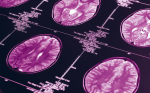
Prior to the development of intra-operative imaging tools, surgery relied on images acquired pre-operatively. However, during the surgical procedure, dynamic changes of the intracranial contents regularly occur, which means that the neurosurgeon is faced with a continuously changing intra-operative field. This phenomenon is often referred to as brain shift . As such, only intra-operatively acquired images will provide the surgeon with the information required to perform real intra-operative image-guided surgery.
With the introduction of intra-operative imaging tools, the ability to image the brain during surgery is now a reality. Although intra-operative ultrasound and intraoperative computed tomography (CT) units have been successfully utilized in neurosurgery, iMRI confers a number of distinct advantages. Its excellent imaging qualities, combined with the avoidance of ionizing radiation, suggest that iMRI will remain the gold standard of neuroimaging for the foreseeable future.
iMRI
MRI was first brought into the operating room (OR) by Peter Black et al. at the Brigham and Women s Hospital, working in concert with General Electric Medical Systems (GEMS). Their device, the General Electric Signa magnetic resonance therapy (MRT) system, employed two superconducting magnets to create a 0.5T magnetic field. The Brigham and Women s Hospital has reported over neurosurgical 800 procedures with the Signa iMRI. These included a wide variety of intracranial operations, aimed mostly at the treatment of patients with brain tumors.
Besides the concept of iMRI itself, the major innovation in the Signa design was the ability to operate within the imaging space itself. Thus, no magnet or patient movement needs to occur in order to acquire an image. The major limitations of this iMRI have been the 56cm gap between the vertically oriented magnets, limiting surgeon access to the operative field, variable image quality, and the need for a fully non-ferromagnetic operating room. The Brigham group and GE, recognizing these concerns, are planning a new generation of iMRI. This system will employ a 3T magnet, provide wider surgical access, and potentially allow for true realtime imaging without the interruption of surgery.
In the wake of this pioneering effort other groups reported on the implementation of other interventional MRI concepts, with a variety of magnetic field strengths, versatility, and ergonomic pros and cons. Another iMRI design that, in essence, required moving an OR to a radiology suite, was developed with Philips Medical Systems at the University of Minnesota. Based on a 1.5T magnet, this unit is housed in the radiology suite, and is converted as needed into an OR.As a dualresource MRI it is used for diagnostic imaging when not used for surgery. Surgical images are of diagnostic quality, and such functional applications as MR spectroscopy, functional MRI (fMRI), and diffusion tensor imaging (DTI) are available. Siemens designed iMRI systems with a relatively narrow horizontal magnet gap. The Siemens system, first described in 1998, featured a 0.2T magnet with a 25cm gap. Early experience with this magnet-involved surgery was carried out in an adjacent room, with the patient moved in for imaging. Later, the concept of operating in the fringe fields, within the same room as the magnet, was described. However, the limitations of this approach were obvious.After a considerable amount of expense and effort, the system provided usable but non-diagnostic quality anatomical images alone. With this impetus Siemens created a 1.5T iMRI. Constructed in a specially designed suite, the fringe fields of this magnet are such that, despite the high field strength, operating with regular instrumentation is possible. The patient is rotated on a special OR table when imaging is required. This system also includes an integrated infrared navigational tool. Using this system, the neurosurgeons at Erlangen- Nurnberg University have operated on over 300 patients, mainly for patients with gliomas and pituitary adenomas. The images are of diagnostic quality and intra-operative DTI has been acquired. These tools share certain disadvantages.They may require the OR team to move to an unfamiliar environment; regular surgical tools may need to be replaced with less durable, non-magnetic copies; the images may be inadequate for surgical decision making; the patient s or the surgeon s position might have to change from the routine; and they are expensive enough so that they cannot be used to define a new paradigm for brain surgery.The author s experience at the New Jersey Medical School began with the first generation 0.12T PoleStar N10 iMRI, developed in Israel by Odin Medical Technologies (now a part of Medtronic Navigation, Louisville, CO). The relatively small (25cm) gap between the vertically oriented magnet poles tended to make positioning difficult, e.g. limiting the degree to which a patient s head could be rotated.Together with a limited field of view (FOV), imaging at or below the skull base was difficult, especially for patients with large shoulders or stout necks.
The first generation system has now been replaced with the PoleStar N20, a compact, mobile iMRI.This system is built around a permanent 0.15T magnet. It is designed to sit under a regular operating table, and is raised as needed for imaging sessions before and during surgery.A portable shield that opens over the table is used, or the entire room is shielded against radiofrequency interference.The PoleStar N20 has an integrated infrared navigational tool, with stereotactic accuracy comparable or better than conventional image guidance systems.
The field of view of the PoleStar N20 is significantly bigger than that of its predecessor, and the magnet gap is 2cm wider. As a result, options for patient positioning are greatly improved, as is image quality. To date, the author has operated on over 160 patients using the N20, for a similar range of procedures as with the N10. Intraoperative images have led to additional resection in 27% of surgeries, with unnecessary dissection being avoided in approximately 12%. The benefits of the improved PoleStar iMRI have come with some cost. The newer unit is some 220kg heavier and sits 8cm higher than its predecessor. These changes are indicative of the tradeoffs inherent in the evolution of iMRI systems. Improved functionality means larger devices.While the PoleStar N20 has achieved a reasonable compromise in this regard, at some point the increasing size of an iMRI may no longer allow it to be a practical surgical adjunct.
Functional Neuroimaging and the Future of iMRI
For patients with brain tumors, surgical resection is often the ideal first treatment; in some tumor types, such as meningiomas, surgery may be curative. However, the proximity of vital brain structures may limit the ideal goal of complete tumor removal with preservation of function. Surgery may not be offered to patients who might benefit from it on the assumption that their tumor is too close to so-called eloquent brain, such as the areas responsible for controlling movement or speech. fMRI, a noninvasive means of identifying cortical areas responsible for such critical functions as movement, vision, or speech, often aids in the surgical decision-making process. DTI, using a different process, can image white matter tracts. By combining these methods a comprehensive functional map of a patient s brain can be created. Intra-operative MRI has the potential to facilitate functional imaging with the ability to routinely acquire fMRI and DTI in the OR, and will provide neurosurgeons with a dynamic functional and anatomical map. This data, properly interpreted, will enhance surgical efficacy and safety. iMRI will have a role in functional neurosurgery as well. When electrodes are to be inserted for the treatment of patients with movement disorders such as Parkinsonism, or for emerging functional neurosurgical applications, iMRI will be a vital means to ensure the correct placement of these or other devices. Increasingly, iMRI will also be used as a tool for patients in whom adjuvant stereotactic radiosurgery (SRS) is planned. Intra-operative images will demonstrate that the surgical goals have been reached in a given procedure, leaving an acceptably small residual target for SRS.
Conclusion
The ability to image the brain in the OR was a logical innovation in the development of neurosurgery. Intraoperative MRI has been marked by a steady decrease in the amount of guesswork . Currently, and for the foreseeable future, MRI is the best means of brain imaging. Ten years after the groundbreaking introduction of iMRI by Peter Black and colleagues, this technology has yet to gain widespread acceptance.
This mainly reflects the cost and effort involved in implementing the rouSince initial reports were published in the 1990s, the use of intra-operative magnetic resonance imaging (iMRI) -guided therapy has continued to grow. While the use of iMRI has been reported in hepatic tumor ablations, endometrial treatment, sarcoma resection, and perirectal disease, it is in neurosurgical procedures that significant advances have been made. Image-guided neurosurgery represents a substantial improvement in the microsurgical treatment of neurological tumors, vascular malformations, and other intracranial lesions.
Prior to the development of intra-operative imaging tools, surgery relied on images acquired pre-operatively. However, during the surgical procedure, dynamic changes of the intracranial contents regularly occur, which means that the neurosurgeon is faced with a continuously changing intra-operative field. This phenomenon is often referred to as brain shift . As such, only intra-operatively acquired images will provide the surgeon with the information required to perform real intra-operative image-guided surgery.
With the introduction of intra-operative imaging tools, the ability to image the brain during surgery is now a reality. Although intra-operative ultrasound and intraoperative computed tomography (CT) units have been successfully utilized in neurosurgery, iMRI confers a number of distinct advantages. Its excellent imaging qualities, combined with the avoidance of ionizing radiation, suggest that iMRI will remain the gold standard of neuroimaging for the foreseeable future.
iMRI
MRI was first brought into the operating room (OR) by Peter Black et al. at the Brigham and Women s Hospital, working in concert with General Electric Medical Systems (GEMS). Their device, the General Electric Signa magnetic resonance therapy (MRT) system, employed two superconducting magnets to create a 0.5T magnetic field. The Brigham and Women s Hospital has reported over neurosurgical 800 procedures with the Signa iMRI. These included a wide variety of intracranial operations, aimed mostly at the treatment of patients with brain tumors.
Besides the concept of iMRI itself, the major innovation in the Signa design was the ability to operate within the imaging space itself. Thus, no magnet or patient movement needs to occur in order to acquire an image. The major limitations of this iMRI have been the 56cm gap between the vertically oriented magnets, limiting surgeon access to the operative field, variable image quality, and the need for a fully non-ferromagnetic operating room. The Brigham group and GE, recognizing these concerns, are planning a new generation of iMRI. This system will employ a 3T magnet, provide wider surgical access, and potentially allow for true realtime imaging without the interruption of surgery.
In the wake of this pioneering effort other groups reported on the implementation of other interventional MRI concepts, with a variety of magnetic field strengths, versatility, and ergonomic pros and cons. Another iMRI design that, in essence, required moving an OR to a radiology suite, was developed with Philips Medical Systems at the University of Minnesota. Based on a 1.5T magnet, this unit is housed in the radiology suite, and is converted as needed into an OR.As a dualresource MRI it is used for diagnostic imaging when not used for surgery. Surgical images are of diagnostic quality, and such functional applications as MR spectroscopy, functional MRI (fMRI), and diffusion tensor imaging (DTI) are available.
Siemens designed iMRI systems with a relatively narrow horizontal magnet gap. The Siemens system, first described in 1998, featured a 0.2T magnet with a 25cm gap. Early experience with this magnet-involved surgery was carried out in an adjacent room, with the patient moved in for imaging. Later, the concept of operating in the fringe fields, within the same room as the magnet, was described. However, the limitations of this approach were obvious.After a considerable amount of expense and effort, the system provided usable but non-diagnostic quality anatomical images alone. With this impetus Siemens created a 1.5T iMRI. Constructed in a specially designed suite, the fringe fields of this magnet are such that, despite the high field strength, operating with regular instrumentation is possible. The patient is rotated on a special OR table when imaging is required. This system also includes an integrated infrared navigational tool. Using this system, the neurosurgeons at Erlangen- Nurnberg University have operated on over 300 patients, mainly for patients with gliomas and pituitary adenomas. The images are of diagnostic quality and intra-operative DTI has been acquired. These tools share certain disadvantages.They may require the OR team to move to an unfamiliar environment; regular surgical tools may need to be replaced with less durable, non-magnetic copies; the images may be inadequate for surgical decision making; the patient s or the surgeon s position might have to change from the routine; and they are expensive enough so that they cannot be used to define a new paradigm for brain surgery.The author s experience at the New Jersey Medical School began with the first generation 0.12T PoleStar N10 iMRI, developed in Israel by Odin Medical Technologies (now a part of Medtronic Navigation, Louisville, CO). The relatively small (25cm) gap between the vertically oriented magnet poles tended to make positioning difficult, e.g. limiting the degree to which a patient s head could be rotated.Together with a limited field of view (FOV), imaging at or below the skull base was difficult, especially for patients with large shoulders or stout necks. The first generation system has now been replaced with the PoleStar N20, a compact, mobile iMRI.This system is built around a permanent 0.15T magnet. It is designed to sit under a regular operating table, and is raised as needed for imaging sessions before and during surgery.A portable shield that opens over the table is used, or the entire room is shielded against radiofrequency interference.The PoleStar N20 has an integrated infrared navigational tool, with stereotactic accuracy comparable or better than conventional image guidance systems.
The field of view of the PoleStar N20 is significantly bigger than that of its predecessor, and the magnet gap is 2cm wider. As a result, options for patient positioning are greatly improved, as is image quality. To date, the author has operated on over 160 patients using the N20, for a similar range of procedures as with the N10. Intraoperative images have led to additional resection in 27% of surgeries, with unnecessary dissection being avoided in approximately 12%. The benefits of the improved PoleStar iMRI have come with some cost. The newer unit is some 220kg heavier and sits 8cm higher than its predecessor. These changes are indicative of the tradeoffs inherent in the evolution of iMRI systems. Improved functionality means larger devices.While the PoleStar N20 has achieved a reasonable compromise in this regard, at some point the increasing size of an iMRI may no longer allow it to be a practical surgical adjunct.
Functional Neuroimaging and the Future of iMRI
For patients with brain tumors, surgical resection is often the ideal first treatment; in some tumor types, such as meningiomas, surgery may be curative. However, the proximity of vital brain structures may limit the ideal goal of complete tumor removal with preservation of function. Surgery may not be offered to patients who might benefit from it on the assumption that their tumor is too close to so-called eloquent brain, such as the areas responsible for controlling movement or speech. fMRI, a noninvasive means of identifying cortical areas responsible for such critical functions as movement, vision, or speech, often aids in the surgical decision-making process. DTI, using a different process, can image white matter tracts. By combining these methods a comprehensive functional map of a patient s brain can be created. Intra-operative MRI has the potential to facilitate functional imaging with the ability to routinely acquire fMRI and DTI in the OR, and will provide neurosurgeons with a dynamic functional and anatomical map. This data, properly interpreted, will enhance surgical efficacy and safety.
iMRI will have a role in functional neurosurgery as well. When electrodes are to be inserted for the treatment of patients with movement disorders such as Parkinsonism, or for emerging functional neurosurgical applications, iMRI will be a vital means to ensure the correct placement of these or other devices. Increasingly, iMRI will also be used as a tool for patients in whom adjuvant stereotactic radiosurgery (SRS) is planned. Intra-operative images will demonstrate that the surgical goals have been reached in a given procedure, leaving an acceptably small residual target for SRS.
Conclusion
The ability to image the brain in the OR was a logical innovation in the development of neurosurgery. Intraoperative MRI has been marked by a steady decrease in the amount of guesswork . Currently, and for the foreseeable future, MRI is the best means of brain imaging. Ten years after the groundbreaking introduction of iMRI by Peter Black and colleagues, this technology has yet to gain widespread acceptance.
This mainly reflects the cost and effort involved in implementing the routine use of iMRI. Those who believe that iMRI is a logical, inevitable progression in image-guided neurosurgery must strive to demonstrate that surgery and patient outcomes are improved by the use of this innovative and exciting technology.tine use of iMRI. Those who believe that iMRI is a logical, inevitable progression in image-guided neurosurgery must strive to demonstrate that surgery and patient outcomes are improved by the use of this innovative and exciting technology.