The author s experience at the New Jersey Medical School has revolved around the PoleStar N20, a compact, mobile iMRI developed in Israel by Odin Medical Technologies. This system is built around a permanent 0.15 Tesla magnet (the first generation of this iMRI contained a magnet with 0.12T strength). It is designed to sit under a regular operating table, and is raised as needed for imaging sessions before and during surgery. A portable shield that opens over the table is used, or the entire room is shielded against radiofrequency interference. The PoleStar N20 has an integrated infrared navigational tool, with stereotactic accuracy comparable or better than conventional image guidance systems.
Case Illustration
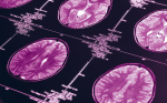
A 35-year-old man complained of headaches and gait difficulty. Diagnostic MRI had revealed an enhancing mass and cyst in the right cerebellar hemisphere. He was positioned prone for surgery and the PoleStar N20 was parked under the head of the operating table (see Figure 1). Reformatted images obtained before surgery showed the lesion in multiple planes (see Figure 2).A hemangioblastoma was removed with an operating microscope. Repeat MRI confirmed that the resection was complete and that the fourth ventricle was opened (see Figure 3).
Discussion
The ability to image the brain in the OR was a logical innovation in the development of neurosurgery, which has been marked by a steady decrease in the amount of guesswork . Currently, and for the foreseeable future, MRI is the best means of brain imaging, although seven years after the groundbreaking introduction of iMRI by Peter Black and colleagues, this technology has yet to gain widespread acceptance. Currently, there are fewer than 50 active iMRI units in the world.A review of the reported experience with various iMRI systems and on iMRI applications may shed light on this issue.
Experience with Other iMRI Units
New Operating Environments
The largest published iMRI experience to date has been with the 0.5T General Electric Sigma iMRI, also known as the double donut . The greatest number of patients (over 800) reported is from the initial site at the Brigham and Women s Hospital. A large series of stereotactic tumor biopsies as well as intracerebral hematoma evacuations in this iMRI at the University of Zurich have also been described.
Besides the concept of iMRI itself, the major innovation in the Sigma design was the ability to operate within the imaging space itself, therefore no magnet or patient movement needs to occur in order to acquire an image. The major limitations of this iMRI have been the 56cm gap between the vertically-oriented magnets, limiting surgeon access to the operative field, variable image quality, and the need for a fully non-ferromagnetic operating room.Two centers that had installed the Sigma iMRI in essence abandoned its use, favoring the PoleStar N-10.The Brigham group and GE, recognizing these concerns, are planning a new generation of iMRI. This will employ a 3T magnet, have wider surgical access, and potentially allow for true realtime imaging without the interruption of surgery.
Another iMRI design that, in essence, required moving an OR to a radiology suite was developed with Philips Medical Systems at the University of Minnesota.
Evidence from another study of 346 operations, including 101 tumor resections and 140 brain biopsies, has been gathered using this system. Based on a 1.5T magnet, this unit is housed in the radiology suite, and is converted as needed into an OR.As a dual-resource MRI it is used for diagnostic imaging when not used for surgery. Surgical images are of diagnostic quality, and such functional applications as MR spectroscopy, functional MRI, and diffusion tensor imaging (DTI) are available.
The PoleStar N20 is under operating table and is covered with a transparent sterile drape.
iMRI in a Neurosurgical OR
Other evidence suggests that iMRIs are designed, like the PoleStar system, to work in a neurosurgical OR, albeit with some modifications. An iMRI built around a superconducting 1.5T magnet, which is housed in an alcove adjoining the OR, has also been developed.
Another design in iMRI – that of a relatively narrow horizontal magnet gap – was developed by Siemens and Hitachi. The former system, first described in 1998, featured a 0.2T magnet with a 25cm gap. Initially designed so that surgery was carried out in an adjacent room, with the patient moved in for imaging, the concept of operating in the ‘fringe fields’, within the same room as the magnet, has been outlined; however, the limitations of this approach were obvious. After a considerable amount of expense and effort, the system provided usable but non-diagnostic quality anatomical images alone.With this impetus the Erlangen group, in conjunction with Siemens, has created a 1.5T iMRI. Constructed in a specially designed suite, the fringe fields of this magnet are such that, despite the high field strength, operating with regular instrumentation is possible. The patient is rotated on a special OR table when imaging is required.This system also includes an integrated infrared navigational tool. They have operated on over 300 patients in this system, mainly for patients with gliomas and pituitary adenomas. The images are of diagnostic quality and intra-operative diffusion tensor imaging has been acquired.
Experience with the Pole Star iMRI
The author worked with the first generation, 0.12T PoleStar N-10 iMRI for 3.5 years. In 184 patients, the great majority of surgery was carried out for tumor resection, with six patients undergoing temporal lobe resection for seizure control and similar numbers having stereotactic biopsies or management of complex hydrocephalus.The relatively small (25cm) gap between the vertically oriented magnet poles tended to make positioning difficult, e.g. limiting the degree to which a patient’s head could be rotated.Together with a limited field of view (FOV), imaging at or below the skull base was difficult, especially for patients with large shoulders or stout necks.
The PoleStar N20 was developed with a 0.15T magnet in order to overcome these limitations. The FOV is significantly bigger, and the magnet gap is 2cm wider. As a result, options for patient positioning were greatly improved, as was image quality.To date, the author has operated on 82 patients using the N20, for a similar range of procedures as with the N10. Intra-operative images have led to additional resection in 25% of surgeries, with unnecessary dissection being avoided in approximately 25%. Additional time resulting from use of the system has been approximately one hour, compared with 80 minutes with the N10.
The benefits of the improved PoleStar iMRI have come with some cost.The newer unit is some 220kg heavier and sits 8cm higher than its predecessor.These changes are indicative of the trade-offs inherent in the evolution of iMRI systems. Improved functionality means larger devices. While the PoleStar N20 has achieved a reasonable compromise in this regard, at some point the increasing size of an iMRI may no longer allow it to be a practical surgical adjunct.
Conclusion
Intra-operative MRI is now a mature technology.Many authors have reported that it is a useful neurosurgical tool for a wide number of applications. The slow pace of the neurosurgical community as a whole in embracing this technology no doubt reflects a view that the benefits of iMRI do not clearly justify the cost and effort involved. Those who believe that iMRI is a logical, inevitable progression in image-guided neurosurgery must strive to demonstrate that surgery and patient outcomes are improved by the use of this innovative and exciting technology.