NF-1
NF-1, also known as von Recklinghausen’s neurofibromatosis, is a common neurocutaneous disorder with an incidence of one in 3,000. Although genetic testing is available, NF-1 remains a clinical diagnosis. Two of seven diagnostic criteria, as developed by the National Institutes of Health (NIH) consensus conference, must be met to make the diagnosis (see Table 1).1,2 Café-au-lait macules are flat, uniformly hyperpigmented lesions, generally with smooth borders, which may be present at birth and typically increase in size and number with age. Other clinical findings develop over the course of childhood and early adolescence, so that 95% of patients with NF-1 have met diagnostic criteria by eight years of age.
NF-1 is an autosomal dominant genetic disorder with complete penetrance; however, as many as 50% of cases are a result of sporadic mutations.3 The NF-1 gene, identified in 1990, is large and has been mapped to chromosome 17q11.2. The types of mutations are diverse, ranging from total deletion of the NF-1 gene to a subtle change of a single base, in 300,000, in the gene. Direct genetic testing now exists and allows confirmation of the diagnosis of NF-1 in patients with only a single clinical manifestation, such as caféau- lait macules.4
One of the hallmarks of NF-1 is that affected individuals develop both benign and malignant tumors at increased frequency. Neurofibromin, the protein product of the gene, is expressed in many tissues.5 Most mutations of the NF-1 gene produce a truncated form of neurofibromin. This has serious implications for tumor formation based on the hypothesis that the NF-1 gene is a tumor suppressor gene. In this role, neurofibromin may downregulate cellular proto-oncogenes that direct cell growth and regulation.6 It is well established that patients with NF-1 have an increased lifetime risk of many other tumor types in addition to malignant peripheral nerve sheath tumors, such as OPGs and brainstem gliomas, pheochromocytomas, and certain sarcomas and leukemias.
OPGs
OPGs are the most common central nervous system (CNS) neoplasm in patients with NF-1. These lesions occur in young children, generally before the age of six, and rarely occur beyond adolescence.7,8 Computed tomography (CT) of asymptomatic patients with NF-1 has shown the prevalence of OPG in NF-1 to be 15% by five years of age, and prospective studies have suggested that the incidence may be even higher. Approximately 47% of patients with NF-1 and a known OPG will develop vision loss.9
OPGs may occur anywhere along the anterior visual pathway including the optic nerves, chiasm, and tracts. Rarely, they may affect the optic radiations.10 However, patients with NF-1 are more likely to experience involvement of the optic nerves and are less likely to develop a chiasmatic tumor compared with OPGs, which occur in non-NF-1 patients.7 Additionally, bilateral optic nerve gliomas (ONGs) without chiasmal involvement are more common in NF-1, comprising 30% of all OPGs in this patient population.11
Most OPGs are benign, low-grade astrocytomas. In the setting of NF-1, and when the neuroradiologic features are typical (see Figure 1 and 2), biopsies of CNS lesions located along the optic pathway are not commonly obtained.11,12 Though often indolent, some OPGs will progress with time; however, progression is less common in patients with known NF-1. One study reported that 87% of children without NF-1 have evidence of tumor progression at a median follow-up of 72 months compared with only 14% of patients with NF-1.13 Unfortunately, the natural history cannot be predicted from pathology and there are no established MRI criteria for the prediction of tumor progression.7 There is a continuous spectrum of growth from a small number of tumors that grow rapidly to a larger majority of tumors, which progress slowly or not at all, to the infrequent, but documented, occurrence of spontaneous regression.12,14,15 Since OPG in the setting of NF-1 is less likely to exhibit rapid progression than in patients who do not have NF-1, treatment is usually reserved for those with evidence of progression of disease or progressive vision loss.16Neuroradiographic Assessment of OPGs i n NF-1
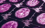
MRI is non-invasive and has a high sensitivity for tissue differentiation and structural definition, making it a useful tool in the diagnosis of OPG in NF-1.17 Demonstration of fusiform dilation of the optic nerves or pathway with or without contrast enhancement is diagnostic of OPGs (see Figure 1). Involvement of the chiasm may also appear fusiform or can appear globular (see Figure 2). The degree of contrast enhancement of OPGs is variable.
Note the right optic nerve nerve is moderately enlarged (arrow) but does not enhance on a) axial and b) coronal images.The left optic nerve is only slightly enlarged.
There has been much debate surrounding the practice of undertaking a screening MRI in asymptomatic patients. A consensus panel convened by the National Neurofibromatosis Foundation (NNFF) did not recommend routine screening and reserved the use of MRI for symptomatic patients or patients found to have an abnormality upon ophthalmologic screen.18 In the contemporary era, many institutions consider young patients with NF-1 to be at such a high risk of OPG that screening MRIs of the brain and orbit is often performed. Additionally, any patient with NF-1 exhibiting signs or symptoms concerning for an intracranial process should be imaged. Whether symptomatic or asymptomatic, many patients with NF-1 will therefore have an MRI performed at some point in childhood.
Neuro-ophthalmologic Assessment
Annual neuro-ophthalmologic or ophthalmic examinations remain the primary screening tool for the early detection of OPGs in children with NF-1. Loss of visual acuity, a visual field deficit, optic disc swelling or atrophy, strabismus, and proptosis are all possible presenting symptoms of an OPG. However, many patients are asymptomatic at diagnosis.The presence of an ophthalmologic complaint brings patients with OPG to diagnosis at an earlier age (median age 1.6 years) than those who have no noticeable clinical symptoms.8 Additionally, patients with an OPG who present with visual pathway symptoms have a higher rate of progression of both tumor growth and subsequent vision loss than asymptomatic patients diagnosed by MRI screening.
While early evidence suggested that the prevalence of vision loss in patients with NF-1 and OPG is approximately 20%, more recent studies indicate that the prevalence is much closer to 50%.8,9,19 Additionally, 28% of children with NF-1 diagnosed with OPG will experience progression of their vision loss within the first year of diagnosis. The likelihood of vision loss is related to both the location and extent of the tumor, with involvement of the chiasmatic and postchiasmatic structures associated with a higher probability of vision loss.9
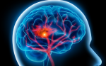
The large heterogenous mass (arrow) contrast enhances on both a) axial and b) coronal T1-weighted MRI sections.
Various methods for assessing visual function have been used as primary outcome measures in published studies of children with OPGs. These include Snellen visual acuity,9 visual fields,20 and visual evoked potentials;21–23 however, visual field testing is notoriously unreliable and poorly reproducible in very young children.Visual field testing should therefore not be a primary outcome measure in this setting.Visual evoked potentials are not widely accepted as an optimal method for following such children because of their relative insensitivity and unreliability.24 It is the authors’ opinion that visual acuity is the most reliable and reproducible measurement in these children.
Serious consideration should be given to the development of an OPG in a young child who has a two-line worsening of visual acuity on Snellen chart examination or an age-appropriate equivalent. In young children who cannot complete Snellen examination, the visual acuities measured from HOTV or Lea testing procedures can be used. In very young children, a preferential looking test (Teller acuity or grating acuity) may be performed. A two octave change is a sufficiently large change in a patient with NF-1 to raise concern over the development of OPG and warrant an MRI examination.
It is currently the authors’ practice to perform neuroophthalmic examinations, including visual acuity measurements, color vision testing, and assessment of pupils, eyelids, ocular motility, fundi, and anterior segments annually in children under the age of eight. If an OPG is present but no visual loss is found, the interval for examinations is six months. After the age of eight, because the likelihood of radiographic and/or visual progression is low, the interval is two years for those without an OPG, and yearly for those with an OPG, until the age of 18. In adults with NF-1, with or without an OPG, no specialized ophthalmic follow-up is necessary, except for routine eye care.
Treatment of OPGs
Patients with an OPG and NF generally have a better prognosis than those without NF-1.25 Additionally, location appears to be prognostic. Children with tumor involvement of the optic nerves have a more favorable prognosis than those with chiasmatic gliomas. Owing to the fact that bilateral optic nerve involvement and chiasmatic location are far more common than unilateral optic nerve glioma, the role of surgical resection is often limited.The authors advocate surgery only for debulking of chiasmal/hypothalamic tumors to relieve obstructive hydrocephalus, or for resection of an optic nerve tumor when vision loss is severe and irreversible, and proptosis is disfiguring or causes corneal exposure.11 The authors have not seen a case of an optic nerve glioma ‘spread’ to involve a previously normal chiasm, so resection of an optic nerve glioma to prevent this is generally unnecessary.
Ideally in NF-1, a period of observation where tumor size and visual function are followed should precede any treatment of young patients with NF-1, due to the previously described natural history. Radiotherapy has been shown to stop the progression of these gliomas and preserve vision.26 Total radiation doses of 5,000–5,400cGy will result in disease stabilization or tumor shrinkage in 80% of patients.27,28 However, in younger patients the long-term cognitive sequelae of radiation can be significant and primary chemotherapy is therefore the preferred option for deferring radiation. A clinical trial involving 58 children with chiasmatic gliomas showed a 58% response rate (RR) to the combination of vincristine and carboplatin.29 While this therapy is never curative, inhibiting or delaying tumor growth may preserve vision and delay use of radiation or aggressive surgical procedures.
Conclusion
OPGs are an important complication of NF-1.Currently, radio- and chemotherapies are helpful but are not always curative.However, it is hoped that future improvements in these techniques will result in better clinical and visual outcomes for patients with NF-1 and OPGs.30–32