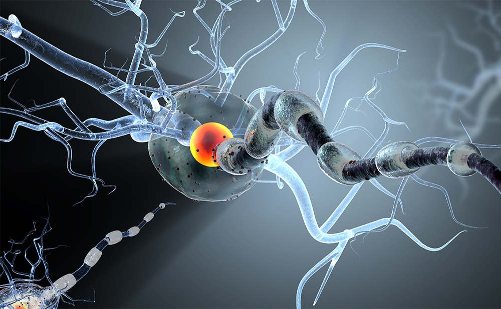
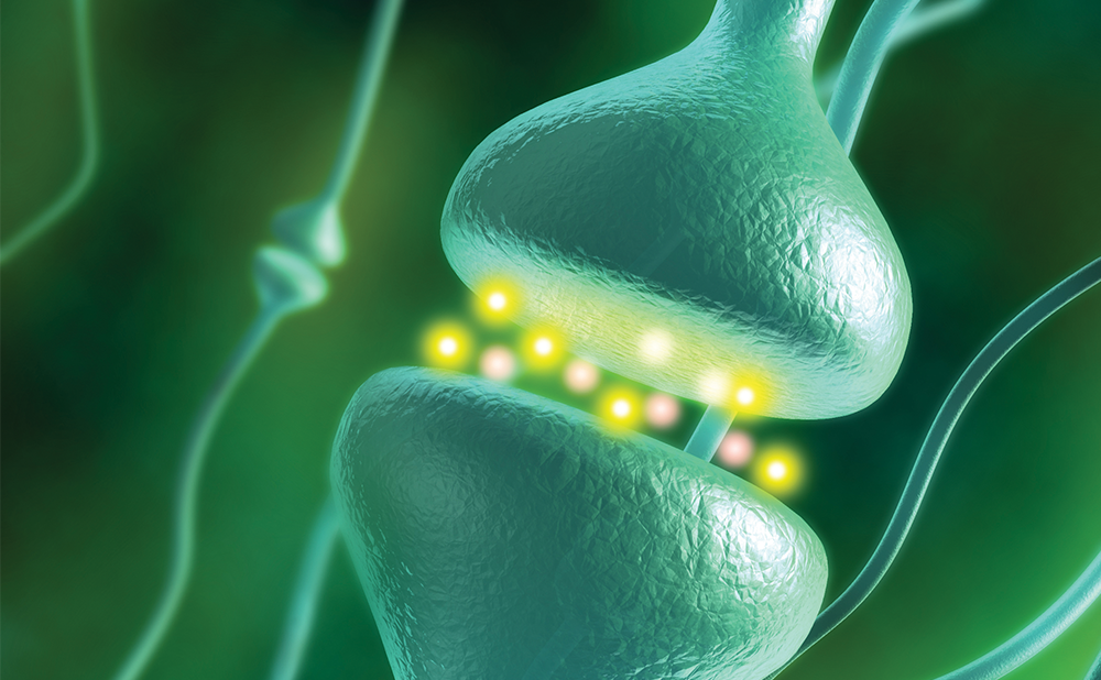
In the adult rodent brain, neurogenesis occurs primarily in the subventricular zone (SVZ) of the lateral ventricle and in the subgranular zone (SGZ) of the dentate gyrus, and neurogenesis persists for the lifetime of the animal.1–9 In the adult human brain, neurogenesis occurs in the hippocampus and SVZ.10 Studies in experimental stroke demonstrate that focal cerebral ischaemia increases neurogenesis in the SVZ and induces SVZ neuroblast migration towards the ischaemic boundary.11–25 Stroke-induced neurogenesis is present in the adult human brain, even in advanced-age patients.26,27 Findings of endogenous neural progenitor cell reservoirs in response to brain injury in the adult brain have raised hopes that amplification of endogenous neurogenesis may replace damaged neurons and stimulate restorative processes in the brain microenvironment, which may subsequently improve neurological outcomes. This article briefly reviews stroke-induced neurogenesis and emerging potential therapies aimed at amplification of endogenous neurogenesis during stroke recovery.
Stroke Induces Neurogenesis
In the rodent, neural stem cells in the adult SVZ generate neuroblasts that travel the rostral migratory stream to the olfactory bulb, where they differentiate into granule and periglomerular neurons.28–30 Neuroblasts generated in the SGZ differentiate into dentate granule cells and integrate into pre-existing neuronal networks. More than 30,000 neuroblasts are generated daily in the rodent SVZ.31,32 Neural stem cells are present in the SVZ of the adult human brain.33,34 Although the cellular composition and cytoarchitecture of the adult human SVZ differ from those of the adult rodent SVZ, the presence of a human rostral migratory stream organised around a lateral ventricular extension to the olfactory bulb has been demonstrated.35
Stroke induces neurogenesis that involves proliferation, differentiation and migration of neural progenitor cells.11–25 Proliferation of neural progenitor cells is tightly controlled by cell cycle kinetics.36,37 Studies in the rodent indicate that stroke reduces the G1 phase of the SVZ neural progenitor cell cycle, resulting in early expansion of a neural progenitor pool in the SVZ.38–40 Neural progenitor cells preferentially differentiate into neuroblasts.38–40 The neuroblasts then migrate out of the SVZ to reach the ischaemic cortex and striatum.41,42 During migration, individual neuroblasts actively change directions by extending or retracting their processes, suggestive of probing the immediate microenvironment.41 Newly arrived neuroblasts in the ischaemic boundary regions exhibit phenotypes of mature neurons.11,12,15,16,18,43 Using the patch-clamp technique, studies show that the new neurons in the ischaemic boundary have electrophysiological characteristics of mature neurons, suggesting that neuroblasts mature into resident neurons and integrate into local neuronal circuitry.44 Ageing decreases neurogenesis in the dentate gyrus and SVZ in the rodent45–48 and stroke primarily occurs in aged patients. Data from the aged rodent show that stroke can augment neurogenesis in aged animals.45–48 However, the degree of stroke-induced neurogenesis in the aged rodent is substantially less than in the young adult rodent.45–48 Stroke-induced neurogenesis has also been demonstrated in the adult human SVZ and ischaemic boundary, even in advanced-age patients.10,26,27,49 The effect of neuroblasts on the ischaemic brain extends beyond the replacement of damaged neurons. Under physiological conditions, neurogenesis in the SVZ and the SGZ of the dentate gyrus occurs within an angiogenic niche.50 Neurogenesis couples with angiogenesis in the ischaemic brain. Neural progenitor cells express an array of angiogenic factors that promote angiogenesis in the ischaemic brain,51,52 while cerebral endothelial cells activated by stroke secrete an array of factors including chemokines and neurotrophic factors that attract migrating neuroblasts to the ischaemic boundary and support the survival and maturation of newly arrived neuroblasts, respectively.50,53,54
Therapies Enhance Endogenous Neurogenesis
Endogenous neurogenesis in response to stroke is limited and only a small population of newly generated neurons survives, while the vast majority of neuroblasts die in the ischaemic boundary regions.16,18,42 There are emerging therapies in experimental stroke which aim to amplify endogenous neurogenesis and to improve the ischaemic microenvironment to be receptive to integration of newly arriving cells within the tissue. These therapies are usually initiated days after stroke, which differ from neuroprotective therapies that start within hours after stroke onset.
Infusion of a variety of neurotrophic and growth factors, including basic fibroblast growth factor (bFGF), epidermal growth factor (EGF) and brain-derived neurotrophic factor (BDNF), into the lateral ventricle of the rodent with stroke further increases neurogenesis.13,55–58 Treatment of stroke in the rodent with bone marrow mesenchymal cells (MSCs) days after stroke stimulates brain parenchymal cells to secrete an array of neurotrophic factors, leading to augmentation of neurogenesis.59,60 A significant improvement in neurological function and enhancement of neurogenesis have been observed even one year after stroke in animals treated with MSCs.61 Patients with ischaemic stroke treated with autologous bone marrow MSCs show no adverse effects and exhibit functional improvement.62 Cerebrolysin is a peptide preparation which has demonstrated robust neurotrophic effects in the rodent.63 Administration of cerebrolysin to the rat 24–48 hours after stroke significantly increases neurogenesis and improves neurological outcome 28 days after stroke.64 Cerebrolysin enhances proliferation and differentiation of SVZ neural progenitor cells and increases numbers of neuroblasts migrating to ischaemic boundary regions.64
Vascular endothelial growth factor (VEGF) is an angiogenic growth factor.65 Intraventricular infusion of VEGF increases neurogenesis in the SVZ and dentate gyrus of adult mice.66 Treatment with VEGF 24 hours after stroke enhances angiogenesis and neurogenesis. 66,67
In addition to its role in erythroid progenitors, endogenous erythropoietin (EPO), through its receptor, EPOR, regulates neurogenesis in the adult rodent brain.68,69 Studies in vivo in the rodent after stroke and in vitro studies with cultured neural progenitor cells and cerebral endothelial cells indicate that exogenous EPO elevates VEGF and BDNF levels in the ischaemic brain, and that EPO-increased VEGF increases angiogenesis. The newly generated vessels produce BDNF which then fosters neurogenesis. In addition, EPO also has direct effects on neurogenesis.68,70–73
The nitric oxide (NO)/cyclic guanosine monophosphate (cGMP) pathway plays dual roles in promoting angiogenesis and neurogenesis in the ischaemic brain. NO is an activator of soluble guanylate cyclase and causes cGMP formation in target cells.74,75 The phosphodiesterase type 5 (PDE5) enzyme is highly specific for hydrolysis of cGMP, and sildenafil citrate and tadalafil are potent inhibitors of PDE5, causing intracellular accumulation of cGMP.76 PDE5 is present in the brain.77 Administration of sildenafil and tadalafil to adult and aged rats one to seven days after stroke increases angiogenesis and neurogenesis and improves neurological outcomes.77,78 Application of sildenafil to a locked-in patient evoked a remarkable recovery.79 A dose-tiered clinical Phase I safety trial of sildenafil in stroke patients is on-going, with patients treated from three to seven days post-stroke. In addition, administration of the 3-hydroxy-3-methyl-glutaryl-coenzyme A (HMG-CoA) reductase inhibitors atorvastatin and simvastatin 24 hours after stroke increases angiogenesis, neurogenesis and brain levels of cGMP.80
Neurogenesis in the adult brain is related to neurological function.81 However, there are currently no data to demonstrate the causality of endogenous neurogenesis to functional recovery after stroke. Neurogenesis enhanced by cell-based and pharmacological therapies is often coupled with angiogenesis. Thus it is likely that improved neurological function observed after these therapies results from a composite of events including angiogenesis, neurogenesis and axonal as well as dendritic plasticity.82 ■


