Recovery of neurological functions after a stroke has long been a puzzling question for clinicians and scientists. On the one hand, clinicians knew from their own practice that partial recovery was very often observed after a stroke and on the other hand, it was well known that neurons, when destroyed after ischaemia, were not restored despite some very localised neurogenesis. In the past two decades, we have learnt from modern neuroimaging techniques, mainly positron emission tomography (PET) scanning and magnetic resonance imaging (MRI), that the human brain is able to spontaneously re-organise after a stroke and that brain re-organisation can be considered as a rational biological basis for recovery of neurological functions. The question of whether lesioned-brain plasticity can be modulated by external factors such as pharmacological agents is now addressed with the aim of improving recovery and reducing the final disability of patients. Preclinical studies, mainly using small animal models, have shown that monoaminergic drugs can modify functional recovery. This is particularly the case for noradrenergic drugs, which have been shown to improve functional recovery, while neuroleptics have been shown to impair it. From this approach, selective serotonin re-uptake inhibitors (SSRIs) were tested and their suspected positive action in the recovery process was recently proved in the Fluoxetine for motor recovery after acute ischaemic stroke (FLAME) trial.
Preclinical Arguments for Direct Action of Monoaminergic Drugs on the Damaged Brain
Studies in laboratory animals clearly show that the rate and extent of functional recovery after focal brain injury can be modulated by drugs affecting certain neurotransmitters in the central nervous system (CNS). Several lines of evidence suggest that motor recovery after injury to the cerebral cortex can be modulated through the effects of norepinephrine on the CNS. For example, in rats, central infusion of norepinephrine hastens locomotor recovery after a unilateral sensorimotor cortex lesion. In contrast, the administration of DSP-4 [N-(2-chloroethyl)-N-ethyl-2-bromobenzylamine], a neurotoxin that leads to the depletion of norepinephrine in the CNS, has the opposite effect and delays the recovery process. In addition, bilateral or unilateral selective lesions of the locus ceruleus, the major source of noradrenergic projection fibres to the cerebral cortex and cerebellum, also impair motor recovery after a subsequent unilateral cortical lesion. Dopaminergic agents also act in damaged brains. They may influence recovery from neglect caused by prefrontal cortical injury. Apomorphine, a dopamine agonist, reduces the severity of experimentally induced neglect, and spiroperidol, a dopamine receptor antagonist, reinstates neglect in recovered animals. Concurrent administration of dopamine-blocking drugs such as haloperidol also blocks amphetamine-promoted recovery and haloperidol, as well as other butyrophenones (fluanisone, droperidol), transiently reinstates the deficits in recovered animals.1–5
The role of antidepressant SSRIs was initially more controversial. Some studies detected little or no significant action on recovery. However, more recent studies have underlined that fluoxetine is active in rat stroke models. Fluoxetine reduces the size of the infarcted zone and demonstrates a strong neuroprotective action through its anti-inflammatory action. Moreover, fluoxetine has been shown to improve cognitive deficits in rats and to stimulate neurogenesis.6,7 From published experimental studies, several main conclusions can be emphasised that have been used in developing SSRI fluoxetine clinical trials.
- First, these studies provide convincing evidence that there is obviously a large interaction between certain drugs and the recovery process in animal models. Norepinephrine and its agonists and antagonists have probably been the most studied drugs but others with potentially fewer side effects, such as SSRIs, could be expected to be beneficial.
- Second, it appears that the cellular mechanisms underlying these significant effects of drugs acting on the CNS is beginning to be better understood. Additional basic research is needed to further investigate such pharmacological actions in the setting of rewiring and cellular growth in the damaged brain.
- Third, drugs can have varying effects according to the dosage and also the dose regimen. For example, animal studies have found that, with increasing dose, amphetamine brings increasing then decreasing benefit.
- Fourth, the timing of drug administration may be crucial. A therapeutic time window probably exists.
- Last, the effects of many drugs are highly dependent on experimental details. For example, drug infusion paired with behavioural training does not have the same behavioural effect as the drug infusion without training.
Monoaminergic Drugs and Motor Recovery After Stroke
Many monoaminergic drugs have been tested in small or middle-sized clinical trials in patients with stroke. Amphetamines were probably the most studied, including a total of 287 patients. Only the first two studies were able to demonstrate beneficial effects. Walker-Batson et al. administered 10 mg D-amphetamine every fourth day, coupled with physiotherapy.8 Changes in motor performance were evaluated with the Fugl–Meyer Motor Scale (FMMS). Subsequent studies failed to show a superiority of D-amphetamine compared with placebo, even though some of these studies used the same protocols as one of the early intervention studies. A recent review summarised that it is currently impossible to draw any definite conclusions about the potential role of D-amphetamine in motor rehabilitation. Methylphenidate produces an increase in dopamine signalling through multiple actions. A prospective, randomised, double-blind, placebo-controlled trial with 21 patients early after stroke indicated that the combination of methylphenidate with physiotherapy over a period of three weeks improved motor function (as measured by the FMMS and a modified version of the Functional Independence Measure) and decreased depression. A subsequent neuroimaging study by Tardy et al. confirmed these findings.9–13
Levodopa gave conflicting results both in single-dose and in repeated-dose trials. A randomised study with stroke patients (n=53) six weeks after stroke onset demonstrated that 100 mg levodopa given once a day over a period of three weeks in combination with carbidopa was significantly better than placebo in reducing motor deficits as measured by the Rivermead Motor Assessment. The improvement persisted over the subsequent three weeks. However, the study results have not been replicated by others up to now and a recent study with subacute stroke patients who received 100 mg levodopa per day for two weeks did not find a stronger improvement of motor functions than in the group treated with placebo.14–18
Very little is known about the mechanism of action of piracetam, but there is some evidence that it enhances glucose utilisation and cellular metabolism in the brain. A Cochrane Review concluded that “treatment with piracetam may be effective in the treatment of aphasia after stroke”.19
Other drugs, such as reboxetine, an inhibitor of the re-uptake of norepinephrine, moclobemide, an inhibitor of monoamine oxidase A, and donepezil, an inhibitor of acetylcholinesterase, have been tested in small series with variable results, which prevent any conclusion being drawn on their efficacy.2,3
Until now, there has been only limited evidence supporting or refuting the use of centrally acting drugs to enhance the effects of neurorehabilitation. Many reasons have been given to explain the difficulties encountered by the investigators: small number of patients, recruitment of patients (25–40 screened for one enrolled), heterogeneity in stroke types, sizes and locations of lesions, concomitant neurological symptoms (within-subject variability in recovery), standardisation of rehabilitation programmes, dose of the drug, specific chemical formulation of the drug under study (D- or DL-amphetamines), time of prescription, duration of treatment, etc. The interpretation is further complicated by conflicting results and the occurrence of side effects (noradrenergic drugs).
Selective Serotonin Re-uptake Inhibitors and Stroke
Selective Serotonin Re-uptake Inhibitors, Stroke and Depression
Post-stroke depression (PSD) is a common disorder, affecting 30–50 % of hemiplegic patients within one year of their cerebral infarction. In the early stage, i.e. during the first three to four months after a stroke, PSD poses serious problems, such as worsened functional and vital prognoses as well as worsened quality of life of the patient and carer. In this context, however, SSRIs were first used as antidepressants and were tested in humans in PSD.
Selective Serotonin Re-uptake Inhibitors as a Treatment for Depressed Stroke Patients
Sixteen trials (17 interventions), with 1,655 participants, were included in a recent Cochrane review. Data were available for 13 pharmaceutical agents. There was some evidence of the benefit of pharmacotherapy in terms of a complete remission of depression and a reduction (improvement) in scores on depression rating scales, but there was also evidence of an associated increase in adverse events. From those series, two studies with the SSRI fluoxetine showed a benefit in depressed patients (Fruehwald et al. and Wiart et al.).20–24
Selective Serotonin Re-uptake Inhibitors Probably Also Prevent Post-stroke Depression
Fourteen trials involving 1,515 participants were included in a recent Cochrane review. Data were available for 10 pharmaceutical trials (12 comparisons) with different antidepressants. There was no clear global effect of pharmacological therapy on the prevention of depression. However, arguments exist for a positive effect of citalopram.25–27
Selective Serotonin Re-uptake Inhibitors and Motor Recovery After Stroke
Small Trials
Few clinical trials with serotonin re-uptake inhibitors have been reported (see Table 1).28–31 They have all included small numbers of patients; all have results that suggest a positive effect on recovery after stroke. In an early trial, fluoxetine and maprotiline were tested against placebo for three months in patients with hemiplegic stroke enrolled one to six months after the stroke. The patients in the fluoxetine group (n=16) had a better outcome than those in the maprotiline or placebo groups.28 Acler et al. confirmed this finding in ten patients in the active-treatment group versus ten in the placebo group.30 In a double-blind, placebo-controlled cross-over trial, Zittel et al. investigated the effects of a single dose (40 mg) of citalopram in eight patients with chronic stroke.29 Dexterity was significantly improved.
Proof of Concept
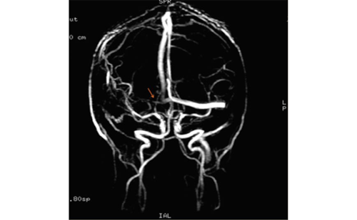
In a double-blind placebo-controlled study by our group, Pariente et al., by combining clinical motor testing and functional MRI motor assessment in patients recovering from post-stroke hemiplegia (n=8), showed that a single dose (20 mg) of fluoxetine improved hand motor function and was correlated with an overactivation of motor cortices on functional MRI (see Figure 1).32 In a subsequent double-blind, placebo-controlled trial in healthy individuals, transcranial magnetic stimulation showed that the intake of a single dose of the serotonin re-uptake inhibitor paroxetine was associated with hyperexcitability of the primary motor cortex, whereas chronic intake was associated with hypoexcitability of the brain motor cortices. Serotonin re-uptake inhibitors increase interneuron-facilitating activity in the primary motor cortex. This study demonstrated that in recovering stroke patients a single dose of 20 mg fluoxetine transiently improved motor deficit and acted directly in overactivating motor cortices through a fluoxetine-induced change in cortical excitability.33
Fluoxetine for Motor Recovery After Acute Ischaemic Stroke Trial
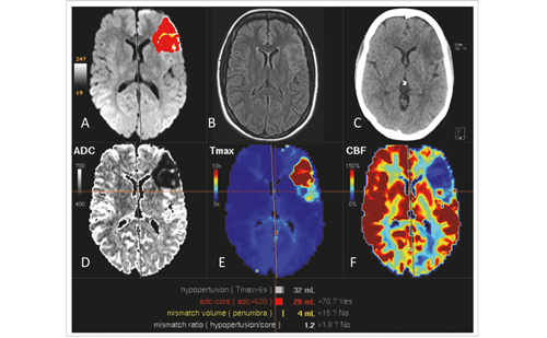
The FLAME trial was then designed to test the efficacy of fluoxetine in motor recovery of patients with ischaemic stroke, as hemiplegia and hemiparesis are the most common deficits caused by stroke.34 Despite the positive small-sized clinical trials and the proof of concept, its clinical efficacy was unknown. The FLAME trial investigated whether fluoxetine would enhance motor recovery if given soon after an ischaemic stroke to patients who had motor deficits (see Figures 2, 3 and 4).
In this double-blind, placebo-controlled trial, patients from nine stroke centres who had suffered an ischaemic stroke, had hemiplegia or hemiparesis, had FMMS scores of 55 or less and were aged between 18 and 85 years were eligible for inclusion. Patients with depression were excluded. Patients were randomly assigned, using a computer random-number generator, in a 1:1 ratio, to fluoxetine (20 mg once per day, orally) or placebo for three months starting five to 10 days after the onset of stroke. All patients had physiotherapy. The primary outcome measure was the change on the FMMS between day 0 and day 90 after the start of the study drug. Participants, carers and physicians assessing the outcome were masked to group assignment. Analysis was of all patients for whom data were available (full analysis set). One hundred and eighteen patients were randomly assigned to fluoxetine (n=59) or placebo (n=59), and 113 were included in the analysis (57 in the fluoxetine group and 56 in the placebo group). Two patients died before day 90 and three withdrew from the study.
FMMS improvement at day 90 was significantly greater in the fluoxetine group (adjusted mean 34.0 points [95 % confidence interval (CI) 29.7–38.4]) than in the placebo group (24.3 points [95 % CI 19.9–28.7]; p=0.003). The drug was well tolerated. Moreover, the number of independent patients (modified Rankin scale [mRS] 0–2 after three months of treatment) was higher in the fluoxetine group. The number of depressions occurring during the three-month treatment period was lower in the fluoxetine group.
The mechanism of action of fluoxetine needs to be discussed. An effect of fluoxetine on mood is likely even in non-depressed people. However, we do not think that fluoxetine acted only through antidepressant mechanisms in this study. As mentioned above, a single dose of fluoxetine improved hand motor function and increased activity in the motor cortex compared with placebo in patients recovering from stroke, showing a specific motor effect, whereas a mood effect is unlikely after a single dose. However, a fluoxetine-mediated attention and/or a fluoxetine-mediated motivation effect cannot be excluded and needs to be investigated in further studies.
Nevertheless, the FLAME trial has limitations. The number of patients included was small. Those who were included were selected for motor deficit and did not represent the general population of stroke patients. Secondly, treatment was stopped after 90 days and we have no idea of the long-term development of patients’ motor function and whether the treatment effect persisted in the months after treatment was stopped. However, the effect of fluoxetine seems to be strong and clinically relevant, and the data of the trial show a global coherence.
In patients with ischaemic stroke and moderate to severe motor deficit, the early prescription of fluoxetine with physiotherapy led to enhanced motor recovery after three months. Modulation of spontaneous brain plasticity by drugs is a promising pathway for treatment of patients with ischaemic stroke and moderate to severe motor deficit.
It is still fair to estimate that no regulatory agency will grant approval for use of such drugs until evidence is also provided by properly powered, formal, phase III clinical trials including a larger number of patients whose characteristics are more similar to those of the general population of patients with stroke. Such trials would probably have to evaluate effects in the long term. ■


