Intravenous (IV) thrombolysis with recombinant tissue plasminogen activator (rt-PA) is one of the most effective stroke therapies yet established. However, current clinical guidelines recommend this therapy only within three hours of the onset of ischaemic stroke symptoms, although, according to a pooled analysis of computed tomography (CT)- based IV thrombolysis stroke trials, the beneficial effect might extend beyond three hours.1 This is supported by the recently published results of the third European Cooperative Acute Stroke Study (ECASS 3), which was Editor’s Choice for The Lancet ‘Paper of the Year’,2 and the Safe Implementation of Treatments in Stroke-International Stroke Thrombolysis Registry (SITS-ISTR),3 which showed the safety and feasibility of IV thrombolysis within a time window of three to 4.5 hours after onset. Nevertheless, this time window is still somewhat limited, so quite a few stroke patients are ineligible for this treatment. Therefore, there is an ongoing need to extend this time window.
The benefit of thrombolysis declines from onset to administration of treatment: the odds ratio (OR) for a favourable outcome with rt-PA is 2.8 (95% confidence interval [CI] 1.8-4.5) for 0–1.5 hours, 1.6 (95% CI 1.1–2.2) for 1.5 to three hours, 1.4 (95% CI 1.1–1.9) for three to 4.5 hours and 1.2 (95% CI 0.995% CI 1.5) for 4.5 to six hours.1 Perhaps counterintuitively, substantial intracranial haemorrhage (ICH), which is a trade-off against a favourable outcome, is not associated with the time between onset and treatment.1 Therefore, in order to extend the time window for thrombolysis, it is important to recruit eligible candidates with the most potential for response to therapy and yet with a relatively low risk of haemorrhage. Modern imaging techniques have made this a realistic possibility.4 Since patients with potentially salvageable cerebral tissue (i.e. those with persistent ischaemic penumbra) are more likely than those without to respond to thrombolytic therapy, established markers that identify patients with this feature are likely to be clinically useful. The mismatch between magnetic resonance (MR) diffusion-weighted imaging (DWI) and perfusion-weighted imaging (PWI) has become the most commonly used technique and is a reasonable marker for the presence of penumbral tissue. In this context, the potential of MR imaging (MRI)- based selection in IV thrombolysis is discussed.
Mismatch Between Diffusion-weighted Imaging and Perfusion-weighted Imaging as a Marker of Penumbra
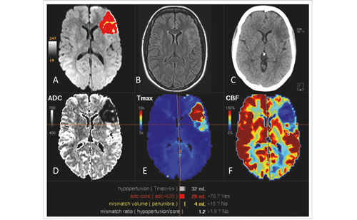
A more complete definition of the ischaemic penumbra is “ischaemic tissue which is functionally impaired and is at risk of infarction but has the potential to be salvaged by reperfusion and/or other strategies. If not salvaged this tissue is progressively recruited into the infarct core, which will expand with time into the maximal volume originally at risk.”5 With the advent of MRI techniques, the concept of mismatch – defined by PWI exceeding DWI lesion volumes – was postulated as a penumbral marker in 1997,6 and has since become the most widely used imaging technique for acute stroke patients.
The DWI image represents the degree of free diffusion of water molecules, which is quantitated by the apparent diffusion co-efficient (ADC) and visualises acute ischaemic lesions within minutes as an increased signal.7 DWI identifies acute infarcts within six hours after onset with high sensitivity and specificity.8 Since the region detected with DWI is irreversible under most clinical conditions, it is a reasonable representative of the infarct core. On the other hand, PWI delineates ischaemic areas as an abnormal perfusion area, which is commonly defined by time–domain parameters on dynamic contrastenhanced imaging, including mean transit time (MTT), time to peak (TTP) or time to maximum plasma concentration (Tmax).9 The current PWI technique involves a bolus injection of a contrast agent (gadolinium), which is rather time-consuming and invasive compared with other non-contrast MRI protocols; although perfusion territory imaging techniques without the used of a contrast agent, such as arterial spin-labelling, have been used in animal models, they are not yet feasible for routine use in humans.10
Uncertainties in Magnetic Resonance Imaging-defined Mismatch
There are several uncertainties with this useful technique. First, DWI-restricted lesions are reversible in some cases receiving thrombolytic therapy,11–14 even for severely reduced ADC values.15 Furthermore, the delayed reappearance of DWI lesions following initial reversal after reperfusion has been reported.16 Second, DWI does not identify acute stroke with 100% sensitivity;17,18 a false-negative DWI study occurs more often in the posterior circulation. Third, PWI shows benign oligaemia as well as true penumbral tissue; therefore, the mismatch could be much larger than the true penumbra. Several thresholds to distinguish between oligaemia and penumbra have been proposed.19–21 Fourth, there are different perfusion thresholds for infarction between grey and white matter;22 hence, tissue-specific rather than whole-brain thresholds may be required to predict the likelihood of infarction. Finally, there is no consensus as to which PWI parameters are the most accurate, reliable or appropriate perfusion measures.23 Despite these uncertainties, mismatch between DWI and PWI remains the most practical tool for identifying at-risk tissue during the early phases of acute ischaemic stroke.
Magnetic Resonance Imaging-based Selection Trials
Several clinical trials with MRI-based mismatch selection have been published or presented; others are in progress or are in the planning stages. In these trials, several outcome measures with MRI are used as clinical surrogates.
The Diffusion and Perfusion Imaging Evaluation for Understanding Stroke Evolution (DEFUSE) trial was a prospective multicentre trial of 74 patients to confirm eligibility for IV thrombolysis with alteplase within three to six hours after onset.24 The results of this trial suggested that this approach was safe and that patients with MRI-based mismatch beyond three hours after onset are likely to benefit from reperfusion therapy, while those without mismatch are rather unlikely to benefit. Therefore, it was considered that MRI-based mismatch might be useful in selecting candidates for IV thrombolysis beyond three hours, and that reperfusion of PWI might be a useful surrogate outcome measure.
The Echoplanar Imaging Thrombolytic Evaluation Trial (EPITHET) was a prospective, randomised, double-blinded, placebo-controlled, multinational trial of 101 patients with mismatch between three to six hours after onset.25 In this trial, reperfusion and infarct growth of MRI were used as surrogate outcome measures between alteplase and placebo. Although the primary outcome of infarct growth showed a trend towards attenuation with alteplase in mismatch patients, this did not reach statistical significance. However, the results of this trial showed that reperfusion was increased by alteplase and was strongly associated with infarct growth attenuation, good neurological outcome and functional outcome. These findings lent strong biological support to the idea that selecting patients with penumbral mismatch beyond three hours might be a useful approach to extending the time window for thrombolytic therapy.
Two phase II trials using the novel plasminogen activator desmoteplase – Desmoteplase in Acute Ischemic Stroke (DIAS) and Dose Escalation Study of Desmoteplase in Acute Ischemic Stroke (DEDAS) – that included patients with PWI-DWI mismatch between three and nine hours after stroke onset have been published.26,27 Both trials showed extremely low rates of symptomatic ICH and better clinical outcome in patients treated with 125μg/kg desmoteplase than placebo. However, DIAS-2, which was a phase III trial to further investigate the clinical efficacy and safety of desmoteplase with a three- to nine-hour time window, showed negative results in the primary outcome of clinical improvement despite the low rates of symptomatic ICH.28 The reasons for the negative results of the DIAS-2 trial are not well understood and we must await the full publication to allow a reasoned analysis. However, possibilities include chance, lack of thresholding for PWI measures, which means that large areas of benign oligaemic tissue may have been included in the selected patients, and limitations of the perfusion measures in terms of the proportion in which CT perfusion was used as a penumbral measure.29
Although door-to-needle time was about 20 minutes longer in MRI than in CT in at least one study,30 the feasibility and safety of MRI-based trials beyond three hours of thrombolysis have been confirmed.4 In planning future trials of IV thrombolysis beyond three hours after onset, MRI-based mismatch and MRI surrogate outcome measures would be useful in selecting patients with adequate penumbra, as well as providing explanatory data about efficacy.
Other Mismatch Models with Magnetic Resonance Imaging Parameters
Several mismatch models with combinations of parameters other than DWI and PWI have been considered. Mismatch between stroke severity, assessed with the National Institutes of Health Stroke Scale (NIHSS) and the DWI lesion (clinical–diffusion mismatch [CDM]), has been suggested as an alternative to PWI–DWI mismatch.31 CDM is based on the assumption that the abnormal PWI volume has a higher correlation with the NIHSS score; however, CDM detected PWI–DWI mismatch with poor sensitivity in several studies.32,33 While patients with CDM tended to have infarct expansion,32 they were less likely to have clinical benefit from reperfusion.33 Identifying the location of the mismatch area rather than the volume may be required to translate the abnormal PWI into clinical deficit.34,35
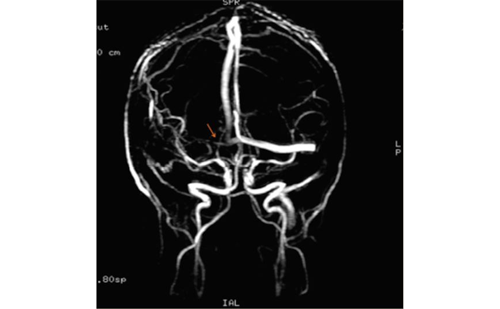
Mismatch between a vessel occlusion detected by MR angiography (MRA) and the DWI lesion (MRA–DWI mismatch) is a newly postulated model and is based on the hypothesis that optimal selection criteria for thrombolytic therapy should include the presence of a vessel occlusion or stenosis and a small lesion.36 In the DEFUSE trial, patients with MRA–DWI mismatch were likely to benefit from early reperfusion.36 This model is less robust in practice than models using PWI, and it selects patients with only moderate sensitivity; that is, there is a risk of excluding patients who might benefit from thrombolytic therapy.
How to Reduce Post-thrombolytic Intracranial Haemorrhage
The risk of ICH increases with thrombolysis,1 and symptomatic ICH has a high mortality rate. However, the majority of patients with rt-PA-related ICH are asymptomatic and benign.37,38 The major purpose of a baseline CT is to exclude those with acute ICH. There were historical concerns that MRI was not sufficiently sensitive to detect acute ICH in the earliest hours after onset. However, with appropriate sequences MRI is superior to CT for the diagnosis of acute ischaemic stroke and chronic haemorrhage, and similar to CT for the detection of acute ICH.18,39
Another purpose of CT for thrombolysis is to detect early ischaemic changes, such as density attenuation, sulcal effacement or ventricular compression, which have been associated with an increased risk of ICH in patients with thrombolysis within six hours.40–42 Although a definite correlation between early ischaemic changes on CT and acute DWI restrictions has not been well established,43,44 large DWI lesions within six hours after onset have been reported as a predictor for subsequent symptomatic ICH.45,46 In the DEFUSE trial, the OR for symptomatic ICH was 1.42 (95% CI 1.13–1.78) per 10ml increase in DWI lesion volume.45
Hypointense lesions on T2*-weighted MRI are thought to represent ‘microbleeds’.47 It is uncertain whether patients with these lesions are at greater risk of post-thrombolytic ICH, but for patients with few lesions this does not seem to be the case.48–50 More recently, some preliminary reports showed that patients with parenchymal enhancement on T1-weighted MRI scan immediately after thrombolysis, which represents blood–brain barrier disruption, had subsequent ICH with 100% specificity.51,52 Currently, there are no validated exclusion criteria for thrombolysis on the basis of MRI alone. Nevertheless, MRI-based trials beyond three hours showed lower risk than CT-based trials for symptomatic ICH.4
Conclusions
The evidence for the clinical effectiveness of MRI-based IV thrombolysis beyond three hours (and now beyond 4.5 hours, following the publication of the ECASS 3 study) has been limited. There is strong biological evidence that the use of imaging techniques, particularly MR using PWI–DWI mismatch, may allow the selection of patients likely to be responsive to thrombolytic therapy beyond these currently established time limits. ■


