Stroke is the leading cause of long-term disability and the second leading cause of death worldwide. According to World Health Organization (WHO) estimates, 15 million people each year suffer from strokes and five million are left permanently disabled.1 Ischaemic stroke is the most common type of stroke, accounting for 65–80% of all strokes. Accumulated data strongly suggest genetic influences in the pathogenesis of ischaemic stroke. Genetic factors could act by modulating the effects of conventional risk factors on the end organs or by a direct independent effect on stroke risk and on infarct evolution in acute phase and outcome.2,3
Apolipoprotein E (ApoE) is a polymorphic glycoprotein associated with the transport of cholesterol and other lipids. In the central nervous system, ApoE is involved in the growth, maintenance and regeneration of both peripheral and central nervous tissue both during development and following an injury.4 Three major isoforms arising from different amino acid substitutions and encoded by the different alleles, ε2, ε3 and ε4, have been identified. The range of ε4 allele frequency is about 5–35% depending on the population studied, and it is approximately 15% in the North American population of European descent.4 Individuals who carry ε4 are at increased risk of various dementias,5 and are also more likely to have impaired recovery of a less specific brain insult following closed head injury,6 intracerebral haemorrhage and subarachnoid haemorrhage.7,8 ε4 has been postulated as a genetic risk factor for human ischaemic stroke in case–control studies9–12 and meta-analyses.13–16 Experimental studies on stroke in ApoE knockout17,18 and transgenic animals19 have shown that the ApoE genotype may affect the infarct volume and outcome. This article provides a review of the literature for magnetic resonance imaging (MRI) evidence of the influence of ApoE on acute stroke and its outcome.
Infarct Volume
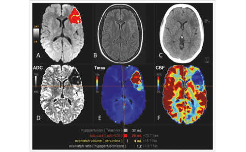
ApoE ε4 is related to a larger infarct volume and worse neurological outcomes in the transgenic mice stroke model.19 In young mice expressing the ε4 isoform, the infarct volume was significantly greater compared with those expressing the ε3 isoform, evaluated 24 hours after middle cerebral artery occlusion for 60 minutes.19 The difference in isoform-specific protections between ε4 carriers and non-carriers in response to focal ischaemia may account for the larger infarct volume; however, the reduced protection effect of ε4 in humans is not straightforward. In a preliminary clinical study,20 the volumes of ischaemic lesions in 31 elderly patients, including nine ε4 carriers and 22 non-carriers, were measured on diffusion-weighted MRI (DWI). The total ischaemic volume was significantly smaller in the ε4 carriers than in the non-carriers at acute stage, but this difference vanished after one week of onset. On average, the total infarct volume increased to 145% of the initial infarct volume in the ε4 carriers, and 84% in the non-carriers. Given the fact that the infarct volume is correlated to clinical condition, a smaller infarct volume at acute stage and statistically equal final infarct volume may explain the findings observed by McCarron et al. that improved survival with increasing ε4 dose21 and functional outcomes are not associated with ApoE genotype.22 On the other hand, the preliminary data of larger infarct growth in ε4 carriers may support the theory that the brain tissue is more vulnerable to damage in ischaemic insult in ε4 carriers.
Perfusion and Collateral Circulation
Clinically, perfusion-weighted MRI (PWI) is usually used to evaluate the perfusion deficit in ischaemic stroke, and the combination of DWI and PWI, the perfusion–diffusion lesion mismatch, can be used to estimate the ischaemic penumbra, i.e. the functionally silent but structurally intact tissue in the hypoperfusion area. Three perfusion parameters on PWI – cerebral blood flow (CBF), cerebral blood volume (CBF) and mean transit time (MTT) – are often used to evaluate the hypoperfusion severity. Reduced relative CBV usually indicates irreversible ischaemic tissue. The brain tissue with reduced CBV at acute stage is believed to have run out of the cerebrovascular reserves.23 The volume of the lesion on the CBV map at acute stage is closest to the final infarct volume in the general population;24,25 however, it seems that this is not true in ε4 carriers. The study by Liu et al. found that although the volume of lesions with reduced CBV at acute stage was significantly smaller in ε4 carriers than in non-carriers, the final infarct volumes had no significant difference between carriers and non-carriers.20 Semi-quantitative measurements showed that under similar ischaemic severity, the brain tissue proceeded to infarction earlier in ε4 carriers than in non-carriers (see Figure 1).26 The observation that in ε4 carriers the CBV viability perfusion threshold is closer to the CBV values of normal uninjured brain than in non-carriers20 provides imaging evidence that the brain tissue is less tolerant of ischaemia in ε4 carriers than in non-carriers.
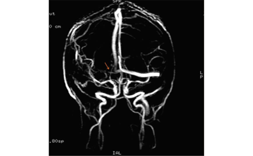
An interesting finding of an MR angiographic (MRA) study in a prospectively selected stroke study population24,26 was that in spite of a more frequent internal carotid artery (ICA) occlusion rate (50%) in the subgroup of ε4 carriers, this group had a high frequency of established collateral middle cerebral artery (MCA) flow. Interestingly, there was actually a significantly higher frequency in establishing MCA flow via collateral circulation in the ε4 carriers than in the non-carriers on day one of onset. All of those ε4 carriers who had an occlusion of the ICA showed collateral MCA flow, while the remaining ε4 carriers showed normal appearance on MRA. On the other hand, among the ε4 non-carriers, one-third had an occlusion of the ICA and only one of them (20%) showed collateral MCA flow.
ApoE ε4 is known to be associated with higher serum cholesterol levels and development of atherosclerosis.4 From this point of view a plausible explanation for the higher frequency in establishing collateral M1 flow in patients with ICA occlusion may be increased cholesterol levels, slow narrowing of the cerebral arteries by atherosclerosis and subsequent gradual development of collateral pathways. Well-established collateral circulation in ε4 carriers may support the observation by McCarron et al. that improved survival with increasing ε4 dose was found in the ischaemic group of acute stroke patients, and that ε4 may also have some favourable effect.21 However, this is contradictory to the findings from stroke models in transgenic mice. This contradiction may account for the species difference or the young age of the animal. In young animals, no haemodynamically significant lesions have been observed, even in ApoE-deficient mice, despite marked hypercholesterolaemia.27–29
Weir et al.30 hypothesised that divergent influences of the ApoE ε4 allele on ischaemic and haemorrhagic stroke survival may result from differences in coagulation profiles. In haemorrhagic stroke patients, ε4 carriers had higher partial thromboplastin time ratios than non-ε4 carriers. Among 529 ischaemic stroke patients, an increasing ε4 allele dose was associated with improved survival after adjusting for baseline National Institutes of Health Stroke Scale (NIHSS) and partial thromboplastin time ratio.
In cerebral amyloid angiopathy-related haemorrhage, a less frequent cause of ICH, the ApoE ε4 allele is thought to enhance amyloid protein deposition in cortical blood vessels, whereas the ε2 allele predisposes amyloid-laden blood vessels to rupture.31 Lemmens et al. obtained brain MRI scans and ApoE genotypes in 334 transient ischaemic attack or ischaemic stroke patients, but found no association between the presence of microbleeds and the ApoE ε4 or ApoE ε2 alleles.32 In the Rotterdam Scan Study with individuals from the general population 60 years of age and over, ApoE ε4 carriers had significantly more strictly lobar microbleeds than non-carriers.33
Clinical Outcome
Evidence has shown that functional outcomes are not associated with ApoE genotype.22 In the McCarron study,22 189 consecutive patients (58 ε4 carriers) with acute ischaemic stroke were prospectively studied. Neurofunctional status and functional independence were not statistically different between ε4 carriers and patients with the ε3/ε3 genotype. In agreement, Liu et al. in their imaging study found that the neurological and functional status at acute stage and at the three-month follow-up did not significantly differ between ε4 carriers and non-carriers, even though the volume of infarcted tissue at acute stage was smaller in ApoE ε4 carriers.20
The detrimental effects of harbouring an ε4 allele can also be observed in the patterns of correlations between imaging findings and neurological and functional status and outcome. Previous studies have suggested that the volume of infarction and haemodynamics at the acute stage of stroke significantly correlate with neurological and functional status and outcome.34–36 DWI lesions have even been proposed as a part of an armoury to predict stroke recovery37 and as a surrogate marker of clinically meaningful lesion progression in stroke clinical trials.38
In a study with DWI and PWI, the measured infarct volumes did not correlate with clinical outcome in the ε4 carriers, either initially or during the follow-up. This was in stark contrast to the non-carriers, in whom strong correlations between the imaging findings and clinical outcome were found. Similarly, in the ε4 carriers the imaging parameters were not significantly correlated with clinical status, such as the NIHSS score at acute stage.20
Prediction of ischaemic lesion growth on the individual level is still challenging,24 and on the basis of the imaging studies mentioned above it seems that such predictions would be especially challenging in individuals who are ε4 carriers. From the clinical point of view, various predictive imaging markers are not reliable to show the clinical outcome profile of an individual patient. Although a well-known confounding factor, ApoE ε4 cannot usually be taken into account at the time of acute stroke to find out the best treatment options or to control the possible side effects of treatments, especially bleeding.
In summary, the ApoE allele ε4 may have both some adverse and some beneficial effects in patients with acute stroke. ApoE ε4 is associated with increased vulnerability of the brain to the ischaemia, which is reflected in relatively greater lesion growth and low threshold for the brain tissue to survive stroke and in the lack of correlations between the imaging findings and clinical outcome in the ε4 carriers. On the other hand, ε4 has at least a seemingly beneficial effect on collateral blood flow, which seems to be better in ε4 carriers compared with non-carriers. In the clinical studies that focus on the relationship between harbouring ε4 and clinical outcomes in ischaemic stroke patients, the adverse and beneficial effects may be compromised. In any case, when possible, ApoE allele ε4 should be classified as a confounding factor when studying stroke either by imaging methods or clinically. ■


