For decades neurology was a specialty field of medicine that was expert in making diagnoses and applying names to esoteric disorders. However, 20–30 years ago a revolution began that included new and exciting imaging techniques, followed by a full armament of medications to treat problems long monitored by clinical neurologists, but rarely treated effectively. A similar revolution is now under way in neurorehabilitation, particularly of stroke patients. Rehabilitation is a relatively new specialty, with the primary origins of the practice only dating back to World War II. During its early stages, the rehabilitation of stroke patients was a ‘high touch’ experience, teaching the stroke survivor how to compensate for their deficits. For example, one-handed shoe tying was taught, long-handled ‘reachers’ and shoehorns were prescribed, and clumsy and uncomfortable splints were applied. For years, conventional wisdom was what I call ‘The Humpty Dumpty Myth’: all the king’s horses and all the king’s men simply could not repair an injured brain. It was thought that the only conceivable option was teaching a patient how to compensate for their deficits. However, the time has now come to forget about these notions and embrace the concept of neural plasticity; that is, the ability of the brain to repair itself.
To comprehend the mechanisms of neural repair it is important to understand two major concepts:
- Collateral sprouting: when nerve fibers (axons) are damaged, they sprout and regrow, similar to a pruned rose bush. The challenge is to direct these new sprouts so that they connect with the correct structures, ultimately leading to functional improvement.
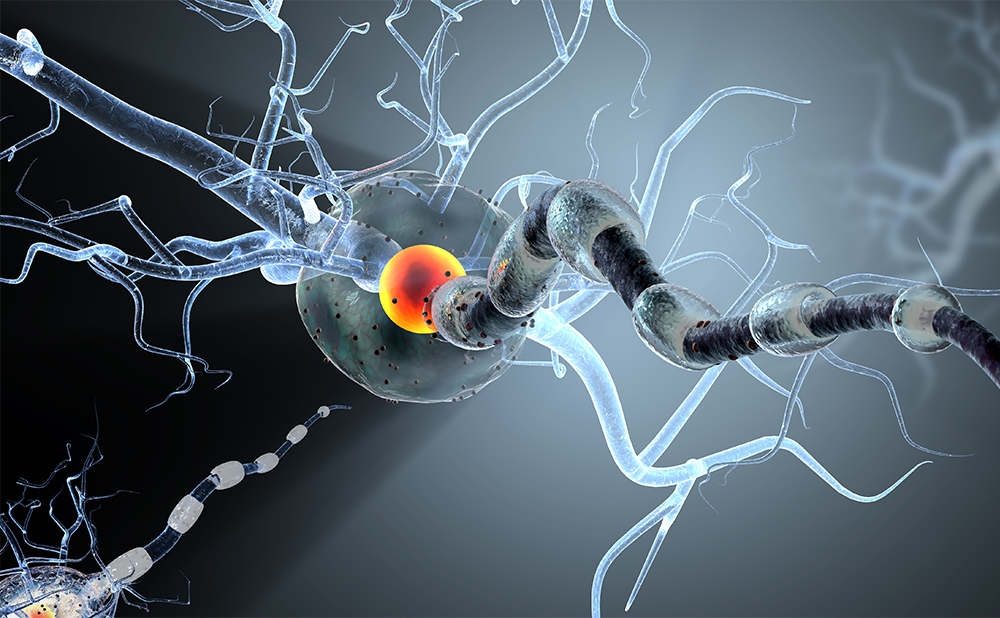
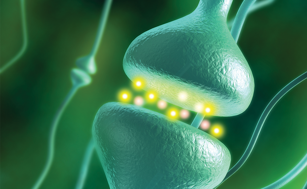
- Neural plasticity: the property of the central nervous system (CNS) to adapt to an injury, lesions, or new environmental demands. After an injury, the CNS attempts to unmask other neural pathways and synapses that can take over from the damaged areas.
In a series of elegant experiments in primates, Randy Nudo showed that neural plasticity and repair depends on the performance of functional tasks and not simply on the use of an extremity.1,2 In his experiments, monkeys that only performed range-of-motion exercises showed minimal improvement, whereas those that performed multiple repetitions of functional tasks made greater functional gains. Nudo also found that adjacent brain areas adopted the function of the damaged brain area in monkeys that received a full rehabilitation program.Use-dependent Plasticity
Nudo’s findings have been termed ‘use-dependent plasticity’, with the concept and key components documented in a paper by Kleim and Jones.3 Neural plasticity is “the mechanism by which the brain encodes experience and learns new behaviors,” and the brain relearns “lost behaviors in response to rehabilitation.”3 Rehabilitation is crucial in the improvement and acquisition of functional abilities. Although the authors describe 10 key components, I summarize three points of importance:
- The task must be functional. Just as Nudo demonstrated, the task performed in rehabilitation must be functional and not just motor use. The learning of specific skills is required to bring about significant changes in neural connectivity. Neural plasticity and repair therefore depend on the performance of specific tasks.
- Dose matters. It is generally accepted that the correct dose of an antibiotic or blood pressure medication is important. It is also generally believed that the dose of exercise matters. In the same vein, the number of repetitions (i.e. dose of rehabilitation) appears to be crucial in driving plasticity and learning/relearning tasks. Kleim and Jones suggest that there is a critical level of rehabilitation and repetition needed for a patient to see continued improvement, and to maintain their functional gains outside of a therapy setting. It is also suggested that there is a prime window of opportunity for optimal neural plasticity, and that early intervention is crucial. Delays in therapy could lead to the development of behaviors that interfere with recovery.
- Motivation. Many physicians and therapists have followed patients who become frustrated and give up on attempting to perform functional tasks. The compensatory strategies taught to them might seem easier, but the patient fails to perform enough repetitions of a functional task. Ways are needed to motivate the patient and to engage them in tasks that have successful outcomes and rewards.
Forced Use/Constraint Therapy
A brief discussion of ‘learned non-use’ and ‘forced-use’ therapy builds upon the aforementioned concepts. Taub and Wolf presented a model in which a person who had had a stroke made unsuccessful motor attempts with their paretic extremity.4,5 This led to negative reinforcement and suppression of the behavior. A therapist then became involved who taught compensatory motor strategies that had positive outcomes and reinforcements. Although not a functional task, the patient was able to complete the task with compensatory strategies. The less effective strategy was strengthened and the ‘potential ability’ was masked. In other words, there was a reservoir of abilities waiting to be tapped. This leads to the next logical question: how does one encourage patients with limited functional movement to perform functional tasks with multiple repetitions, particularly during the early stages of injury? The marriage of ‘high tech’ and ‘high touch’ provides a solution.
The Challenge—Flaccid Extremities, Paraplegia, and Quadriplegia
One can see how one might design tasks for patients who have enough residual function to perform functional tasks. For years, therapists have been limited in their ability to replicate functional tasks in patients with no movement or limited voluntary movement. As discussed above, it takes a certain number of repetitions of a functional task to drive neural repair. However, the range of motion of an upper extremity on a table (or a lower extremity on a mat) does not achieve this goal. This led to the development of a neuroprosthesis, which enables the patient to perform functional tasks with high repetitions, even when they have minimal voluntary movement.Functional Electrical Stimulation
Electrical stimulation has been used for over a century to treat neural conditions. Low levels of electrical current are used to stimulate physical or bodily functions lost through nervous system impairment.
For years, electrodes were hooked up to single muscles or groups of muscles to facilitate increased movement. The therapist turned on the current while asking the patient to extend their wrist. This treatment operated under the idea that repeated use would strengthen the muscle. Conventional surface neuromuscular electrical stimulation (NMES) is limited by the difficulty in placing the electrodes consistently in the right places, and the failure to perform a coordinated task. What is needed is NMES that will enable the patient to perform a coordinated functional task with the critical number of repetitions (i.e. dose). The repeated movements induced by NMES will reinforce network patterns and lead to enhanced synaptic connections and neural plasticity.
Technological Advances in Upper Extremity Rehabilitation
Functional electrical stimulation (FES) enables therapists to combine NMES with task-specific training. FES can be delivered through a neuroprosthesis that allows a patient with limited or no movement to perform functional tasks over and over again. A new, noninvasive neuroprosthesis, the Bioness H200® Hand Rehabilitation System, provides reproducible, synchronized electrical stimulation of the flexor and extensor muscles of the affected arm so that the patient can perform a variety of functional tasks (see Figure 1). The NESS H200 is useful in promoting motor recovery not only in patients with stroke, but also in those with traumatic brain and spinal cord injury. The NESS H200 has five electrodes that come in different sizes, enabling therapists to ‘custom fit’ a patient. The electrodes are positioned over the extensor digitorum, extensor pollicis brevis, flexor digitorum superficialis, flexor pollicis longus, and thenar muscles. By customizing the size and location of the electrodes, the patient receives a consistent level of stimulation every time they use the device. The NESS H200 is programed to alternate between finger/wrist extension and finger/wrist flexion.
Once the patient has been properly fitted with the NESS H200, the therapist designs a task-specific program, which might include:
- grasping, holding, and releasing large objects, such as soft Nerfballs;
- picking up and moving small objects on a table;
- pinch grips to stack or lift, or performing overhead activities; and
- dressing, grooming, eating, opening bottles, and self-feeding.
One is limited only by the imagination of the therapist and the patient. The real breakthrough is that the NESS H200 meets the three crucial goals of function, dose, and motivation. Even a patient with a flaccid hand can pick up an object and perform functional tasks with enough repetitions to drive neural repair. The positive reinforcement of successfully completing these tasks over and over again provides motivation that I have not seen before in my clinic; an increasing number of studies are also confirming these results.6–9 Patients are also able to use the device at home, enabling them to increase their repetitions and incorporate treatment into their daily activities.
Technological Advances in Lower Extremity Rehabilitation
Orthotics and braces have been the mainstay for therapists and physicians in compensating for weakness in the lower extremity. The progression from a metal brace to a lightweight plastic brace was seen as a major accomplishment. However, the traditional ankle–foot orthosis (AFO) has many drawbacks. The patient is typically ‘fitted’ with an off-the-shelf AFO to use at the hospital, but then has to wait until discharge to be ‘fitted’ again with a custom AFO that can be placed inside their shoe. It is often necessary to have to buy two pairs of shoes because the AFO requires a half-size larger. The patient also walks in an unnatural, stiff manner, with a fixed ankle. FES once again solves the problem in the lower extremity. Another neuroprosthesis, the NESS L300® Foot Drop System, again from Bioness, is useful for individuals suffering from the effects of stroke as well as those with traumatic brain injury and multiple sclerosis. The NESS L300 includes an electronic orthosis, a control unit and a gait sensor (see Figure 2).A single, attractive unit wraps around the leg just below the knee, with stimulating electrodes over the peroneal nerve and the anterior tibialis muscle (see Figure 3). The gait sensor consists of a lightweight pad that is placed under the patient’s heel and is connected to a small sending unit. When the patient advances their leg and pressure comes off the heel switch, a signal is sent to the stimulating electrodes, causing dorsiflexion of the ankle. As the leg swings through the gait cycle and the heel strikes the ground, heel switch contact causes stimulation to cease, and the foot returns normally to the ground. Patients quickly develop a more normal gait pattern and, given the choice between the FES neuroprosthesis or a regular AFO, they consistently prefer the NESS L300. They have the ability to walk in similar-sized shoes, walk further and more frequently, and, most importantly, are more likely to avoid falls.10–12
There Is Always More to Do
New technologies are only just beginning to scratch the surface of what can be achieved in rehabilitation. Robotics, mechanized ambulation, virtual reality, and mental practice all enable the patient to meet the criteria of task-specific therapy. Physicians and therapists consistently give up too soon on their patients, and leave them with a reservoir of untapped abilities. Remember the old commercial that stated “It is not your father’s Oldsmobile”? This is not your ‘father’s rehabilitation’, either. New technological advances offer patients opportunities that did not exist even five years ago. One must be certain that they get that opportunity.


