Alzheimer’s disease (AD) is a complex, heterogeneous age-related disorder and the most common form of dementia. It is characterised clinically by a decline in cognitive and functional ability and the development of behavioural and psychological symptoms. At a cellular level, it comprises degeneration of neurons and synapses, formation of amyloid plaques and intracellular neurofibrillary tangles.1
Alzheimer’s disease (AD) is a complex, heterogeneous age-related disorder and the most common form of dementia. It is characterised clinically by a decline in cognitive and functional ability and the development of behavioural and psychological symptoms. At a cellular level, it comprises degeneration of neurons and synapses, formation of amyloid plaques and intracellular neurofibrillary tangles.1
Diagnosis of AD requires neuropsychological testing, limited laboratory tests and brain imaging. Since pathology generally precedes symptoms, AD is diagnosed after clinical onset of the disease, at which stage the patient is already suffering from cognitive defects such as mild cognitive impairment (MCI).2 Moreover, the diagnostic accuracy of neurodegenerative disorders is low, ranging from 50 to 85%.3
Current therapeutic options for AD are symptomatic treatments that target dysfunctional neurotransmitters associated with the disorder. The only approved therapies are cholinesterase inhibitors and one N-methyl-D-aspartate receptor antagonist. Use of these agents is widespread and long-term;4 however, they confer only moderate clinical effects in patients with mildly to moderately severe symptoms and do not affect the fundamental pathology of AD.5 The majority of recent research has focused on therapeutic strategies that inhibit the production and aggregation of amyloid beta protein (Aβ) in plaques and increase its clearance from the brain. Such strategies are likely to be most effective at pre-clinical stages of the disease, before widespread synaptic and neuronal loss occurs.1
Therefore, there is a need for biomarkers that predict the disease course and outcome and monitor disease progression and treatment efficacy. Biomarkers could also identify AD at an early stage and improve diagnosis. Biomarkers could also predict the disease course and outcome. Disorders such as diabetes have identifiable biomarkers that not only can be followed easily and repeatedly for diagnosis, but also can monitor therapeutic response. The development of such biomarkers for AD is critical to translating the efficacy of new therapies.
This article will outline the challenges and limitations of the current knowledge of AD biomarkers and discuss the validity of biomarker candidates as surrogate end-points in clinical trials.
Requirements of Biomarkers
Biomarkers have typically been selected by a ‘candidate’ approach in which a hypothesis about the disease is tested in order to identify a gene or molecular component of the disease process.3 Established criteria exist for potential biomarkers of AD: they should reflect a neuropathological characteristic of AD and should be validated in patients with independently diagnosed AD pathology. Ideally, they should have sensitivity of ≥85% and specificity (to differentiate from controls and other forms of dementia) of ≥75%.6
Neurochemical Biomarkers
It is widely considered that the deposition of Aβ peptides in the brain is a key event in the pathogenesis of AD, and Aβ is secreted by cells, therefore it is a logical choice of biomarker. It is the main component of AD plaques and generally has a length of 42 or 40 amino acids (Aβ1–42 and Aβ1–40). A number of studies, involving a total of around 2,000 patients and controls, demonstrate a 50% reduction of Aβ1–42 in the cerebrospinal fluid (CSF) in AD patients compared with controls of the same age, with sensitivity and specificity levels ranging from 80 to 90%. The full reason for the reduced levels in AD is unclear,7–10 but it is speculated that Aβ1–42 aggregates within plaques reducing the freely available fraction detectable in CSF assays.
The main marker of neuronal pathology is the microtubule-associated tau protein, which is often present in a hyperphosphorylated form in AD patients. Most studies have focused on total tau (T-tau) and hyperphosphorylated tau (P-tau), particularly the subtypes hyperphosphorylated at threonine 231 (P-tau231) and at threonine 181 (P-tau181). Approximately 50 studies involving 5,000 patients have shown that CSF levels of T-tau are increased by approximately 300% in AD patients compared with controls, with sensitivity and specificity levels similar to those observed for Aβ1–42.7,10 However, levels of T-tau in control groups typically increase with age, therefore it is a better discriminator in patients <70 years of age.11 Increases in P-tau in AD patients have also shown high sensitivity and specificity, and are particularly useful in distinguishing AD from other forms of dementia.10
Clinical trials of anti-AD agents require participants with mild symptoms who are likely to display measurable cognitive decline in the course of a study. Neurochemical biomarkers have the potential to identify such individuals and predict AD. Tau protein and Aβ peptides have been used as predictors of AD in those with MCI, considered a transitional clinical state between normal ageing and mild AD.12 Recent studies of CSF in patients exhibiting mild AD have found lowered Aβ1–42 levels, high T-tau or P-tau181 levels or high T-tau:Aβ1–42 ratios, suggesting that these biomarkers may quantitatively predict progression of cognitive deficits and dementia.13 A previous study found that Aβ1–42, but not T-tau or P-tau, may be used to predict cognitive decline in healthy elderly individuals.14
Combined biomarker levels have been shown to be better diagnostic predictors of AD than single measurements. The ratio of Aβ1–42 to Aβ1–40 is more accurate than Aβ1–42 level alone, and the combination of this ratio with T-tau levels further improves accuracy.15,16 AD has been distinguished from dementia with Lewy bodies (DLB) by using the ratios of Aβ peptides of varying lengths (Aβ1–42:Aβ1-38 and Aβ1–42:Aβ1–37) and T-tau.17 Very high sensitivity and specificity of 92 and 89%, respectively, have been reported using combined biomarker measurements.18 Furthermore, clusters of biomarker levels have been correlated to cognitive profiles of AD patients. A study of 177 patients identified a subgroup displaying a distinct cognitive profile with severe impairment of memory, mental speed and executive functions. This was associated with low levels of Aβ1–42 and extremely high levels of T-tau and P-tau181, and showed no link to disease duration or severity. These findings have important implications: AD is a heterogeneous disease and future research is likely to focus on individualised therapy.19
In order to be clinically meaningful, the intra-individual variation of biomarker levels over time must be low. A two-year study of 83 patients with MCI analysed levels of T-tau, P-tau181 and Aβ1–42 and found that intra-individual levels of these biomarkers were highly stable over this time.20 In another study including 105 patients attending a memory clinic, the levels of Aβ1–42, tau and ptau-181 in CSF were measured at baseline and at 21±9 months later (50 patients had AD, 38 had MCI and 17 had subjective complaints [SC]).21,22 Over time, in each of the three patient groups there was a slight increase in Aβ1–42 and tau (4–72 and 49–143pg/ml, respectively), but there was little change in ptau-181. The differences between patient groups in these parameters exceeded the changes over time within each group. Therefore, the authors concluded that repeatedly monitoring these biomarkers is not useful because they are insensitive to disease progression. Further work showed that change in CSF biomarker levels was not related to change in Mini-Mental State Examination (MMSE) or to atrophy rate.23
One disadvantage of CSF biomarkers is the requirement for a lumbar puncture, a time-consuming, invasive procedure that could restrict the use of CSF biomarkers in large clinical trials and has the side effect of post-lumbar-puncture headache (PLPH). However, the use of smaller needles and modern atraumatic techniques has reduced this risk, and the incidence of PLPH has been shown to be lower, particularly in elderly subjects with cognitive disturbances.24 While blood or urine sampling would be more convenient, the much larger volumes of liquid involved mean that T-tau and P-tau levels are diluted and become undetectable by enzyme-linked immunosorbent assay (ELISA). Aβ peptides can be detected in plasma at concentrations 20–30-fold lower than in CSF, but there is no correlation between Aβ levels in plasma and CSF, and the reduction of CSF Aβ1–42 in AD patients is not reflected by a similar reduction in plasma levels.25
A recent worldwide multicentre comparison of assays for CSF biomarkers in AD highlighted another limitation of their use, namely the high intercentre coefficient of variance (CV). The intercentre CVs were 30% for Aβ1–42, 21% for T-tau and 13% for P-tau in 2004, although by 2008 they had fallen to 21, 15 and 9%, respectively. High intracentre CVs were also observed, at 25, 18 and 7% for Aβ1–42, T-tau and P-tau, respectively. Therefore, there is a high variation in test results both between and within centres. In order to reliably demonstrate differences between groups, a CV of <10% is required. There is a need for standardisation of the analytical procedures employed.26
Several other candidates for AD biomarkers have been investigated but none have as much clinical data as Aβ peptides and Tau. These include pro-inflammatory cytokines,27 isoprostanes28 and β secretase.29 A further useful biomarker in AD is serum amyloid P component (SAP). In a study on 241 patients (67 with AD, 144 with MCI and 30 controls), SAP was determined in CSF samples at baseline and at follow-up (2.6±1.0 years for AD patients and 2.1±0.8 years for MCI patients).30 There were no differences in SAP between the AD and MCI groups. Patients with MCI who had developed dementia at follow-up had lower SAP levels (13mg/l, range 3.3–199.3mg/l) than those not developing it (20.2mg/l, range 7.0–127.7 mg/l; p<0.05). In addition, low CSF SAP levels were shown to be correlated with a two-fold increased risk of progression to AD (hazard ratio 2.2). It was suggested that SAP could be used to identify AD patients among a population with MCI.
Neuroimaging Techniques
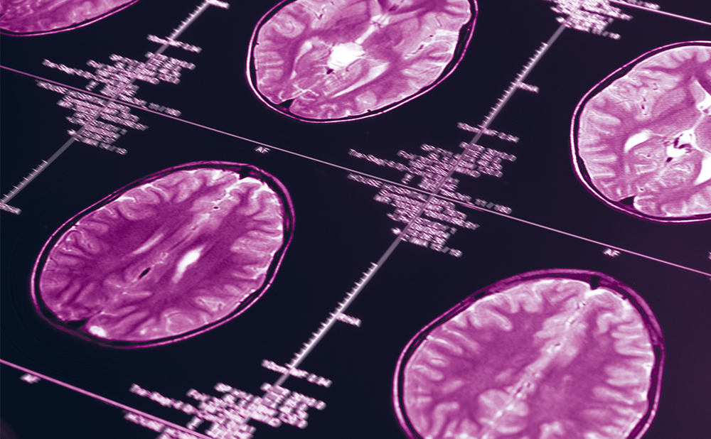
In vivo neuroimaging techniques have become valuable candidate biomarkers in the determination of structural changes in the brain which aid diagnosis of AD. The two most widespread neuroimaging techniques are magnetic resonance imaging (MRI) and positron emission tomography (PET).
Magnetic Resonance Imaging
Brain atrophy starting in the medial temporal lobe and ultimately resulting in global atrophy is characteristic of AD. MRI has shown that there is significant atrophy of the hippocampal formation in pre-clinical stages of AD and can predict later development of AD with about 80% accuracy.31,32 The entorhinal cortex, a structure adjacent to the hippocampus, has also been the focus of imaging studies, as it is thought to undergo degenerative changes at an early pre-clinical stage.33 MRI has also been used to identify a pattern of regional atrophy in MCI patients that is predictive of AD development.34 Such methods currently require manual analysis, which is time-consuming and unlikely to become routine, although automated analysis tools are becoming available.35
One automated method that is well established is the measurement of whole brain volume over time. There is an atrophy rate of around 2.5% in AD patients over one year compared with 0.4–0.9% in controls, which has been used as a secondary end-point in some clinical trials. However, the method is limited by its inability to show regionally differentiated effects.10 Other automated methods currently under investigation include: voxel-based volumetry, which measures a reduction in cortical grey matter in the mediotemporal lobes and lateral temporal and parietal association areas in AD patients;36 deformation-based morphology, which has potential as a technique for predicting risk of converting from MCI to AD;37 and analysis of cortical thickness, which has been shown to distinguish between AD and healthy controls with 90% accuracy.38
Positron Emission Tomography
Reduction in the cerebral metabolic rate of glucose, a measure of neuronal function, is a consistent feature of AD. In vivo brain fluorodeoxyglucose-PET (18FDG-PET) imaging demonstrates consistent and progressive cerebral glucose metabolism reductions in AD patients, the extent and topography of which are associated with symptom severity. Increasing evidence suggests that these reductions occur at the pre-clinical stages of AD and may predict cognitive decline.39 Significant reduction in cerebral glucose metabolism in the parietal lobes have been demonstrated in mild to moderate AD.40 PET is expensive and not yet widely available. Although no large multicentre trials have involved PET as yet, several studies have employed the technique in the evaluation of systemic therapies, including one that employed the technique in measuring progression AD in both untreated patients and those treated with rivastigimine.41 A further trial determined the effects of treatment of AD with donepezil on cortical metabolism using 18FDG-PET.42
PET may be used to image intracerebral amyloid, which not only has important diagnostic implications but may also have applications in clinical trials of amyloid-related agents.43 Studies using the PET tracer Pittsburgh Compound B (PIB) have demonstrated increased brain uptake of PIB in MCI cases, indicating the presence of early AD.44–46 PET has also been used to image anticholinesterase activity in AD.47
Combined Biomarker Approaches
Since combined measurements of CSF biomarkers improve accuracy in diagnosis of AD, combinations of neurochemical measurements and imaging parameters may achieve a more accurate early and differential diagnosis than individual methods. Combined measurements of the CSF T-tau, Aβ1–42, P-tau profile and regional cerebral blood flow or mediotemporal lobe atrophy have proved to be greater predictors of AD development in MCI patients than either technique alone.21,48 The value of using combined biomarkers was also demonstrated in the Development of Screening guidelines and Criteria for Pre-dementia in Alzheimer’s disease (DESCRIPA) study.49 In this European study, an abnormal Aβ:tau ratio was termed an ‘AD profile’ and occurred more frequently in patients with SC (31 of 60 [52%]), non-amnestic MCI (naMIC, 25 of 37 [68%]) and amnestic MCI (aMCI) (56 of 71 [79%]) than in healthy controls (28 of 89 [31%]).1–42 The AD profile was associated with different aspects of cognitive decline in both naMIC and aMIC patients. In aMIC patients an AD profile was also seen to be predictive of AD-type dementia.
Apolipoprotein E Genotype
The apolipoprotein E (APOE) β4 genotype is an important risk factor for AD and is associated with increased deposition of Aβ peptides and tau.50,51 Association between CSF biomarkers and APOE genotype is modified by age in both controls and AD patients, suggesting that cognitively healthy APOE β4 carriers are more prone to developing AD pathology with ageing.52 This article does not discuss genetic parameters; however, the relevance of APOE should be mentioned, since it can affect activity or level of expression of biomarkers, and therefore is often included as a covariable in biomarker studies. A recent Dutch study investigated whether patient age affects the relationship between APOE genotype and the CSF biomarkers amyloid-β1-42, tau and P-tau 181 both in AD patients (n=302) and in healthy controls (n=174).52 Among controls, older age and APOE β4 were associated with lower β1–42 but higher tau and ptau-181 levels (p<0.05). Older carriers had higher levels than older non-carriers of tau and ptau-181, but this was not the case in younger controls. In AD patients, the strongest effect on β1–42 was the APOE β4 genotype; older carriers had lower β1–42 than older non-carriers; but this was not the case in younger AD patients (p<0.05). The study indicated that cognitively unimpaired APOE β4 carriers are more likely to develop AD pathology as they become older and supported the existence of subtypes within the disease.
Surrogate End-points and Clinical Trials
Currently, the primary end-points for clinical trials of AD drugs are clinical outcomes. However, measurements of the severity of cognitive decline present difficulties as markers of progression. Recent trials with disease-modifying treatments have shown negative results, including the amyloid-lowering agent tarenflurbil53 and rosiglitazone.54 Negative results could prove inefficacy of the drug studied, but may reflect inadequacies of the study designs: poor choice of end-point, late initiation and/or short duration.55 A systematic review of clinical trials conducted on cholinesterase inhibitors found that the methodological quality of many trials was poor, and cited numerous reasons for this, notably use of multiple primary end-points, incomplete data due to drop-outs and subjective assessment measures. It concluded that the recommendation of anticholinesterase inhibitors does not seem to be evidence-based.56
There is clearly a need for biomarkers to act as surrogate end-points, i.e. a well-characterised biomarker that can act as a primary end-point in clinical trials. Although surrogate end-points are by definition biomarkers, not all biomarkers meet the requirements of a surrogate end-point, namely ease of measurement, accuracy, reproducibility and representation of clinical benefit.57 The effect of therapy on a surrogate end-point must reliably predict the effect on a clinical outcome. It is highly unlikely that a single surrogate end-point will capture all of the pharmacological benefits and adverse effects of a drug in a diverse population, and combinations of biomarkers will probably be required.
In addition to their use as surrogate end-points, biomarkers may be employed in clinical trials as markers of disease progression, in which case treatment would be expected to slow the rate of change of the biomarker, e.g. hippocampal atrophy. Since individual biomarkers correlate to specific brain pathology, they may be used to classify subjects into groups based on the underlying AD pathology.58 Furthermore, they could potentially shorten the time-frame of clinical trials from years to months, an attractive prospect given the slow rate of clinical progression of AD. Another useful function is as predictors of future AD or identification of Alzheimer’s pathology in non-demented patients, allowing high-risk individuals to be selected for trials and therefore enriching the sample cohort. In previous trials based on MCI selection, half of the patients did not develop AD, impairing identification of drug efficacy.59 Diagnosis of AD based on biomarkers is superior to using the intrinsically heterogeneous MCI state, and could eventually make it superfluous as a diagnostic state.60 Most importantly, biomarkers are the only means of demonstrating that a therapeutic intervention reaches the pathological mechanism of the disease.
An example of the successful use of biomarkers in clinical trials was a recent randomised, double-blind, placebo-controlled pilot study in which 36 patients with moderate AD symptoms were given memantine 20mg/day or placebo. Patients were evaluated at baseline and 26 and 52 weeks using a number of outcome measures, including global and regional glucose metabolism measured by PET and total brain and hippocampal volumes measured using MRI. These imaging techniques proved useful in demonstrating the efficacy of the drug and were considerably more reliable than chemical shift-imaging-derived global and regional N-acetylaspartate and myoinositol concentrations.61
Large-scale international controlled multicentre trials, such as the US, European, Australian and Japanese Alzheimer’s Disease Neuroimaging Initiative62 and the German Dementia Network, are involved with phase III development of the key CSF biomarker and imaging candidates in AD. Furthermore, biomarkers are in the process of being implemented as primary outcome variables into regulatory guideline documents regarding study design and approval for compounds claiming to modify the disease.10
Summary and Conclusions
Currently, clinical assessment is the standard means of diagnosing and assessing AD progression. However, mounting evidence supports the fact that the analysis of CSF biomarkers such as Aβ1–42, T-tau and P-tau, together with neuroimaging techniques such as MRI and PET, performs well in the diagnosis of AD, even in its early clinical stages. Analysis of a combination of biomarkers has improved sensitivity and specificity and allowed differential diagnosis between AD and related complaints.
Each step in the series of physiological events leading to dementia in AD may be linked to a unique biomarker. It is likely that advances in research will result in new, more specific biomarkers.
Research is currently most advanced at the diagnostic stage, but it is anticipated that these methods will become routine procedures in evaluating patients with cognitive symptoms. Their use in clinical practice is likely to become widespread, especially if the disease-modifying therapies currently under investigation prove successful. Clinical research should diminish the technical difficulties encountered in accurate measurement of neurochemical biomarkers in biological fluids and may discover improved assays for Aβ in plasma that more accurately reflect the neurochemical metabolism in the brain.
The ultimate validation of the use of biomarkers in AD will be achieved by clinical trials using imaging/biomarkers to select high-risk subjects and as surrogate end-points to assess therapeutic benefits. Multiple therapeutic interventions must result in changes in biomarkers that can be correlated to clinical outcomes. Biomarkers will then be able to stand alone as surrogate end-points in clinical trials. Long-term monitoring of AD patients using biomarkers will be valuable in developing new treatments and, ultimately, preventing AD. ■


