The term ‘dementia’ is used to describe a decline in intelligence, memory and judgement as a result of brain disorders. The following cognitive deficits are common in dementia: impaired judgement (decline in intellectual performance and critical thinking), deficits in logical thinking and deductive reasoning, inability to understand or process information, memory deficits and loss of orientation to people, time and places. In some forms of dementia, such as frontotemporal dementia, personality changes are also present, and almost all forms are associated with a number of other deficits involving higher cortical functions, such as recognition of objects (agnosia), speech disorders (aphasia) and inability to perform learned purposeful movements (apraxia). This wide spectrum of neurological and psychiatric symptoms leads to markedly impaired cognitive and social function.
The most common cause of dementia is Alzheimer’s-type dementia (AD, which represents about 60% of all forms of dementia), followed by vascular dementia (VD, 15% of all dementia) and Lewy body dementia (LBD; also about 15%). Pre-senile dementias are characterised by much more heterogeneous clinical manifestations and a higher number of potentially reversible conditions (see Table 1).
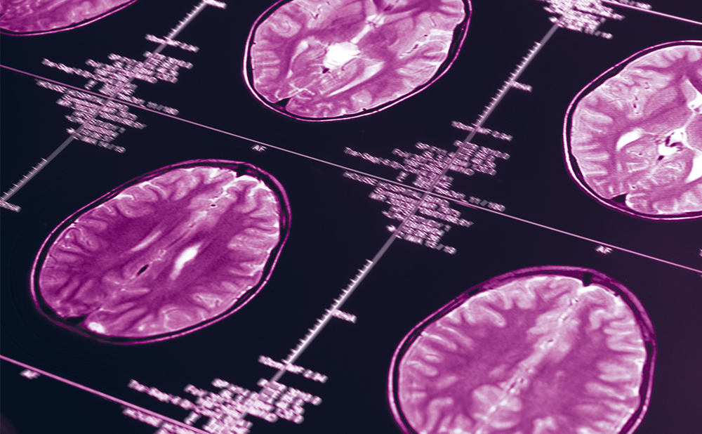
In recent years there have been tremendous advances in our knowledge of the pathogenesis of neurodegenerative dementias, but for most dementias no tests are yet available that allow a definite diagnosis while the patient is still alive. There is also a lack of pre-clinical tests, in particular tests that could help predict the course of the disease. An early diagnosis is essential to maximise the efficacy of potential therapies, both because the likelihood of treatment success is better the earlier the diagnosis is made and because early differentiation between the different forms of dementia is important if we are to provide more sophisticated therapies in the future. Currently, the diagnosis of dementia is based on clinical criteria. Not only are imaging techniques important in excluding certain structural brain lesions or cerebral space-occupying lesions, but with the continuous improvement of magnetic resonance imaging and functional examination techniques, they are becoming more and more important for enabling clinicians to diagnose both neurodegenerative and reversible dementias. Cerebrospinal fluid (CSF) testing is another important diagnostic tool, details of which are discussed below. A combination of clinical presentation, neurological symptoms and technical findings can lead to a correct diagnosis (see Table 2).
Pre-senile dementia is defined as dementia with an onset of symptoms before 65 years of age. Since dementia is frequently caused by neurodegenerative diseases such as Alzheimer’s disease, it predominantly affects people around 70 years of age and has an increasing prevalence with older age. However, younger patients are also at risk. The prevalence of pre-senile dementia has been estimated at 67–81 per 100,000 in those between 45 and 65 years of age1,2 and pre-senile dementia accounts for about 10% of all dementia patients.3
As in older patients, of all the neurodegenerative disorders, Alzheimer’s disease is the most frequent underlying disease in pre-senile dementia, accounting for about one-third of cases, followed by vascular and frontotemporal dementia. By contrast, LBD is uncommon. Furthermore, some diseases appear typically in younger patients, such as variant Creutzfeldt-Jakob disease (CJD), which has a median age at onset of 28 years. However, in young patients secondary causes of dementia are quite frequent, most of which are due to various forms of autoimmune inflammatory brain disorders or vasculitis. This subgroup of patients suffers from potentially treatable disorders, thus a detailed examination and a variety of tests from laboratory work-up and brain imaging up to brain biopsy4 should be performed.
Alzheimer’s-type Dementia
In 1906, Alois Alzheimer was the first to describe in detail the symptoms and pathological changes in his patient Auguste D. Auguste D suffered from a pre-senile form of dementia, being 51 years of age at disease onset. In 1911, Emil Kraepelin introduced Alzheimer’s dementia for the first time into the scientific literature. The term was used to describe pre-senile dementia for many years. Only in later years was the term Alzheimer’s dementia extended to include senile dementias of a primary degenerative nature with similar neuropathological changes. Alzheimer’s dementia is characterised by progressive cognitive impairment and prolonged disease duration (seven years on average). Alzheimer’s disease is the most frequent cause of dementia.5 In most cases, the progression of the disease is slow, with disease duration of approximately 10 years, although rapid progression is observed in some cases. Early occurrence of focal signs has been reported to indicate poor prognosis.6–9 Motor signs and their frequency in Alzheimer’s disease patients have previously been reported.10–13 The frequency increases during the course of the disease. Tremor is observed in 11% of the Alzheimer’s disease population, rigidity in 26% and posture instability/gait disturbance/falls in 29%.12 Occurrence of motor signs seems to be predictive of a poor outcome in Alzheimer’s disease.9 Early-onset Alzheimer’s disease shows a more rapid progression, more generalised cognitive deficits and greater cortical atrophy and hypometabolism compared with late-onset patients at a similar disease stage.14,15 Furthermore, atrophy in early-onset Alzheimer’s disease is localised predominantly occipital and parietal, whereas late-onset Alzheimer’s disease is remarkably atrophic in the hippocampus.16
The brains of patients with Alzheimer’s disease show marked atrophy of cortical structures and hippocampal formation. Histologically, two types of lesion are important diagnostic hallmarks: senile (neuritic) plaques and neurofibrillary tangles (NFTs). The amyloid core of senile or neuritic plaques contains an amyloid-like substance formed by peptides that originate through proteolytic cleavage of the membrane-associated precursor protein (amyloid precursor protein [APP]). One of these, termed Aβ1–42, shows the highest propensity for aggregation of plaques, thus an important role in the pathogenesis of Alzheimer’s disease was attributed to the plaque formation by Aβ1–42. Intracellular NFTs, which are neuronal inclusions consisting of abnormal cytoskeletal elements of hyperphosphorylated tau protein, are another characteristic pathological feature of AD. These tangles are found throughout the neocortex, in the nucleus basalis Meynert, in the thalamus and in the mammillary bodies. The formation of hyperphosphorylated tau protein in Alzheimer’s disease is hypothesised to result in disruption of binding to microtubules. Tau protein is phosphorylated at 21 sites, a process that leads to modification of its physiological properties. In pre-senile dementia, synaptic loss is more pronounced than in late-onset dementia, and there are more neuritic plaques and NFTs in the frontoparietal lobes.17
Both Aβ1–42 and tau (and its phosphorylated forms) became important biomarkers in the diagnosis of dementia. Various studies demonstrated an increase in tau protein and a decrease in Aβ1–42 in Alzheimer’s disease patients compared with controls. The value of these changes will be discussed later in the article. Three genes with pathogenic mutations have been identified so far (APP on chromosome 21, presenilin 1 on chromosome 14 and presenilin 2 on chromosome 1). APP is the precursor protein of Aβ. Presenilin 1 and 2 play a role in the function of β-secretase, which cleaves APP (to produce Aβ). A current concept regarding the cause of Alzheimer’s disease is that the mutations in all three genes lead in different ways to increased formation of pathological Aβ and senile plaques. In line with these findings, patients with Down’s syndrome, in whom chromosome 21 is present in triplicate, are at an increased risk of Alzheimer’s disease. The clinical presentation is similar to that of sporadic AD, apart from age at onset, but some mutations can present with characteristic clinical features such as early behavioural change,18 speech production deficit19 or spastic paraparesis with white-matter changes.20 In addition, the apolipoprotein E (ApoE) polymorphism on chromosome 19 has been identified as a risk factor. There are three alleles of ApoE (2, 3 and 4), and about two-thirds of the general population has the ApoE3 form of the gene. However, ApoE4 is known to be a relative and gene-dosage-dependent risk factor for AD.
Lewy Body Dementia
In contrast to other neurodegenerative dementias, pre-senile onset of LBD is very rare, comprising only 4% of early-onset dementias. LBD is characterised by deposits of so-called Lewy bodies, which are eosinophilic cytoplasmatic inclusion bodies consisting of ubiquitin, neurofilament, α-synuclein and other proteins in cortical and subcortical structures. The clinical picture of LBD consists of dementia in combination with Parkinson’s disease-like symptoms (extrapyramidal motor disorders). Clinical criteria in support of the diagnosis are visual hallucinations, recurrent falls and pronounced fluctuations in symptoms.21 Magnetic resonance imaging (MRI) scans showed only non-specific atrophy, but single-photon-emission computed tomography (SPECT) with 123I-ioflupane (DaTSCAN®) improved LBD diagnostics, with high sensitivity for differentiation from Alzheimer’s disease. Immune therapies for LBD are being developed that aim at eliminating or preventing synuclein deposits. Recently, some antibodies have been reported.22 Correct diagnosis is important as neuroleptics are obsolete in LBD, causing marked reduction of vigilance.
Vascular Dementias
There is agreement in the literature that dementia can be caused by cerebrovascular diseases. However, the most common concepts and classifications are heterogeneous. In general, a distinction is made between macroangiopathy and microangiopathy, cortical and subcortical multi-infarct dementia and dementia due to strategic infarcts. One common classification distinguishes between dementia after stroke (post-stroke dementia), subcortical vascular dementia (cerebral microangiopathy has to be present here) and dementia associated with vascular pathology and Alzheimer’s disease-specific changes. The known cardiovascular risk factors such as increased blood pressure, diabetes, abuse of nicotine, disorders of fat metabolism and cardiac arrhythmia are believed to be causes of vascular dementia. The clinical symptoms are as heterogeneous as the underlying causes. Multiple infarcts result in extensive neuronal cell loss, eventually leading to multi-infarct dementia. This form is primarily associated with word-finding deficits, impaired object recognition and loss of attention and judgement, in addition to the infarct-typical, position-dependent symptoms such as hemiplegia. By contrast, strategic infarcts affect important brain structures such as the thalamus, striatum or head of the caudate nucleus, and may result in dementia after only one infarct. This form of dementia is mainly characterised by deficits in memory, orientation and naming. Microangiopathic vascular dementia (Binswanger’s disease) is associated with marked apathy, cognitive slowing and attention and memory deficits. Recurrent ischaemic events lead to a gradual worsening of symptoms, and in the case of Binswanger’s disease also to a continuous progression of symptoms.
Diagnostic criteria (National Institute of Neurological Disorders and Stroke and Association Internationale pour la Récherche et l’Enseignement en Neurosciences [NINDS-AIREN] or Alzheimer’s Disease Diagnostic and Treatment Centers [ADDTC]) were proposed to differentiate vascular dementias from AD.23,24 Previous studies showed that by using these criteria it was possible to discriminate between these forms in 70–80% of cases. Current therapeutic strategies are based on an appropriate therapy in conjunction with secondary prophylaxis of vascular events, but there are currently no specific pharmaceutical therapies.
As cardiovascular risk factors are the major cause of vascular dementia, this form of dementia typically occurs in older age, at around 70 years of age, but vascular dementia has also been described in younger patients. Cerebral autosomal-dominant arteriopathy with subcortical infarcts and leukencephalopathy (CADASIL) is caused by mutation of Notch3 on chromosome 19. The clinical presentation is characterised in the early stages by migraine; during the clinical course psychiatric problems occur, followed later by dementia after repeated infarctions. Furthermore, various hereditary amyloid angiopathies have been described (familial British and Danish dementias associated with mutations in the BRI gene on chromosome 13) that produce complex neurological syndromes.
Frontotemporal Dementias
Unlike other neurodegenerative diseases, frontotemporal dementias occur earlier in life: at 57 years of age on average. In accordance with the clinical criteria,25 there are three clinical forms: frontotemporal dementia, primary progressive aphasia and semantic dementia. The characteristic features of frontotemporal dementia early in the disease course are personality changes, impaired social contact and emotional indifference. Only later, when the disease has progressed to the point where memory deficits occur, is dementia, not a psychiatric cause, which is considered as a differential diagnosis. Primary progressive aphasia, by contrast, is characterised by slowed speech, impaired repetition of speech and grammatically incomplete sentences. Semantic aphasia, like primary progressive aphasia, is also essentially characterised by speech disorders, but although spontaneous speech is fluent, it is confused. On MRI, frontotemporal atrophy is seen in all three forms of frontotemporal dementia, but in the early stages of the diseases it may not be very pronounced.
There is a subform of frontotemporal dementia called Pick’s disease, which is characterised by deposition of Pick bodies. In about 40% of these cases there is an increased familial occurrence, even in the absence of pathogenic mutations. Depending on the disease entity, Pick bodies (visualisation of ballooned neurons with tau deposits by means of silver staining) are seen on neuropathological examination. Ubiquitin-positive cellular inclusions, which are typical of motor neuron diseases, are also found.
Pre-senile Dementia
The underlying cause of pre-senile dementia spans a wide range, with a higher frequency of familial, autoimmune and metabolic reasons. Furthermore, toxic exposure plays a more important role in the differential diagnosis than in older people. A list of mutations associated with dementia is given in Table 3. A typical pre-senile neurodegenerative disorder is Huntington’s disease, which is caused by autosomal-dominant expansion of CAG trinucleotide repeats in the huntingtin gene on chromosome 4. The prevalence in Europe is up to 8/100,000. The cognitive decline starts typically in middle age with neuropsychiatric symptoms, followed by subcortical dementia and extrapyramidal signs such as choreatiform hyperkinesias. In addition to familial disorders, a higher frequency of metabolic disorders is ascertained in the differential diagnosis of pre-senile dementia. Mitochondrial disorders (incidence 2/10,000), Wilson’s disease (incidence 1/30,000–300,000) and lysosomal storage diseases such as Fabry’s disease or Niemann-Pick type C (incidence 1/8,000) are examples of this entity, some of which have known causative mutations. Usually, metabolic disorders cause a subcortical dementia characterised by disturbance of vigilance and attention; memory deficits appear typically later in the course of the disease. Five per cent of all patients with multiple sclerosis develop dementia, while cognitive symptoms (memory, attention, processing speed, executive functions) are present in around 50%. The profile of dementia corresponds to subcortical dementia. The degree of atrophy on MRI scans correlates with the degree of cognitive dysfunction. Treatment strategies are similar to those for neurodegenerative dementia, and a clinical trial of donezepil showed improved learning.26
Another cause of presenile dementia is AIDS–dementia complex (ADC). Ten to thirty per cent of all HIV-positive patients develop a dementia during the course of their disease, usually years after disease onset but sometimes in the early stages. Interestingly, the amount of virus in the brain does not correlate with the severity of dementia, suggesting a secondary mechanism. The envelope protein GP120 has been shown to inhibit the cell-cycle signal cascade. Furthermore, it inhibits neurogenesis from progenitor neuronal cells in the brain.27 The standard for HIV therapy, highly active antiretroviral therapy (HAART), has reduced the incidence in favour of increasing prevalence. It has also been shown that HAART can delay the onset of dementia and even improve already existing ADC. Recently, ADC patients treated with memantine showed neuropsychological improvement.28
Chronic abuse of alcohol can result in multiple cognitive changes. The well-known Wernicke-Korsakow syndrome is characterised by amnestic disturbances associated with other neurological problems (polyneuropathy, oculomotor signs, ataxia and vegetative symptoms). It is believed to be caused by malnutrition, especially thiamine deficiency. Thus, therapy demands early substitution of thiamine. Nevertheless, clinical improvement is uncommon due to advanced changes in brain. Additionally, a primary alcohol dementia due to direct neurotoxic effects has been described, but seems to be rare.29 It typically presents as combined frontal and subcortical dementia. It has been shown that symptoms stabilise or even improve after several months of strict alcohol abstinence.
Potentially Reversible Dementias
Dementia syndromes may have a neurodegenerative aetiology, but they can also be caused by potentially reversible diseases. The percentage of potentially reversible dementias, according to investigations, is 10% of all cases in memory clinics. This proportion is even higher (about one-third) among patients with rapidly progressive dementia.30
Autoimmune diseases are caused by a misguided immune response to central nervous system (CNS) structures. In general, these diseases can be treated by immunosuppression. One important disease in this group is cerebral vasculitis, which may occur as a result of either systemic diseases or isolated CNS vasculitis. The symptoms are caused by inflammatory changes and cerebral circulatory disorders (decreased blood flow caused by inflammatory and swollen vessels), explaining the combination of neurological deficits, such as hemiplegia, and cognitive disorders. In about 50% of all cases, inflammatory changes are found in the CSF.
Another autoimmune CNS disease is Hashimoto encephalitis, whose more precise designation has been steroid-responsive encephalopathy associated with autoimmune thyroiditis (SREAT). This disorder is characterised by rapid progressive dementia, myoclonus and seizures. A rapid improvement after administration of high-dose steroids is another clue for diagnosis and treatment. Independent of impaired thyroid function, thyroid autoantibodies are detectable in the blood. One hypothesis for the pathogenesis of Hashimoto encephalitis is that a cross-reaction of thyroid autoantibodies with neuronal tissue could result in neurological impairment. Inflammatory changes may be detectable in the CSF.
Coeliac disease is another systemic autoimmune disorder that occurs predominantly in the small intestine. It is believed to be caused by a reaction to nutritional gliadin. Typically, it presents with chronic diarrhoea and fatigue. In addition to the gastrointestinal symptoms, malabsorption due to changes in the bowel (truncation of the villi in the small intestine) and its sequelae also occur. No causative mutation is known, but a genetic predisposition (as in many other autoimmune disorders) has been confirmed. As well as the predominantly intestinal disease, antigliadin antibodies are described without intestinal changes, causing neurological symptoms such as cerebellar ataxia, schizophrenia and autism. This is presumed to be caused by a cross-reaction of antibodies to the neuronal tissue. Treatment is mainly based on a gluten-free diet. In diet-resistant patients, immunosuppression (steroids or azathioprine) can be used.
Autoimmune limbic encephalitis is associated with antibodies against voltage-gated potassium channels and glutamate decarboxylase (GAD). In addition to cognitive changes, epileptic seizures, myoclonus, autonomic failure, psychosis and sleep disturbances are frequent. Cancer, mainly lung cancer, can be associated with this form of dementia, causing paraneoplastic neurological symptoms. Dementia associated with NMDA antibodies have been described, with a similar clinical presentation. Most patients had an associated teratoma and strongly improved after surgery. In addition to treatment of the potentially associated cancer, immunosuppression is the standard therapeutic regimen.
Infections of the nervous system can cause rapid progressive cognitive decline. Confusion, hallucinations and psychosis are frequent encephalitic symptoms. Thus, a CSF test for CNS infection is essential for differential diagnosis, especially in patients with rapid cognitive decline. In addition to primary CNS infections, systemic and chronic infections can cause dementia. Whipple’s disease is caused by infection with Tropheryma whipplei, predominantly in intestine, causing malabsorption and weight loss. Frequently, joint pain is described. Fifteen per cent of all patients do not present with classical intestinal symptoms, thus hampering diagnosis. The malabsorption is usually slow and progressive, and diagnosis is made only after years. The hallmark associated neurological symptoms are cognitive decline, nystagmus and oculomasticatory myorhythmia. Diagnosis is made by intestinal biopsy and detection of PAS-positive macrophage inclusions, which can also be found in the CSF. Furthermore, antibodies can be detected. As this is a bacterial infection, therapy is based on antibiotic therapy for at least one year.
Cerebrospinal Fluid Tests
The CSF is the main component of the brain’s extracellular space and participates in the exchange of many biochemical products in the CNS. Consequently, CSF contains a dynamic and complex mixture of proteins that reflects the physiological or pathological state of the CNS.31 CSF analysis is extremely important for the identification of autoimmune disorders and inflammatory conditions that might lead to dementia. Although changes are non-specific (for example pleocytosis, elevated protein content, increased albumin ratio and oligoclonal immunoglobulin G [IgG] in the CSF), their presence clearly differentiates inflammatory and autoimmune diseases from neurodegenerative dementia. On the other hand, changes in the CSF proteome have been studied in various neurodegenerative disorders. The most important and best validated to date are tests for tau and phosphorylated isoforms and Aβ peptides.
Aβ1–42 levels were initially studied in CSF samples from Alzheimer’s disease patients and controls, demonstrating high potential for this biomarker for discrimination between the two groups. However, decreased levels were later reported in other degenerative conditions too. Aβ1–42 levels are reduced in patients with LBD and Alzheimer’s disease compared with non-demented controls32–35 as well as in patients with CJD,36 frontotemporal dementia37 and normal-pressure hydrocephalus (NPH).38–40 Our own data revealed significantly lower Aβ1–42 levels in all dementias tested compared with controls.40
High CSF tau levels were reported first in patients with Alzheimer’s disease, and later also in other conditions such as CJD.32,41 Conflicting results were obtained for frontotemporal dementia: whereas some studies reported increased CSF tau in this form of dementia,42,43 normal levels44 or even significantly reduced level45 were observed in others. A potential explanation for this might be the heterogeneity of the definitions of frontotemporal dementia used. Conflicting data are available also for LBD, with either increased46 or normal47–49 concentrations compared with non-demented groups. Significantly decreased tau concentrations in LBD patients compared with Alzheimer’s disease have also occasionally been reported.50,51
One study described that the total tau level in CSF from NPH patients was significantly higher than that in controls, with a correlation between tau levels and dementia or urinary incontinence.52 Our own data demonstrated increased tau protein concentrations in CJD, Alzheimer’s disease, LBD and frontotemporal dementia, but not in NPH, while only patients with CJD dissociated significantly from the other dementias.40 To summarise, these markers have become standard CSF tests in the routine dementia work-up. Although some data point towards a limited value of these markers in the clinical differential diagnosis between dementia entities, their specificity can be increased if they are used in a defined clinical context. Their value is high when they are used as a part of a multimodal approach together with neuropsychological test batteries and brain imaging. In addition, the determination of ratios and isoforms is helpful. The ratio calculated from the pathological Aβ1–42 and from the less aggregating form Aβ1–40 is of considerable value in the diagnosis of Alzheimer’s disease: it has been found that ratios <1.0 are indicative of Alzheimer’s disease. With respect to tau, its phosphorylated form at T181 has been identified as indicating hyperphosphorylation, and it is thus considered to be a disease-specific marker for Alzheimer’s disease. The ratio calculated from tau protein phosphorylated at T181 and total tau has also increased its diagnostic utility.
Recent studies on the value of tau and Aβ as potential markers for differentiation of AD from other dementias have also shown that similar results might be obtained for other dementias, too. Thus, due to their non-specificity and low discrimination levels between different neurodegenerative dementia types, the search for a CSF (or blood) biomarker is still ongoing.
Summary
The clinical symptom dementia is characterised by a variety of changes in memory, planning, orientation and processing speed. Many diseases have been described as underlying causes of dementia. Primary dementia disorders are usually of neurodegenerative origin such as Alzheimer’s disease, LBD and frontolobar degeneration.
Pre-senile dementia is defined as symptom onset before 65 years of age. An estimated 10% of all patients with dementia have this early onset. The most frequent cause, as in senile dementia, is Alzheimer’s disease. Frontolobar degeneration is more frequent at a younger age, while LBD only rarely has a pre-senile onset. In addition to neurodegenerative causes, other diseases such as autoimmune, toxic, genetic and metabolic disorders are common factors behind early-onset dementia. Some of these conditions are potentially reversible and therefore a correct diagnosis is important.
The diagnostic approach to dementia includes clinical examination (looking for associated neurological or systemic symptoms), neuropsychological testing (differentiating frontal, cortical or subcortical profile) and a detailed medical history about the onset and course of symptoms. Cerebral MRI can detect specific focal atrophy, white-matter changes or other clues as to underlying disease. Positron-emission tomography and SPECT can improve diagnostic certainty. In each case, a lumbar puncture should be performed to detect inflammatory signs (infectious or autoimmune disorders), and dementia markers, especially Aβ1–42 and tau protein, should be measured. In young-onset dementia in particular, genetic testing should be part of the diagnostic approach. ■


