Homocysteine – A Marker for Vitamin Deficiency and a Risk Factor for Neurological Diseases
Homocysteine – A Marker for Vitamin Deficiency and a Risk Factor for Neurological Diseases
Homocysteine (Hcy) is the demethylated product of methionine. This toxic amino acid can be removed by remethylation to methionine via methionine synthase, which utilises 5-methyl tetrahydrofolate as a methyl donor and methyl cobalamin as a co-factor. An alternative remethylation pathway for Hcy depends on betaine-Hcy-methyltransferase, which utilises betaine as a methyl donor. Another method of Hcy catabolism is via two vitamin-B6-dependent enzymes, cystathionine beta synthase and cystathioninase, to cysteine. The catabolism of Hcy in the brain depends mainly on its remethylation to methionine, utilising folate and methyl cobalamin. Because B vitamins (folate, vitamin B12, vitamin B6) are important co-factors for Hcy catabolism, an elevated concentration of Hcy can indicate B-vitamin deficiency.1 An important role of Hcy-methionine metabolism is to provide S-adenosylmethionine (SAM), the methyl donor for numerous biological reactions. SAM donates its methyl group to a methyl acceptor and is transformed to S-adenosylhomocysteine (SAH). SAH is hydrolysed to Hcy by SAH-hydrolase. The SAH-hydrolase reaction is reversible, but favours SAH formation in the presence of increased Hcy. Hcy metabolism in the brain is an important source of SAM.2 This methyl donor plays important roles in the formation and catabolism of neurotransmitters and phospholipids (phosphatidylcholine is the methylated product of phosphatidylethanolamine), DNA methylation and activation of several enzymes that have essential roles in the brain (e.g. protein phosphatase 2A [PP2A]). Elevated levels of Hcy can therefore cause damage to several key pathways in the central nervous system, either directly or by changing the methylation potential (SAM/SAH).
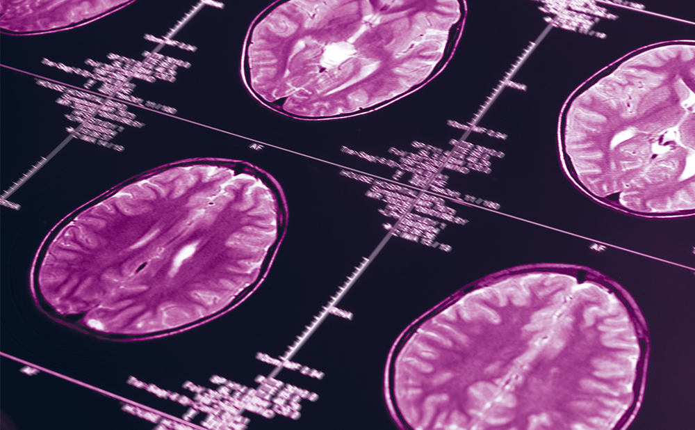
Changes in brain volume and intensity or the presence of small infarcts are early signs of dementia. These conditions can be detected by magnetic resonance imaging (MRI) of the brain. Changes in these MRI parameters indicate increased risk of stroke, dementia and Alzheimer’s disease (AD). Numerous studies have found an association between plasma concentrations of Hcy and qualitative or quantitative MRI analyses.3,4 Additionally, several longitudinal studies have documented an association between baseline Hcy, folate, vitamin B12 or vitamin B6 concentrations and decline in cognitive function with age.
Evidence has suggested that increasing intake of the B vitamins may have a protective effect on the central nervous system. This effect can be related either to lowering Hcy or to a direct effect of the vitamins. An improvement in cognitive impairment or delaying its progression by Hcy-lowering vitamins may support a causal role for hyperhomocysteinemia (HHcy) in neurodegenerative diseases. In this article we discuss recent findings from longitudinal, observational and treatment studies on the role of Hcy in neurodegeneration.
Homocysteine in Dementia and Cognitive Decline
Dementia and AD are the most common age-associated diseases in western countries. The pathophysiological mechanisms in dementia involve degenerative (cellular) or vascular mechanisms, or both. Degenerative dementia constitutes 80% of all dementias in elderly people, whereas approximately 10% are vascular dementias. Sporadic age-associated dementia is a long-standing disease that extends over several decades and usually starts as mild cognitive impairment (MCI). MCI is characterised by memory impairment and/or only a mild decline in cognitive abilities. Patients with MCI are at markedly increased risk of developing AD: approximately 20% of patients with MCI develop AD or progressive dementia within two years of follow-up.5 On the one hand, neuronal degeneration and loss in AD is reflected by reduced brain volume, hippocampal volume and lobar volume; on the other hand, the majority of dementia patients show microvascular and macrovascular injuries that are reflected by the presence of silent brain infarct and extensive white matter hyperintensity, respectively.
The association between Hcy and dementia has been shown in several longitudinal studies. In the MacArthur Studies of Successful Aging,6 370 subjects 70–79 years of age were followed for a mean of seven years. The mean change in total cognitive scores over seven years was -4.3 points. Patients who were in the highest quartile of Hcy (14.4–40μmol/l) lost ≥9 points. Low folate had also significant effect on cognitive delay in this follow-up study.
Other follow-up studies documented that Hcy predicted cognitive decline7 and AD8 after adjustment for low serum folate. In blood samples collected before death, higher concentrations of Hcy were related to confirmed AD compared with age-matched control subjects.9 Moreover, in a three-year follow-up, progression of the disease was worse in AD patients who had higher Hcy at entry.9 In the Rotterdam Scan Study, the association between Hcy and neuropsychological test scores was assessed. Lower scores for psychomotor speed, memory function and global cognitive function were found in subjects with Hcy >14μmol/l compared with those with Hcy <8.5μmol/l.10
Several studies have observed an association between cognitive decline with age and vitamin B12 status. In the Banbury B12 Study, cognitive decline was related to low holotranscobalamin and elevated methyl methacrylate (MMA) and Hcy in elderly people.11 In the Medical Research Council Cognitive Function and Aging Study, elevated MMA (indicating vitamin B12 deficiency) was related to bad cognitive scores in elderly non-demented people.12 In a subgroup from the OPTIMA Study, baseline vitamin B12 status, indicated by total B12 (<308pmol/l) and holotranscobalamin (<54pmol/l), was a significant predictor of decreased brain volume on MRI in healthy, non-demented elderly people after five years of follow-up.13
Therefore, available studies suggested a causal relationship between HHcy and cognitive decline with age. Low vitamin B12 status may be one important risk factor for dementia, and the role of vitamin B12 seems to be only partly related to lowering Hcy. To confirm this causal link, treatment studies have been initiated. However, vitamin supplementation studies in patients with dementia or those at risk of dementia because of their high age were inconclusive.
The effect of folate 2.5mg, vitamin B12 0.5mg and vitamin B6 25mg on cognitive function was tested in a three-month randomised, doubleblind, placebo-controlled study using various combinations of the vitamins. Elderly people with vascular disease were the target population. Each treatment arm included 23–24 participants.14 In this study, Stott et al. failed to detect any improvement in cognitive function.14 In another study, Lewerin et al. observed a significant association between cognitive and movement performance in elderly people (mean age 76 years) and Hcy and MMA.15 Nevertheless, in a placebo-controlled, double-blind study, a daily dose of folic acid 0.8mg plus vitamin B12 0.5mg and vitamin B6 3mg for four months failed to improve movement or cognitive function despite lowering Hcy and MMA.15 Despite the fact that the study by Lewerin et al. included almost three to five times more participants than that by Stott et al., the short duration of both studies might be a limiting factor.
The hypothesis that lowering Hcy concentrations will improve cognitive function in healthy elderly people has been tested for a longer duration. In a two-year double-blind, placebo-controlled, randomised study, subjects treated with folic acid 1mg plus vitamin B12 0.5mg and vitamin B6 10mg showed no improvement in their cognitive scores compared with the placebo group, despite their Hcy being lowered by an average of 4.4μmol/l.16 In a three-year randomised, double-blind study, 818 participants were treated with either folic acid 0.8mg or placebo.17 Hcy was lowered by a mean of 26%, and a significant improvement in some cognitive domains that decline with age has been reported. Memory scores were also improved after folate supplementation, and the improvement was positively related to the severity of folate deficiency.18 There are several confounding factors that might override the effect of the B vitamins on cognition and memory (see Table 1). These factors are critically important for judging available results or for future studies. Hcy-lowering trials should aim at prevention and not treatment of dementia. Similarly, trials using omega-3 supplementation in patients with dementia documented improvement in patients with less severe dementia, but not in severely demented patients.
Among other factors responsible for variations between studies are baseline Hcy concentrations and the presence of sub-normal vitamin status. The definition of sub-optimal vitamin status or deficiency also depends on the marker used. For example, low concentrations of holotranscobalamin, the active vitamin B12, is common in subjects with total vitamin B12 concentrations below 350pmol/l.
Homocysteine in Cerebrovascular Diseases
Stroke is a clinical syndrome resulting from different underlying vascular pathologies. This heterogenous disease includes different types. Ischaemic stroke constitutes 80% of all stroke types, intracerebral haemmorhage 15% and subarachnoid haemmorhage 5%. Transient ischaemic attack (TIA) is a disorder that is similar to stroke but is not fatal and lasts for less than 24 hours. Ischaemic stroke is caused by atherothromboembolism in 50% of cases and by intracranial small-vessel disease (lacunar infarction) in 25% of cases. A cardiac embolism explains approximately 20% of ischaemic stroke cases.
HHcy is very common in patients with stroke and is suggested to be an independent risk factor for the disease. Systematic reviews of epidemiological and prospective studies revealed a consistent positive dose-dependent relationship between Hcy and stroke risk. A recent study confirmed the association between HHcy and stroke in a black population, and a strong association was detected with lacunar infarction with leukoaraiosis compared with those without such an infection.19 The risk of stroke increased by 6–7% in the Rotterdam Scan Study for each 1μmol/l increase in plasma Hcy concentration.20 Moreover, a graded positive association was also shown between concentrations of Hcy and the risk of stroke in the British Regional Heart Study.21 In the Physicians Health Study, a 1.4-fold increase in the risk of stroke was observed in subjects with Hcy >12.7μmol/l compared with subjects with Hcy lower than this limit.22 Similar results were reported by the Northern Manhattan Study, where the adjusted hazard ratio for a Hcy level ≥15μmol/l compared with <10μmol/l was 2.01 (95% confidence interval [CI] 1.00–4.05) for ischaemic stroke.23 An odds ratio (OR) for stroke of 2.3 was reported in subjects from the National Health and Nutrition Examination Survey (NHANES)
Epidemiologic Follow-up Study III in the highest quartile of Hcy compared with the lowest quartile.24 In the Framingham Offspring Study, elevated concentration of Hcy was a risk factor for silent cerebral infarcts in 2,040 people free of clinical stroke who were tested by MRI.25 The OR for silent cerebral infarcts for plasma Hcy was 2.23 (p<0.001), which exceeded that for hypertension, cholesterol, diabetes and other common risk factors.25
Because the relationship between Hcy elevation and stroke suggests a causal relationship, several trials have been started to test the effect of Hcy lowering on stroke prevention. Several studies have shown that risk of stroke may be slightly but significantly reduced by B vitamins. In a population-based study, Yang et al.26 reported a decline in stroke-related mortality after folic acid fortification in the US and Canada that was stronger than that found in England and Wales during the same period.26 Moreover, folic acid and vitamin B6 lowered Hcy and caused slight improvement in cerebrovascular and cerebral indices.27 Furthermore, the risk of stroke was lowered in a five-year treatment trial on subjects receiving either placebo or folic acid 2.5mg plus vitamin B6 50mg and B12 1mg (relative risk 0.75, 95% CI 0.59–0.97; p=0.03).28 In a two-year trial, high doses of B vitamins (vitamin B6 25mg, vitamin B12 0.4mg and folic acid 2.5mg) were compared with low doses (vitamin B6 200μg, vitamin B12 6μg and folic acid 20μg) in secondary prevention of stroke.29 The effect of treatment on stroke risk was not significant during this trial. Nevertheless, a 21% reduction in the combined risk of stroke, coronary disease and death was observed in a subgroup of this study population that included patients who were likely to benefit from treatment.30 In the Heart Outcomes Prevention Evaluation (HOPE) 2 study, 5,522 patients with vascular disease or diabetes were randomised to receive placebo or folic acid 2.5mg plus vitamin B6 50mg and vitamin B12 1mg for five years; one of the primary outcomes was stroke. An approximately 25% reduction in the risk of stroke was found in the supplement group compared with the placebo group (relative risk 0.75, 95% CI 0.59–0.97; p=0.03).28
There is currently no consensus on using B vitamins for primary or secondary prevention of stroke. Nevertheless, the American Stroke Association Stroke Council reiterates the importance of meeting current recommended daily intakes of the vitamins.31
Homocysteine and Other Brain Lesions
In a recently published report from the Framingham Offspring Study, 1,965 middle-aged adults (mean age 61 years) who were free of stroke, dementia or neurological disorders were tested for their Hcy and MRI brain measurements.25 Higher plasma Hcy levels were strongly and independently associated with lower brain volume and the presence of silent brain infarcts in healthy subjects.32
Homocysteine has been related to several brain lesions that were estimated quantitatively or semi-quantitatively by MRI. In the Rotterdam Scan Study, Hcy was related to white matter lesions.33 Moreover, in non-demented people 60–90 years of age, lower hippocampal volumes were found in subjects with higher Hcy compared with those with lower Hcy.34 This was confirmed by another study on elderly people.35 Moreover, the degree of cortical atrophy increased with increasing Hcy.34 These results suggest that keeping Hcy low might delay brain shrinkage with age and the progression of silent brain infarcts.
Homocysteine in Patients with Parkinson’s Disease
Parkinson’s disease (PD) is the second most common neurodegenerative disease in elderly people, after dementia. PD is characterised by depletion of dopamine, dysfunction and death of dopaminergic neurons. Acquired factors that enhance the risk of PD are of great interest. The association between PD and HHcy has been reported in numerous clinical studies.36,37 The increase in plasma Hcy in PD patients depends on folate and vitamin B12 status and genetic polymorphisms that influence Hcy.37 HHcy can also arise secondary to PD itself, or owing to drugs commonly used for treating the disease.
A significant increase in plasma concentration of Hcy occurs in PD patients after starting L-dopa treatment.38 L-dopa is methylated by catechol-O-methyltransferase (COMT), a SAM-dependent enzyme. L-dopa may increase the requirements for SAM.39 This is supported by the finding that L-dopa induces activity of COMT and methionine adenosyl transferase.40 If food intake of labile methyl groups (methionine, choline) remains unchanged, L-dopa will cause SAM depletion. SAM, or its precursor methionine, can ameliorate the neurotoxicity of L-dopa to dopamine neurons by providing sufficient methyl groups for COMT.41 The effect of combining COMT inhibitors with L-dopa to avoid HHcy is inconclusive.42–44 The available evidence suggests that folate, vitamin B6 and vitamin B12, but not COMT inhibitors, are the most important modifiers of the effect of L-dopa on Hcy levels.45–47
It seems that higher concentrations of Hcy in PD patients are generally associated with poor progression of the disease. An association between plasma Hcy concentrations >14μmol/l and depression, as well as cognitive impairment, has been reported in PD patients.48 Moreover, an increased risk of vascular disease in PD patients with Hcy ≥17.7μmol/l has been reported.49 Elderly women with PD and high baseline Hcy (>21.0μmol/l) were more likely to develop hip fracture during 4.9 years of follow-up than those with lower Hcy.50 Hip fracture is frequent in PD patients,51 and HHcy is a risk factor for hip fracture.52,53 Other studies failed to detect an association between vitamin intake or plasma Hcy and the incidence or clinical course of PD.54,55 However, these studies were limited by a low number of participants, a short-term follow-up or the fact that vitamin intake does not reflect vitamin status, especially in elderly subjects, in whom malabsorption commonly occurs.
Taken together, the above evidence suggests that reducing plasma Hcy may be an important measure for secondary prevention in PD patients. Plasma concentrations of Hcy should be maintained at low levels in PD patients, especially in those patients receiving L-dopa. A causal role for Hcy in the onset of PD has not yet been tested in large studies, despite the consistent association between PD and HHcy. There are several vitamin intervention studies in patients with PD,46,47 but there is no information currently available about clinical outcome in treated patients.
Homocysteine in Patients with Epilepsy
Several in vivo studies have suggested that Hcy can cause epilepsy.56 In addition, most classic antiepileptic drugs (carbamazepin, phenytoin, valproate, phenobarbital) can induce HHcy in approximately 13–40% of patients.57–59 Furthermore, multidrug treatment and longer duration of the therapy increase the risk of HHcy.59
Antiepileptics can increase concentrations of Hcy, probably by reducing concentrations of the B vitamins.57,59 Accordingly, antiepileptics may impair folate absorption and gastrointestinal transport by altering gastrointestinal pH. Moreover, increased activity of liver enzymes that catabolise folate (i.e. cytochrome P450) can result from chronic use of antiepileptics. For example, phenytoin, phenobarbital and carbamazepine induce and valproate inhibits cytochrome P450.60 Antiepileptic drugs that do not induce P450 are not associated with a low concentration of folate. Valproate’s effect on folate seems to be related to inhibiting glutamate formyl transferase and changing the balance between various forms of folate.61 Finally, valproate may induce methionine synthase and methylenetetrahydrofolate reductase (MTHFR) activity in the liver and can inhibit serine hydroxymethyltransferase activity, thus causing lower folate status and increased concentrations of Hcy. Valporate has also been found to cause DNA hypomethylation and to show teratogenic effects.62
Folic acid supplementation (1mg/day orally) significantly reduced plasma Hcy after six and 12 weeks in children with elevated Hcy (>10.4μmol/l).59 Treating epileptic patients who were folate-deficient with 5mg folic acid daily for one to three years caused a marked improvement in cognition, mood and social behaviour.63 Folate deficiency is known to be associated with depressive syndrome. In line with this, patients treated with antiepileptics developed not only low folate status, but also depressive symptoms.64,65 There is strong evidence suggesting that Hcy concentrations should be tested in patients treated with antiepileptics.59 Folate supplementation for subjects on chronic treatment with such medications has been recommended.59
Mechanisms of Homocysteine Neurotoxicity
The mechanisms by which Hcy may damage the brain are not fully understood. Data from experimental in vitro studies have shown that Hcy may induce neurological dysfunction via oxidative stress.66,67 Hcy is a glutamate receptor agonist that actvates N-methyl-D-aspartate (NMDA) receptor.68,69 HHcy is associated with hypomethylation, which in turn affects DNA methylation, phospholipid homeostasis and activity of several SAM-dependent methyltransferases. In addition, some studies documented that the effect of folate or vitamin B12 deficiency may be independent of HHcy in the aetiology of neurological diseases. Therefore, it is highly recommended to test vitamin B status in patients at risk of certain neurological diseases. Because Hcy is more sensitive for folate deficiency, this marker may not be sufficient to rule out vitamin B12 deficiency, at least in the early stages. For this reason, testing holotranscobalamin and/or methylmalonic acid may be essential for the prevention and treatment of vitamin B12 deficiency.
Conclusions
To sum up, retrospective and prospective studies have shown a consistent association between HHcy and several neurological diseases. Treatment with B vitamins is an important, safe and inexpensive approach to modulate the risk. Results from a few treatment trials have shown slight but significant risk reduction after vitamin treatment. We presented several confounding factors and issues to be considered when planning and analysing future trials. Longer duration (>5 years), selection of the patients and standard medications might be considered. B-vitamin trials should aim at primary prevention and not treatment of dementia and other central nervous system diseases. Until the results of such studies become available, everyone is recommended to meet at least the daily intake of the B vitamins. ■


