touchNEUROLOGY touchNEUROLOGY
Alzheimer's Disease & Dementia
Read Time: 5 mins

From Normal to Pathological Ageing – The Spectrum of Mild Cognitive Impairment and Alzheimer’s Disease

Copy Link
Published Online: Jun 4th 2011 European Neurological Review, 2006;(2):74-77 DOI: http://doi.org/10.17925/ENR.2006.00.02.74
Authors: Ruediger Mielke, Joseph Kessler
Quick Links:
Article
Article Information
Article:
Introduction

Human ageing is joined by various physical, social and cognitive-mnestic changes, which can differ considerably both inter- and intra-individually. Cognitive-mnestic changes are caused by processes in the central nervous system, but they are also determined by genetic factors, education, profession, lifestyle, intellectual and physical activity and, especially, general physical condition.
Reliable norms about cognitive and mnestic functions in older people are usually lacking. Many tests just indicate norms for a group older than 60 years. In geronto-psychological literature, a differentiation is sometimes made between ‘young olds’ (65–75 years), ‘old olds’ (75–85 years) and ‘eldest olds’ (greater than 85 years) in orderto define the clinical heterogeneity of ageing in the elderly.

Study Design

There are also problems in study design. In cross-sectional studies, age-dependent impairments are often overestimated, while in longitudinal studies they are underestimated. Since the neurons of the central nervous system lose their ability to replicate, irreversible destructive processes cumulate over the course of the years.1
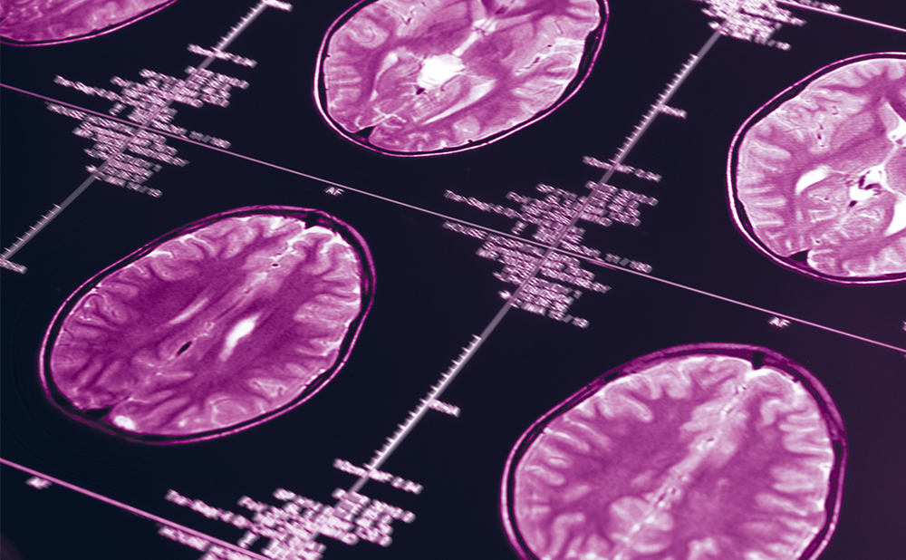
After the age of 30, the human brain begins to lose weight; this can be demonstrated in computed tomography (CT) and magnetic resonance imaging (MRI). The enlargement of cortical sulci and ventricles increases 20% per decade after the age of 50 in men and after the age of 60 in women.
Not only shrinkage and loss of neurons, but also changes in the energy metabolism of the neurons can restrict cell function.
A reduction of density of the synapses and the ‘dying back’ of axonal branching, increases of plaques and tangles and changes in the cholinergic and dopaminergic transmitter systems have all been described. There is little experience in the age-sensitivity of the approximately 50 different transmitter systems.
Cognitive-mnestic changes are described in the elderly.2 Intelligence deficits can be ascertained in distinguishing between crystalline and fluid intelligence.3 Components of the crystalline intelligence can remain preserved or even increase until late age, while fluid intelligence decreases.4 This means that older people act successfully in routine situations and that their knowledge and vocabulary remains stable. In contrast, there is a successive loss in the processing speed of new information.
The flexible adaptation to new situations and problem-solving can become difficult. Older people frequently complain about memory deficits. The extent of mnestic problems largely depends on age, the material to learn and the task to solve.
Memory performances are hampered more strongly in abstract material than in everyday familiar material. There are also problems in free recall. Memory performance in recognition tasks worsens later, but to a small extent only.
Attention task performance, especially processes in selective and divided attention, is impaired. Verbal and communicative abilities may pass as being relatively stable in age, but communication can be disturbed by sensory deficiencies like hearing deficits.
A decline in abstraction ability and cognitive flexibility, as well as an increased susceptibility to interference can be found in older people.
Deceleration of reaction time in cognitive information processing is a universal phenomenon in older people and is one of the main causes of cognitive dysfunction. Reaction time in 60-year-olds compared to 30-year-olds is thought to be reduced by 20%.
Except for some subtypes of vascular dementia, cognitive decline in dementia starts slowly and sneakily, and the leap from normal cognitive decline in age and the beginning of dementia is often very difficult to define. In different classification schemes, the diagnostic uncertainty is described in terms of ‘age-related cognitive decline’, ‘mild neuro-cognitive disorder’, ‘age-associated memory impairment’ and ‘mild cognitive impairment’ (MCI).

Mild Cognitive Impairment

MCI is a heterogeneous phenomenon characterised by a loss of cognitive abilities beyond normal aging.5 According to Petersen et al., patients with MCI can be classified into those with amnestic MCI or non-amnestic MCI on the basis of their leading symptoms.6 The criteria for amnestic MCI are specified by Petersen et al. as:
• memory complaints (preferably corroborated by an informant);
• objective memory impairment on a delayed recall test;
• relatively normal general cognitive function, except for memory (other cognitive domains may be impaired but only to a minimal degree);
• normal or only minimally impaired activities of daily living (ADLs) (below the threshold required for a diagnosis of dementia); and
• not demented (that is, in the opinion of the investigator, the patient does not meet criteria for dementia).
Non-amnestic MCI can be further classified by the impairment of a single domain (language, executive function, visuo-spatial relations) or in multiple domains (combination of cognitive dysfunctions).
Patients with amnestic MCI are more likely to develop Alzheimer’s disease (AD) than non-amnestic MCI patients.
The conversion rate is approximately of 15% per year.7 AD pathology has already been demonstrated in these patients.8 Currently discussed predictors of dementia development are typical signs of AD, including the type and extension of memory of executive dysfunction,9 cerebrospinal biomarkers such as beta-amyloid or tau,10-11 genetic analysis, especially the apolipoprotein E (APOE) allele,4-12 reduced volume of the hippocampus,13-14 hypometabolism in AD relevant (especially entorhinal and tempoparietal) cortical areas and reduced cortical cholinergic activity.15-17
Episodic memory impairment in MCI has primarily been attributed to hippocampal atrophy and to cholinergic dysfunction.18-19 In contrast, executive impairment in MCI may rely more on the impairment of prefrontal functions, which are suspected to be predominantly dependent on dopaminergic neurotransmission.5,20
One major ‘Petersen criteria’ is that MCI-patients have normal or minimal impairment in ADLs. This separates MCI from dementia. Recent studies have shown that impairment of ADLs may already be present in MCI. Intact ADLs cannot be used as a criterion to define the syndrome of MCI.21 MCI is also associated with characteristic neuropsychiatric symptoms, such as dysphoria (39%), apathy (39%), irritability (29%) and anxiety (25%). The definition of MCI varies considerably, depending upon the test used for case definition.22

Alzheimer’s Disease

‘Dementia syndrome’ is a generic term describing symptoms of chronic or progressive impairment of cortical and sub-cortical function that results in complex cognitive decline. These cognitive changes are commonly accompanied by disturbances of mood, behaviour and personality.
At the beginning of the disease, a correct differential diagnosis is necessary to separate age-associated cognitive changes, mild cognitive impairment and dementia syndrome. Clinical estimation must be focused on neuropsychological assessment of disturbed function. Table 1 provides an overview of common tests and scales used for the early diagnosis of memory impairment and the stages of dementia.
AD is the most common cause of dementia and is characterised by an insidious onset and a slow deterioration in cognition, functional ability (e.g. ADLs), behaviour and mood.
While in the early stage of AD, assistance may be necessary with complex tasks, such as managing financial problems and calculations, behavioural disturbances are not evident until the middle stages of AD.
Behavioural disturbances can be characterised by a mini-mental-state examination (MMSE) score of between 20 and 10 points. The need for help by relatives and professionals often increases and makes independent living impossible. It poses a considerable burden on carers, which often results in secondary morbidity.
Valuable therapeutic options are provided by the three approved cholinesterase inhibitors donepezil, rivastigmine and galantamine, as well as the partial glutamate antagonist memantine.
In randomised clinical trials, these drugs have shown considerable results in reducing cognitive decline and behavioural disturbances and improving ADLs. ■

References

  1. McGeer E F, McGeer P L, “Aging, neurodegenerative disease and the brain”, Can J Aging (1997); 16: pp. 218–236.
  2. La Rue A, Aging and neuropsychological assessment, (1992); Plenum Press, New York.
  3. Cattell R B, Intelligence: Its Structure, Growth, and Action, (1987); New York: Elsevier Science Pub. Co.
  4. Schaie K W, “The course of adult intellectual development”, Am Psychol(1994);49: pp. 304–313.
  5. Nordahl C W, Ranganath C, Yonelinas A P, et al., “Different mechanisms of episodic memory failure in mild cognitive impairment”, Neuropsychologia (2005);43: pp. 1688–1697.
  6. Petersen R C, Smith G E, Waring S C, et al., “Mild cognitive impairment: clinical characterization and outcome”, Arch Neurol (1999);56: pp. 303–308.
  7. Petersen R C, Doody R, Kurz A, et al., “Current concepts in mild cognitive impairment”, Arch Neurol (2001);58: pp. 1985–1992.
  8. Markesbery W R, Schmitt F A, Kryscio R J, et al., “Neuropathologic substrate of mild cognitive impairment”, Arch Neurol (2006);63: pp. 38–46.
  9. Arnaiz E, Almquist O, “Neuropsychological features of mild cognitive impairment and preclinical Alzheimer’s disease”, Acta Neurol Scand (2003);179: pp. 34–41.
  10. Maccioni R B, Lavados M, Maccioni C B, et al., “Biological markers of Alzheimer’s disease and mild cognitive impairment”, Cur Alzheimer Res (2004);4: pp. 307–314.
  11. Schoonenboom N S, Pijnenburg Y A, Mulder C, et al., “Amyloid beta(1-42) and phosphorylated tau in CSF as markers for early onset Alzheimer’s disease”, Neurology (2004);62: pp. 1580–1584.
  12. Devanand D P, Pelton G H, Zamora D, et al., “Predictive utility of apolipoprotein E genotype for Alzheimer disease in outpatients with mild cognitive impairment”, Arch Neurol (2005);62: pp. 975–980.
  13. Chetelat G, Landeau B, Eustache F, et al., “Using voxel-based morphometry to map the structural changes associated with rapid conversion in MCI: a longitudinal MRI study”, Neuroimage (2005);27: pp. 934–946.
  14. Mosconi L, De Santi S, Rusinek H, et al., “Magnetic resonance and PET studies in the early diagnosis of Alzheimer’s disease”, Expert Rev Neurother (2004);4: pp. 831–849.
  15. Anchisi D, Borroni B, Franceschi M, et al. “Heterogeneity of brain glucose metabolism in mild cognitive impairment and clinical progression to Alzheimer disease”, Arch Neurol (2005); 62: pp. 1728–1733.
  16. Herholz K, “PET studies in dementia”, Ann Nucl Med (2003);17: pp. 79–89.
  17. Herholz K, Weisenbach S, Zundorf G, et al., “In vivo study of acetylcholine esterase in basal forebrain, amygdala, and cortex in mild to moderate Alzheimer’s disease”, Neuroimage (2004);21: pp. 136–143.
  18. Chetelat G, Desgranges B, de la Sayette V, et al., “Dissociating atrophy and hypometabolism impact on episodic memory in mild cognitive impairment”, Brain (2003);126: pp. 1955–1967.
  19. Grön G, Brandenburg I, Wunderlich A P, “Inhibition of hippocampal function in mild cognitive impairment: targeting the cholinergic hypothesis”, Neurobiol Aging (2006);27: pp. 78–87.
  20. Nieoullon H E, “A modified card sorting test sensitive to frontal lobe defects”, Cortex (2002);67: pp. 53–83.
  21. Perneczky R, Pohl C, Sorg C, et al,. “Complex activities of daily living in mild cognitive impairment: conceptual and diagnostic issues”, Age and Ageing (2006);35: pp. 240–245.
  22. Alladi S, Arnold R, Mitchell J, et al., “Mild cognitive impairment: applicability of research criteria in a memory clinic and characterization of cognitive profile”, Psychol Med (2006);36: pp. 507–515.
  23. Reisberg B, Ferris S H, de Leon MJ, et al., “The Global Deterioration Scale for assessment of primary degenerative dementia”, Am J Psychiatry (1982);139: pp. 1136–1139.
  24. Morris J C, “The Clinical Dementia Rating (CDR): Current version and scoring rules”, Neurology (1993);43: pp. 2412–2414.
  25. Folstein M, Folstein S, MMST – Mini Mental Status Test, (1990); Weinheim: Beltz-Verlag.
  26. Folstein M, Folstein S, McHugh P, “Mini-Mental state: a practical method for grading the mental state of patients by the clinician”, Journal of Psychiatric Research (1975);12: pp. 189–198.
  27. Kalbe E, Kessler J, Calabrese P, et al., “DemTect: a new sensitive cognitive screening test to support the diagnosis of mild cognitive impairment in early dementia”, International Journal of Geriatric Psychiatry (2004);19: pp. 136–134.
  28. Solomon P R, Hirschoff A, Kelly B, et al., “A 7 minute neurocognitive screening battery highly sensitive to Alzheimer’s disease”, Archives of Neurology (1998);55: pp. 349–355.
  29. Delis D C, Kramer J H, Kaplan E, et al., “California Verbal Learning Test (CVLT). Adult Version (Research ed.)”, (1987);San Antonio, TX: Psychological Cooperation.
  30. Rey A, L’examen clinique en psychologie, (1964);Paris : Presses Universitaire de France.
  31. Rey A, “L’examen psychologique dan les cas d’encephalopathie traumatique”, Archives de Psychologie (1941);28: pp. 286–340.
  32. Wechsler D, Wechsler Memory Scale. Third edition manual, (1997);San Antonio: The Psychological Corporation.
  33. Wilson B A, Cockburn J, Baddeley A, The Rivermead Behavioural Memory Test-II, (2003) Bury St. Emdunds, UK: Thames Valley Test.
  34. Baller G, Kalbe E, Brand M, et al., “Inventar zur Gedächtnisdiagnostik (IGD)”, (2006); Göttingen: Hogrefe Verlag.
  35. Morris J C, Heyman A, Mohs R C, et al., “The Consortium to Establish a Registry for Alzheimer’s Disease (CERAD), Part I. Clinical and neuropsychological assessment of Alzheimer’s disease”, Neurology (1989);39: pp. 1159–1165.
  36. Rosen W G, Mohs R C, Davis K L, “A new rating scale for Alzheimer’s disease”, American Journal of Psychiatry (1984);141: pp. 1356–1364.
  37. Mattis S, “Mental status examination for organic mental syndrome in the elderly patient”, In: L. Bellak and T.B. Karasu (eds.), Geriatric Psychiatry (1976);New York: Grune and Straton.
  38. Kessler J, Denzler P, Markowitsch H J, Demenz Test, (1988);Weinheim: Beltz-Test-Verlag.

Further Resources

Share this Article
Related Content In Alzheimer's Disease & Dementia
  • Copied to clipboard!
    accredited arrow-down-editablearrow-downarrow_leftarrow-right-bluearrow-right-dark-bluearrow-right-greenarrow-right-greyarrow-right-orangearrow-right-whitearrow-right-bluearrow-up-orangeavatarcalendarchevron-down consultant-pathologist-nurseconsultant-pathologistcrosscrossdownloademailexclaimationfeedbackfiltergraph-arrowinterviewslinkmdt_iconmenumore_dots nurse-consultantpadlock patient-advocate-pathologistpatient-consultantpatientperson pharmacist-nurseplay_buttonplay-colour-tmcplay-colourAsset 1podcastprinter scenerysearch share single-doctor social_facebooksocial_googleplussocial_instagramsocial_linkedin_altsocial_linkedin_altsocial_pinterestlogo-twitter-glyph-32social_youtubeshape-star (1)tick-bluetick-orangetick-red tick-whiteticktimetranscriptup-arrowwebinar Sponsored Department Location NEW TMM Corporate Services Icons-07NEW TMM Corporate Services Icons-08NEW TMM Corporate Services Icons-09NEW TMM Corporate Services Icons-10NEW TMM Corporate Services Icons-11NEW TMM Corporate Services Icons-12Salary £ TMM-Corp-Site-Icons-01TMM-Corp-Site-Icons-02TMM-Corp-Site-Icons-03TMM-Corp-Site-Icons-04TMM-Corp-Site-Icons-05TMM-Corp-Site-Icons-06TMM-Corp-Site-Icons-07TMM-Corp-Site-Icons-08TMM-Corp-Site-Icons-09TMM-Corp-Site-Icons-10TMM-Corp-Site-Icons-11TMM-Corp-Site-Icons-12TMM-Corp-Site-Icons-13TMM-Corp-Site-Icons-14TMM-Corp-Site-Icons-15TMM-Corp-Site-Icons-16TMM-Corp-Site-Icons-17TMM-Corp-Site-Icons-18TMM-Corp-Site-Icons-19TMM-Corp-Site-Icons-20TMM-Corp-Site-Icons-21TMM-Corp-Site-Icons-22TMM-Corp-Site-Icons-23TMM-Corp-Site-Icons-24TMM-Corp-Site-Icons-25TMM-Corp-Site-Icons-26TMM-Corp-Site-Icons-27TMM-Corp-Site-Icons-28TMM-Corp-Site-Icons-29TMM-Corp-Site-Icons-30TMM-Corp-Site-Icons-31TMM-Corp-Site-Icons-32TMM-Corp-Site-Icons-33TMM-Corp-Site-Icons-34TMM-Corp-Site-Icons-35TMM-Corp-Site-Icons-36TMM-Corp-Site-Icons-37TMM-Corp-Site-Icons-38TMM-Corp-Site-Icons-39TMM-Corp-Site-Icons-40TMM-Corp-Site-Icons-41TMM-Corp-Site-Icons-42TMM-Corp-Site-Icons-43TMM-Corp-Site-Icons-44TMM-Corp-Site-Icons-45TMM-Corp-Site-Icons-46TMM-Corp-Site-Icons-47TMM-Corp-Site-Icons-48TMM-Corp-Site-Icons-49TMM-Corp-Site-Icons-50TMM-Corp-Site-Icons-51TMM-Corp-Site-Icons-52TMM-Corp-Site-Icons-53TMM-Corp-Site-Icons-54TMM-Corp-Site-Icons-55TMM-Corp-Site-Icons-56TMM-Corp-Site-Icons-57TMM-Corp-Site-Icons-58TMM-Corp-Site-Icons-59TMM-Corp-Site-Icons-60TMM-Corp-Site-Icons-61TMM-Corp-Site-Icons-62TMM-Corp-Site-Icons-63TMM-Corp-Site-Icons-64TMM-Corp-Site-Icons-65TMM-Corp-Site-Icons-66TMM-Corp-Site-Icons-67TMM-Corp-Site-Icons-68TMM-Corp-Site-Icons-69TMM-Corp-Site-Icons-70TMM-Corp-Site-Icons-71TMM-Corp-Site-Icons-72