This article examines the possible involvement of nutrients in the pathogenesis, prevention, or management of Alzheimer’s disease (AD). Nutrients are defined here as food constituents that are essential for preventing or treating the clinical syndromes that develop when they are deficient. In general, they act as substrates or co-factors for enzymes and thereby sustain growth and metabolism.
This article examines the possible involvement of nutrients in the pathogenesis, prevention, or management of Alzheimer’s disease (AD). Nutrients are defined here as food constituents that are essential for preventing or treating the clinical syndromes that develop when they are deficient. In general, they act as substrates or co-factors for enzymes and thereby sustain growth and metabolism.
Some nutrients can also produce pharmacological effects if administered in doses higher than those needed to prevent their deficiency syndromes, or if given without the other compounds that usually accompany them in food sources.1 These effects might, for example, involve enzymes, transport proteins (e.g., in brain capillaries through which leucine suppresses the passage of blood tryptophan into the brain and thus diminishes serotonin synthesis),2 antagonism of receptors, synthesis of second messengers, ion channels, or even non-enzymatic processes (e.g., acting as antioxidants).
This article first considers the evidence that deficiencies involving individual nutrients, for example folic acid or docosahexaenoic acid (DHA), might be risk factors for AD, or that administering each such compound might be useful in treating this disease. It then describes the pharmacological effects of a mixture of nutrients on synapse formation and behaviour in experimental animals and on memory functions in patients with early AD and, presumably, deficiencies in cortical and hippocampal synapses.3–5
The course of AD, a very common and poorly treated neurodegenerative disease, is currently thought to involve three characteristic phases.6
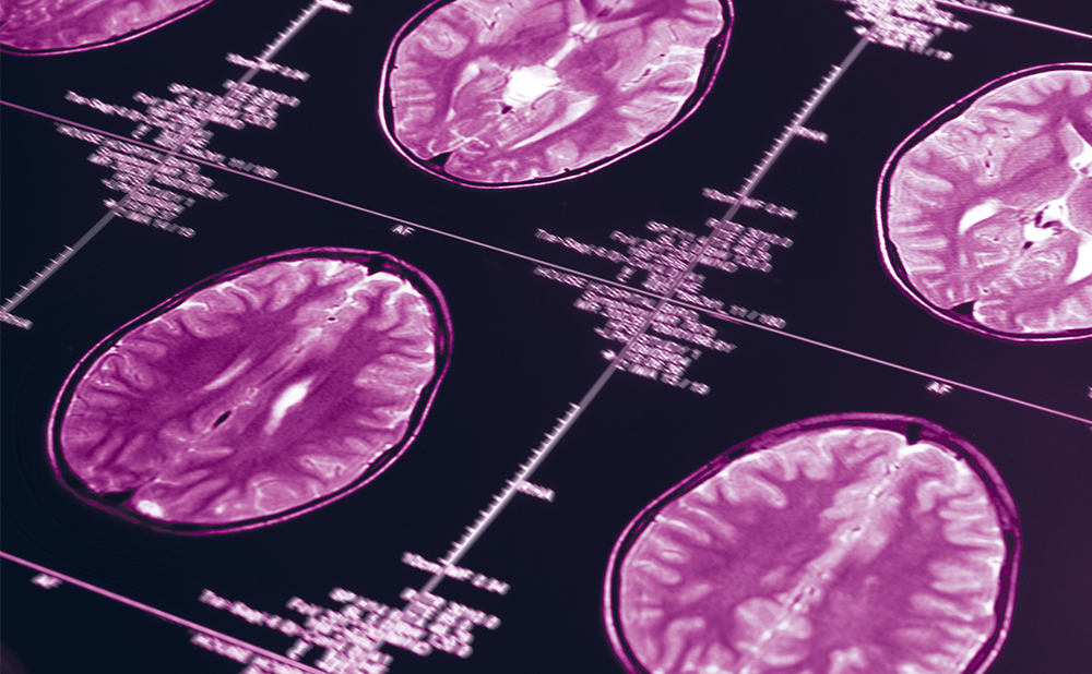
In the earliest phase, memory disturbances are minor or even absent and there is no evidence of dementia; however, neurochemical abnormalities presumably exist which might be assessed by measuring putative biomarkers.6 These include subnormal levels of a characteristic peptide, A-beta 1–42, in cerebrospinal fluid (CSF) and abnormal brain scans exhibiting, by magnetic resonance imaging (MRI), medial temporal lobe atrophy with loss of hippocampal, entorhinal cortical, and amygdalar volume, or, by positron emission tomography (PET), reduced temporoparietal glucose utilisation, or very high beta-amyloid levels revealed using exogenous ligands. In the second, ‘minimal cognitive impairment’ (MCI) phase, mild behavioural problems are observed, particularly involving memory and cognition. However, patients are not demented and can still perform most quotidian activities. This stage is associated, at autopsy, with abundant accumulation of amyloid and loss of synapses in the hippocampus, cerebral cortex, and elsewhere in the brain. In the third phase, the patient is overtly demented and brain size is diminished, reflecting the loss of neurons and synapses and damage to white matter and blood vessels.
Each of these phases is usually associated with characteristic abnormalities in the metabolism of the brain protein amyloid precursor protein (APP) and its A-beta subunits. During the first phase of AD, CSF levels of A-beta 1–42, the most amyloidogenic APP subunit, are, perhaps paradoxically, depressed; during the second and third phases, large amounts of highly insoluble beta-amyloid, formed by the aggregation of A-beta subunits, are deposited, particularly in ‘senile plaques.’ Soluble oligomers of A-beta subunits are thought to be neurotoxic and to cause the degeneration of dendritic spines, the anatomical precursor of glutamatergic synapses;7–9 then degeneration of the synapses themselves; and ultimately, the loss of hippocampal and cortical neurons. The insoluble amyloid formed by the aggregation of A-beta subunits had, until recently, been conceived as neurotoxic; however, more recent observations suggest that amyloid formation may provide neuroprotection by removing toxic A-beta oligomers from solution.10 Attempts to slow the course of AD by administering agents which suppress A-beta formation or remove it from the bloodstream have, to date, been largely unsuccessful.
An intervention based on nutrients which act pharmacologically to enhance synaptogenesis might be expected to slow the course of AD, or even to partially restore memory functions if it also enhances neurotransmission in affected brain areas. Providing this intervention very early in the disease, while abundant neurons are still available to form new dendritic spines and synapses might, theoretically, be optimally effective. Such an intervention might require many years of administration, hence the use for this purpose of nutrients – compounds which are normally present in, and readily metabolised by, the body – might confer particular benefit.
Do Nutrient Deficiencies Contribute to the Pathogenesis of Alzheimer’s Disease?
Does adequate epidemiological or experimental evidence exist that deficiencies in individual nutrients can constitute risk factors for developing AD, or that supplementation of each such compound can prevent AD or slow its course?
Blood levels of several nutrients in AD patients, particularly the omega-3 fatty acid DHA and the vitamins required for methyl group synthesis (folic acid, B12, B6) have been described as subnormal by some investigators but not by others and there is at present no consensus as to whether these deficiencies actually exist. Similarly, some studies have demonstrated significant correlations between the amounts of these nutrients present in plasma (e.g., the DHA content of plasma phosphatidylcholine [PC]),11 or provided by the diet and an individual’s cognitive scores or his/her risk of developing AD,12 while other studies have failed to do so.13,14 The possibility remains that deficiencies in folic acid, vitamin B12, or vitamin B6 manifested as elevated plasma homocysteine levels may be contributory to AD, e.g., in subsets of patients with relatively minor deficiencies in folate levels; however, such subsets have yet to be described.
Apparently, there is agreement that AD brains contain lower levels of free and esterified DHA than brains from control subjects.15–18 Circulating DHA in humans can derive from the pre-existing DHA present in some foods and from the conversion of dietary alpha-linolenic acid to DHA in the liver.19,20 The gene for one of the enzymes that mediates this conversion is reportedly deficient in AD,19 hence this endogenous source of circulating DHA is compromised.
Possible Utility of Nutrient Mixtures that Promote Synaptogenesis in the Management of Alzheimer’s Disease
The loss of cortical and hippocampal synapses,3–5 probably reflecting both impaired synaptogenesis (perhaps consequent to a deficiency in dendritic spines)7–9 and accelerated synaptic degeneration, is an early neuropathological correlate of AD and is probably the finding that best correlates with early memory impairment.4,5 An animal model of AD which overproduces A-beta peptides exhibits a similar early decrease in brain synapses21 and oligomers representing aggregates of such peptides can, when applied locally to the brain, damage synapses, distort neurites, and decrease the formation of the dendritic spines needed to form glutamatergic synapses.10 These observations support the widely held view that a treatment that blocked A-beta synthesis, or removed the peptide from the circulation, might slow the loss of synapses in AD and thereby sustain cognitive functions in patients. A generation of efforts by diligent researchers has provided abundant information about A-beta’s synthesis, fates and toxic effects, and this information is now being used to generate drug candidates for slowing A-beta production or removing it from the circulation. Some of these agents have been shown to lower A-beta levels in body fluids. However, none to date has been able to improve or even sustain memory or other cognitive functions. Perhaps a future drug candidate that reduces brain A-beta might succeed in slowing the course of AD; however, in the interim, an alternative therapeutic strategy might provide patients with more benefit than that presently obtained from the acetylcholinesterase inhibitors or metabotropic glutamate receptor (mGluR) antagonists currently in use – or might amplify the benefits produced by those drugs.
One such therapeutic strategy might entail finding a treatment that accelerates synaptogenesis, thus diminishing the net loss of synapses caused by AD.22,23 There are various loci at which such a treatment might work. For example, it could increase the flux of free calcium into stimulated dendrites, activate receptors that control the formation of dendritic spines, increase the synthesis of pre- or post-synaptic proteins, or generate new synaptic membrane, the main constituent of synapses. One such treatment, which has been shown to improve cognition in studies on experimental animals24 and is being tested in patients,25 is based on administering nutrients which promote synaptogenesis. The nutrients are phosphatide precursors; in test animals they increase the production of synaptic membrane26 and dendritic spines.27 The three required compounds – uridine, an omega-3 polyunsaturated fatty acid such as DHA and choline – are all normally present in the bloodstream and readily cross the blood–brain barrier.22
Synaptic membrane is composed principally of phospholipids, including the phosphatides PC, phosphatidylethanolamine (PE), phosphatidylserine (PS) and phosphatidylinositol (PI), and characteristic pre- and post-synaptic proteins. Conversion of the three circulating precursors to phosphatides is mediated by enzymes that have relatively low affinities for these substrates, and thus are unsaturated at the precursor concentrations normally present in the blood and brain. The choline in blood is obtained from various foods or is synthesised in the liver22 via a pathway that requires methionine and thus is accelerated when vitamins B12, B6 and folic acid are administered.28 Circulating uridine, in humans, also derives from hepatic synthesis.22 Uridine is also present in most foods in the form of RNA; however, there is no satisfactory evidence that this particular potential source of uridine can provide significant quantities of uridine to the circulation (in contrast to the uridine present as uridine monophosphate [UMP] in mothers’ milk and infant formulas).22 As described above, DHA, an essential fatty acid, is obtained from dietary DHA and from the hepatic conversion of alpha-linolenic acid to DHA, a process described as disturbed in AD.19 Because the enzymes that initiate the conversion of these compounds to PC are unsaturated, their activities are readily enhanced when their substrates are administered. Thus, administration of choline increases brain levels of its phosphorylated product phosphocholine; giving uridine (as UMP) increases brain levels of uridine triphosphate (UTP) and cytidine triphosphate (CTP); and giving DHA increases the proportion of diacylglycerol (DAG) molecules that contain this omega-3 fatty acid and thus are preferentially used for synthesising phosphatides instead of triglycerides.22
The phosphocholine and CTP formed in the brains of animals given uridine plus choline then combine to form cytidine diphosphate (CDP)-choline, which in turn combines with DHA-containing DAG molecules to form PC. PE is similarly formed from this pathway, termed the ‘Kennedy Cycle’, except that phosphoethanolamine replaces phosphocholine; PS is formed by substituting a serine molecule for the choline in PC or ethanolamine in PE (‘base exchange’). Treatment of rats or gerbils with supplemental UMP, DHA and choline for several weeks is associated with substantial increases in levels of PC per brain or per brain cell, as well as with roughly proportionate increases in the other phosphatides.26 Moreover, the effects of giving all three precursors tend to be greater than the sum of the effects of giving each separately. Animals so treated also exhibit significant improvements in memory functions, as assessed using the Morris water maze or T- or Y-mazes.24
Administration of the three phosphatide precursors also increases brain levels of pre- and post-synaptic proteins, including, for example, synapsin, synaptophysin, post-synaptic density protein-95 (PSD-95), and mGluR,22,26 as well as the numbers of hippocampal dendritic spines.27 These effects are due in part to an additional action of uridine: besides serving, indirectly, as a CTP precursor in cytidine-dependent phosphatide synthesis, uridine and its phosphorylated products (e.g., UTP) are also agonists for P2Y receptors on brain neurons, which promote neurite outgrowth29 and affect neuronal protein synthesis.22 These receptors, parenthetically, are deficient in the parietal cortex of patients with AD.30 DHA, acting alone, also stimulates neurite outgrowth and dendritic spine formation somewhat, possibly by activating other receptors. Existing techniques have not enabled the direct measurement of cortical or hippocampal synapse formation in animals receiving the three phosphatide precursors. However, the consensus among synaptologists seems to be that an increase in dendritic spines as produced by the precursors virtually always leads to an increase in synapses, i.e., more than 90 % of the time.7–9
In an initial clinical trial in which 110 drug-naive patients with very mild or mild AD received a medical food (Souvenaid®) containing the precursors and co-factors (e.g., vitamin B12, vitamin B6, folic acid) daily for twelve weeks and an equal number served as controls, those in the treated group – particularly those with very mild AD – exhibited a statistically significant improvement in memory compared with control subjects.25
Three additional large-scale trials are under way or have recently been completed; these also include measurements of biomarkers for assessing synaptic density (i.e., electroencephalography [EEG] and magnetoencephalography [MEG]). In one of these studies – designed to determine whether the findings of the initial trial could be confirmed – 259 patients, also drug-naive and with early AD (mini-mental state examination [MMSE] = 25 + 2.8) received Souvenaid or placebo daily for 24 weeks and the memory domain score of a neuropsychological test battery (NTB) – the primary outcome parameter – was measured initially and after 12 and 24 weeks.31 Secondary outcomes tested, using NTB data, included executive function and the individual items that had been scored. A total of 91.9 % of the subjects completed the study, compliance was 97 %, and no differences were noted between the groups in the frequency of adverse events. Souvenaid again significantly improved the primary endpoint, memory performance (p=0.025).


