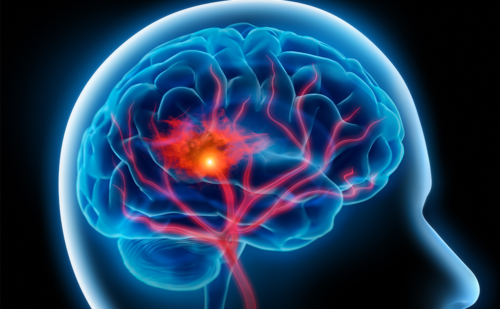
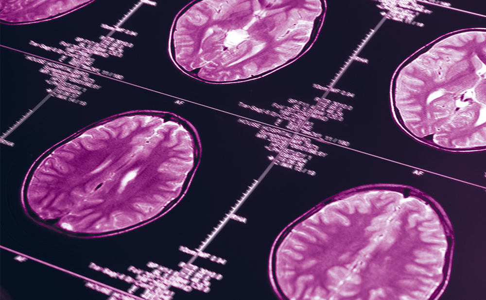
Alzheimer’s disease (AD) is the most common form of dementia and affects millions of people worldwide. The disorder is characterised by severe memory loss, with episodic memory being particularly impaired during the initial phases. Most AD cases occur sporadically, although inheritance of certain susceptibility genes enhances the risk. Familial AD represents the minority of AD cases and is caused by mutations in genes encoding for the amyloid β precursor protein (APP), presenilin 1 (PS1) or presenilin 2 (PS2). Two pathological hallmarks are observed in AD brains at autopsy: intracellular neurofibrillary tangles (NFTs) and extracellular senile plaques (SPs) in the neocortex, hippocampus and other subcortical regions essential for cognitive function (see Figure 1). NFTs are formed from paired helical filaments composed of neurofilaments and hyperphosphorylated tau protein. In turn, plaque cores are formed mostly from the deposition of amyloid β (Aβ) peptide that results from the cleavage of APP.
The literature shows that mitochondrial dysfunction and oxidative stress play important roles in the early pathology of AD.1–3 Indeed, there are strong indications that oxidative stress occurs prior to the onset of symptoms in AD and oxidative damage is found not only in the vulnerable regions of the brain affected in disease,4–6 but also peripherally.7–10 Moreover, it has been shown that oxidative damage occurs before Aβ plaque formation,4 supporting a causative role of mitochondrial dysfunction and oxidative stress in AD. This review is devoted to discussing evidence showing that mitochondrial dysfunction and oxidative stress are intimately involved in AD pathophysiology.
The Dual Role of Brain Mitochondria
Although the brain represents only 2% of bodyweight, it receives 15% of cardiac output and accounts for 20% of total body oxygen consumption. This energy requirement is largely driven by neuronal demand for energy to maintain ion gradients across the plasma membrane, which is critical for the generation of action potentials. This intense energy requirement is continuous; even brief periods of oxygen or glucose deprivation result in neuronal death.
Mitochondria perform pivotal biochemical functions necessary for homeostasis and are arbiters of cell death and survival, in addition to being a main source of adenosine triphosphate (ATP). They represent a convergence point for death signals triggered by both extracellular and intracellular cues. As such, the mitochondria sit at a strategic position in the hierarchy of cellular organelles to either promote the healthy life of the cell or to terminate it.11–12 Mitochondria are essential for neuronal function because the limited glycolytic capacity of neurons makes them highly dependent on aerobic oxidative phosphorylation (OXPHOS) for their energetic needs.13 However, OXPHOS is a major source of toxic endogenous free radicals, including hydrogen peroxide (H2O2), hydroxyl (•OH) and superoxide (O2 -•) radicals, which are products of normal cellular respiration. When the electron transport chain (ETC) is inhibited, electrons accumulate in complex I and co-enzyme Q where they can be donated directly to molecular oxygen to yield O2 -•, which can be further detoxified by the mitochondrial manganese superoxide dismutase (MnSOD) producing H2O2, which in turn can be converted to H2O by glutathione peroxidase (GPx). However, O2 -• in the presence of nitric oxide (NO•), formed during the conversion of arginine to citrulline by nitric oxide synthase (NOS), can lead to the formation of peroxynitrite (ONOO-). Furthermore, H2O2 in the presence of reduced transition metals can be converted to the toxic product •OH via Fenton and/or Haber–Weiss reactions.
It is well recognised that reactive oxygen species (ROS) and reactive nitrogen species (RNS) play a dual role since they can be either harmful or beneficial to living systems.14 Beneficial effects of reactive species occur at low to moderate concentrations and involve physiological roles in cellular responses to noxia, as for example in defence against infectious agents and in the functioning of a number of cellular signalling systems. One further beneficial example of ROS at low to moderate concentrations is the induction of a mitogenic response.14 However, oxidative stress occurs if the amount of free radical species produced overwhelms the cell’s capacity (enzymatic and non-enzymatic antioxidant defences) to neutralise them, which is followed by mitochondrial dysfunction and neuronal damage. Reactive species generated by mitochondria have several cellular targets, including mitochondrial components themselves (lipids, proteins and DNA). The lack of histones in mitochondrial DNA (mtDNA) and diminished capacity for DNA repair render mitochondria an especially vulnerable target of oxidative stress events.15
The central nervous system (CNS) is particularly susceptible to reactive species-induced damage16 because: it has a high consumption of oxygen; it contains high levels of membrane polyunsaturated fatty acids susceptible to free radical attack; it is relatively deficient in oxidative defences (poor catalase activity and moderate SOD and GPx activities); and a high content in iron and ascorbate can be found in some regions of the CNS, enabling generation of more reactive species through the Fenton/Haber–Weiss reactions.
Mitochondria also serve as high-capacity Ca2+ sinks, which allows them to stay in tune with changes in cytosolic Ca2+ loads and aids in maintaining cellular Ca2+ homeostasis, which is required for normal neuronal function.17 Conversely, excessive Ca2+ uptake into mitochondria has been shown to increase ROS production, inhibit ATP synthesis, induce mitochondrial permeability transition pore (PTP) and release small proteins that trigger the initiation of apoptosis, such as cytochrome C and apoptosis-inducing factor (AIF), from the mitochondrial intermembrane space into the cytoplasm. Released cytochrome C binds apoptotic protease activating factor 1 (Apaf-1) and activates the caspase cascade.18 Such alterations in mitochondrial function have been proposed as a causative mechanism in the pathogenesis of AD (see Figure 2).
Mitochondrial Cascade Theory of Alzheimer’s Disease
The amyloid cascade hypothesis has been evoked to explain the pathology that underlies AD. This hypothesis claims that deposition of Aβ is the causative agent of AD pathology and that NFT, cell loss, vascular damage and dementia follow as a direct result of this deposition.19 Accumulating evidence suggests that although the amyloid cascade hypothesis is potentially viable in familial AD cases, it may not apply in its current form to the sporadic type of the disease.20 First, persons with sporadic AD generally lack mutations in APP, PS1 and PS2 genes, so it is unclear what initiates plaque formation in such cases. Second, plaques are a relatively common finding in the nondemented elderly.21,22 Third, pathways through which plaques generate NFTs and other recently described AD pathophysiological processes are unknown. These include neuronal apoptosis, neuronal aneuploidy and cerebral/extracerebral mitochondrial dysfunction.20,23–25 So, important questions remain concerning late-onset sporadic AD: what triggers deposition of Aβ?; and what lies upstream?
Swerdlow and Khan26 proposed the mitochondrial cascade hypothesis, which attempts to connect all the pathological features of the disease. In this model, the individual’s genetics determine the basal rates of ROS production by the ETC, which determine the pace at which acquired mitochondrial damage accumulates. In turn, oxidative alterations induced in mitochondrial nucleic acids, lipids and proteins amplify ROS production and trigger three events: a reset response, in which cells respond to elevated ROS by generating Aβ, which further perturbs mitochondrial function; a removal response, in which compromised cells are purged via programmed cell death mechanisms; and a replace response, in which neuronal progenitors unsuccessfully attempt to re-enter the cell cycle, with resultant aneuploidy, tau phosphorylation and NFT formation.26 In summary, this hypothesis postulates that mitochondrial dysfunction represents a primary pathology in sporadic, late-onset AD, and drives both SP and NFT formation. It further provides a rationale for how mitochondrial dysfunction surpassing certain thresholds triggers compensatory mechanisms that cause the various pathological hallmarks of AD.26
Mitochondrial (Dys)function, Oxidative Stress and Cell Death Are Interlinked in Alzheimer’s Disease
Several in vitro, in vivo and human studies indicate that AD is characterised by mitochondrial dysfunction, increased oxidative stress and neuron death (see Figure 2). Since this is a very extensive topic to discuss, some relevant data are presented following a simple scheme where findings are grouped according to the experimental system through which data were obtained.
In Vitro Studies
Several in vitro studies have provided compelling evidence that Aβ might cause mitochondrial dysfunction. It was previously shown that Aβ requires functional mitochondria to induce toxicity.27 Furthermore, Hansson et al.28 identified an active γ-secretase complex in rat brain mitochondria. Being composed of nicastrin (NCT), anterior pharynxdefective 1 (APH-1) and presenilin enhancer protein 2 (PEN2), this γ-secretase complex cleaves, among other substrates, APP, generating Aβ and APP-intracellular domain. Furthermore, the presence of APP was detected in mitochondrial membranes of PC12 cells bearing the Swedish double mutation in the APP gene.29 Together these studies placed mitochondria in a privileged position concerning APP processing and answered the ‘old question’ of how Aβ interacts with mitochondria.
Studies from our laboratory showed that Aβ peptides in the presence of Ca2+ exacerbate PTP opening.30,31 The PTP induction, a phenomenon characterised by a sudden increase in the permeability of the inner mitochondrial membrane, plays a key role in apoptotic cell death by facilitating the release of apoptogenic factors. We observed that Aβ in the presence of Ca2+ decreases the mitochondrial transmembrane potential and the capacity of brain mitochondria to accumulate Ca2+, and induces a complete uncoupling of respiration and an alteration of the ultrastructural morphology of mitochondria characterised by swelling and disruption of mitochondria cristae.30,31 Altogether these results suggest a clear association between Aβ, mitochondrial dysfunction and alteration of Ca2+ homeostasis. Du and collaborators32 showed that the interaction of cyclophilin D, an integral part of the PTP, with mitochondrial Aβ potentiates mitochondrial, neuronal and synaptic stress. It was also observed that cyclophilin D deficiency substantially improves learning and memory and synaptic function in an AD mouse model and alleviates Aβ-mediated reduction of long-term potentiation.32
We also observed that diabetes-related mitochondrial dysfunction is exacerbated by ageing and/or by the presence of Aβ, supporting the idea that diabetes and ageing are risk factors for the neurodegeneration induced by this peptide.33–35 Ageing of diabetic rats induces an impairment of the respiratory chain and a decrease in OXPHOS efficiency and in the capacity of mitochondria to accumulate Ca2+. In the presence of Aβ25–35 or Aβ1–40, the age-related mitochondrial effects are potentiated.33 Additionally, brain mitochondria isolated from diabetic rats in the presence of Aβ1–40 produce higher levels of H2O2.34 However, insulin and co-enzyme Q10 (CoQ10) treatments prevent the decline in mitochondrial OXPHOS efficiency and avoid an increase in oxidative stress induced by Aβ.35 It was also shown that cultured neurons from transgenic (Tg) mice that overexpress a mutant form of APP and Aβ-binding alcohol dehydrogenase (ABAD) (Tg mAPP/ABAD) display spontaneous generation of H2O2 and O2 -•, decreased ATP levels, release of cytochrome C and induction of caspase 3-like activity followed by DNA fragmentation and loss of cell viability. Furthermore, generation of ROS is associated with a dysfunctional cyclo-oxygenase.36
Other studies from our laboratory also showed that pheochromocytoma cells (PC12) exposed to Aβ1–40 and Aβ25–35 present mitochondrial dysfunction characterised by the inhibition of complexes I, III and IV of the mitochondrial respiratory chain.37 Recently, Rhein and collaborators38 evaluated the mitochondrial respiratory functions and energy metabolism in control and in human wild-type APP stably transfected SH-SY5Y cells. The authors observed that complex IV activity is significantly reduced in APP cells. By contrast, a significant increase in the activity of complex III is observed. The authors interpreted this increase as a compensatory response in order to balance the defect of complex IV. However, this compensatory mechanism does not prevent the strong impairment of total respiration in APP cells. As a result, the respiration together with ATP production decreases in the APP cells in comparison with the control cells.38
We have previously shown that AD fibroblasts present high levels of oxidative stress and apoptotic markers compared with young and age-matched controls.39 Furthermore, AD-type changes could be generated in control fibroblasts using N-methyl protoporphyrin to inhibit cytochrome oxidase (COX) assembly, indicating that the observed oxidative damage is associated with mitochondrial dysfunction. The effects of N-methyl protoporphyrin are reversed or attenuated by both lipoic acid and N-acetyl cysteine.39 These results suggest that mitochondria are important in the oxidative damage that occurs in AD and that antioxidant therapies may represent promising therapeutic strategies. Recently, Wang and collaborators40 showed that sporadic AD fibroblasts present alterations in mitochondria morphology and distribution.40 Mitochondrial abnormalities are due to a decrease in dynamin-like protein 1 (DLP1), a regulator of mitochondrial fission and distribution. Further, Aβ overproduction causes abnormal mitochondrial dynamics via differential modulation of mitochondrial fission/fusion proteins.41 In the same line, Cho and collaborators found that NO• produced in response to Aβ triggers mitochondrial fission, synaptic loss and neuronal damage, in part via S-nitrosylation of DLP1.42
Interestingly, Abramov et al.43 reported that Aβ causes a loss of mitochondrial potential in astrocytes but not in neurons. Since this effect is prevented by antioxidants and reversed by provision of glutamate and other mitochondrial substrates to complexes I and II, they suggested that the depolarisation reflects oxidative damage to metabolic pathways upstream of mitochondrial respiration. However, Paradisi et al.44 demonstrated that astrocytes can protect neurons from Aβ neurotoxicity, but when they interact directly with Aβ, the protection is undermined and the neurotoxicity is enhanced.
Mitochondria in Ntera2 human teratocarcinoma (NT2 rh0+) cells exposed to Aβ25–35 release cytochrome C, with subsequent activation of caspases 9 and 3.45 Marques et al.46 investigated the effect of the APP Swedish double mutation (K670M/N671L) on oxidative-stressinduced cell death mechanisms in PC12 cells. They observed an increased activity of caspase 3 due to an enhanced activation of both intrinsic and extrinsic apoptotic pathways, including activation of the Jun N-terminal kinase (JNK) pathway and an attenuation of apoptosis by SP600125, a JNK inhibitor, through protection against mitochondrial dysfunction and reduction of caspase 9 activity. These results support the idea that the massive neurodegeneration at an early age in familial AD patients could be a result of an increased vulnerability of neurons through the activation of different apoptotic pathways as a consequence of elevated levels of oxidative stress. Yamamori and colleagues47 reported that sub-toxic concentrations (100–500nM) of Aβ1–42 can downregulate the expression of the X-linked inhibitor of apoptosis (XIAP) in human SH-SY5Y neuroblastoma cells and that the vulnerability to oxidative stress caused by Aβ1–42 is attenuated by overexpression of XIAP, suggesting that XIAP expression in response to sub-toxic, more physiological concentrations (100–500nM) of Aβ1–42 increases vulnerability to oxidative stress. Song et al.48 investigated the possibility that overexpression of Bcl-2 may prevent Aβ-induced cell death through the inhibition of pro-apoptotic activation of p38 mitogen-activated protein kinase (MAPK) and the transcription factor nuclear factor-kappa B (NF-κB) in nerve growth factor (NGF)-induced differentiated PC12 cells. These results suggest that Bcl-2 overexpression protects against Aβ-induced cell death of differentiated PC12 and its protective effect may be related to the reduction of Aβ-induced activation of p38 MAPK and NF-κB.
Tamagno et al.49 used differentiated SK-N-BE neurons to investigate molecular mechanisms and regulatory pathways underlying apoptotic neuronal cell death elicited by Aβ1–40 and Aβ1–42 peptides as well as the relationship between apoptosis and oxidative stress. They observed that Aβ peptides, used at concentrations able to induce oxidative stress, elicit a classic type of neuronal apoptosis involving mitochondrial regulatory proteins and pathways (i.e. affecting Bax and Bcl-2 protein levels as well as release of cytochrome C in the cytosol), poly-(adeonosine diphosphate [ADP] ribose) polymerase (PARP) cleavage and activation of caspase 3. This pattern of neuronal apoptosis is significantly prevented by α-tocopherol and N-acetyl cysteine and completely abolished by specific inhibitors of stress-activated protein kinases (SAPK) such as JNKs and p38 MAPK, involved in the early increase of p53 protein levels. These results suggest that oxidative-stress-mediated neuronal apoptosis induced by Aβ operates by eliciting a SAPK-dependent regulation of pro-apoptotic mitochondrial pathways involving both p53 and Bcl2.
Animal Studies
Anandatheerthavarada et al.50 reported that APP, due to its chimeric NH2-terminal signal, is targeted to cortical neuronal mitochondria in a Tg mouse model of AD and the accumulation of full-length APP in the mitochondrial compartment in a membrane-arrested form causes mitochondrial dysfunction and impaired energy metabolism. These results are in accordance with previously discussed in vitro studies and suggest that APP is targeted to neuronal mitochondria under some physiological and pathological conditions. It was also reported that Tg mAPP/ABAD mice display reduced brain levels of ATP and COX activity, diminished glucose utilisation and electrophysiological abnormalities in hippocampal slices compared with Tg mAPP mice.51 By contrast, neither Tg ABAD mice nor non-transgenic littermates show similar changes in ATP, COX activity, glucose utilisation or electrophysiological properties.51 These findings link ABAD-induced oxidant stress to critical aspects of AD-associated cellular dysfunction, suggesting a pivotal role for this enzyme in the pathogenesis of AD. Previous studies also demonstrated that the mitochondrial abnormalities appear to be key features during the maturation of AD-like pathology in YAC and C57B6/SJL Tg mice. A higher degree of amyloid deposition, overexpression of oxidative stress markers, mtDNA deletion and mitochondrial structural abnormalities in the vascular walls were observed in YAC and C57B6/SJL Tg mice compared with age-matched controls.52,53 Hauptmann and collaborators54 reported that mitochondrial dysfunction is an early event in mice bearing the human Swedish and London mutations and these mitochondrial defects accumulate with age. Recently, Fu et al.55 also reported that ageing potentiates the mitochondrial abnormalities occurring in PS1 Tg mice.
Recently, Takuma et al.56 performed a comparative study using mice deficient in caspase 3 versus wild-type mice. They microinjected Aβ1–40 into the hippocampal region of the brains of adult mice and found a significant cellular loss in the hippocampal regions of wild-type mice and a dramatic rescue of neuronal cell death in caspase-3-deficient mice, with gene dosage effect. Furthermore, they observed that Aβ induces a small amount of cell death in cultured neurons prepared from the foetal brain of caspase-3-deficient mice; however, cells from wild-type mice suffer a drastic decrease in cell viability. These results suggest that Aβ-induced neuronal death is mediated by caspase 3 apoptotic cascade. Kaminsky and Kosenko57 investigated the in vivo effects of Aβ peptides on mitochondrial and non-mitochondrial enzymatic sources of ROS and antioxidant enzymes in the rat brain. The authors observed that the continuous intracerebroventricular infusion of both Aβ25–35 and Aβ1–40 for up to 14 days stimulates H2O2 generation in isolated neocortex mitochondria. Infusion of Aβ1–40 leads to an increase in MnSOD activity and a decrease in activities of catalase and glutathione peroxidase in mitochondria, leading to an increase in Cu/ZnSOD and aldehyde oxidase activities, and promotes the conversion of xanthine dehydrogenase to xanthine oxidase leading to an increase in the rate of H2O2 formation in the cytosol.57 Resende and collaborators58 reported that the triple Tg mouse model of AD presents decreased levels of glutathione (GSH) and vitamin E and increased levels of lipid peroxidation. Additionally, the authors observed an increased activity of the antioxidant enzymes GPx and SOD.58 These alterations are evident during the Aβ oligomerisation period, before the appearance of Aβ plaques and NFTs, supporting the view that oxidative stress occurs early in the development of the disease.
David et al.59 observed that Tg mice overexpressing the P301L mutant human tau protein present alterations of metabolism-related proteins, including mitochondrial respiratory chain complex components, antioxidant enzymes and synaptic proteins that are associated with increased oxidative stress. Furthermore, the authors observed that mitochondria from these Tg mice display increased vulnerability towards Aβ insult, suggesting a synergistic action of tau and Aβ pathology on the mitochondria. The authors suggest that tau pathology involves a mitochondrial and oxidative stress disorder possibly distinct from that caused by Aβ.
Human Studies
It was previously demonstrated that ABAD is a direct molecular link from Aβ to mitochondrial toxicity.60 The authors created a crystal form composed of ABAD and Aβ that demonstrates that both molecules interact and accumulate inside mitochondria of AD patients as well as in mitochondria of Tg mice overexpressing ABAD, reinforcing the idea that ABAD links Aβ to mitochondria.
Using in situ hybridisation to mtDNA, immunocytochemistry of COX and morphometry of electron micrographs of biopsy specimens, it was demonstrated that neurons showing increased oxidative damage in AD possess also a striking and significant increase in mtDNA and COX.61,62 Moreover, it was found that much of the mtDNA and COX is localised in the neuronal cytoplasm and, in the case of mtDNA, in vacuoles associated with lipofuscin, whereas morphometric analysis showed that mitochondria are significantly reduced in AD. Interestingly, the cellular expression of COX subunit II and IV is reduced during ageing and these age-related changes are more marked in AD,63 suggesting that ageing is a major risk factor for this disease. However, Cottrell and collaborators64 observed that the distribution of amyloid plaques is anatomically distinct from the COX-deficient hippocampal pyramidal neurons, and the neurons containing NFT or apoptotic labelling are COX-positive. The authors concluded that COX-deficient, succinatedehydrogenase- positive hippocampal neurons indicative of high mtDNA mutation load do not appear to be prone to apoptosis or to directly participate in the overproduction of tau or Aβ.64
Recently, we observed that the immunoreactivity of the two mitochondrial markers, lipoic acid and COX, is increased in the cytoplasm of pyramidal neurons in AD compared with control cases.65,66 Of significance, lipoic acid is strongly associated with granular structures, and ultrastructure analysis shows localisation to mitochondria, cytosol and, importantly, in organelles identified as autophagic vacuoles and lipofuscin in AD but not in control cases. COX immunoreactivity was limited to mitochondria and cytosol in both AD and control cases. These data suggest that mitochondria are key targets of increased autophagic degradation in AD.65,66
Wang et al.67 quantified multiple oxidised bases in nuclear DNA and mtDNA of frontal, parietal and temporal lobes and cerebellum from short post-mortem interval AD brain and age-matched control subjects using gas chromatography/mass spectrometry with selective ion monitoring and stable labelled internal standards. It was found that levels of multiple oxidised bases in AD brain specimens are significantly higher in frontal, parietal and temporal lobes compared with control subjects and that mtDNA has approximately 10-fold higher levels of oxidised bases than nuclear DNA. These data are consistent with higher levels of oxidative stress in mitochondria. Furthermore, 8- hydroxyguanosine, a marker of DNA oxidation, is approximately 10-fold higher than other oxidised base adducts in both AD and control subjects. DNA from the temporal lobe shows the most oxidative damage, whereas cerebellum is only slightly affected in AD brains.67 Altogether these results suggest that oxidative damage to mtDNA may contribute to the neurodegeneration of AD. More recently, Baloyannis68 reported that in AD cases mitochondrial pathology correlates substantially with the dystrophic dendrites, the loss of dendritic branches and the pathological alteration of the dendritic spines.
Altogether these studies indicate a clear involvement of mitochondria dysfunction, oxidative stress and neuronal damage/death during AD evolution.
Conclusions
Crucial to the task of preventing or potentially reducing neuronal injury in AD is the ability to elucidate the mechanisms that precipitate neuronal degeneration and death. There is overwhelming evidence implicating impaired mitochondria and oxidative damage in AD. The fact that mitochondria are the major sources of energy and ROS in cells placed mitochondria at the centre of interest, and much evidence has accumulated implying that mitochondrial defects play a key role in the pathogenesis of the disease. Alterations in mitochondrial function, increased oxidative stress and neurons dying by apoptosis have been observed in AD. These findings support the idea that mitochondria and oxidative stress may trigger the abnormal onset of neuronal degeneration and death that occur in this disease. ■


