touchNEUROLOGY touchNEUROLOGY
Alzheimer's Disease & Dementia
Read Time: 7 mins

Post-mortem Magnetic Resonance Imaging as an Additional Tool of the Neuropathological Examination of Neurodegenerative and Cerebrovascular Diseases

Copy Link
Published Online: May 11th 2016 European Neurological Review, 2016;11(1):22–5 DOI: http://doi.org/10.17925/ENR.2016.11.01.22
Authors: Jacques L De Reuck
Quick Links:
Abstract
Article
Article Information
Abstract:
Overview

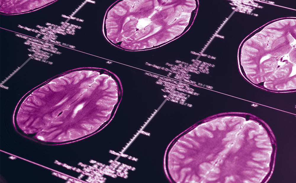
Neuropathological examination of post-mortem brains of patients with dementia due to neurodegenerative and cerebrovascular changes remains important, as the family wants to be sure about the clinical diagnosis and the risk of a hereditary disease. 7.0-tesla magnetic resonance imaging (MRI) can be applied as an additional tool to examine post-mortem brains of patients with neurodegenerative and cerebrovasular diseases. It allows examination of serial coronal sections of a cerebral hemisphere and horizontal sections of brainstem and cerebellum and comparison with the neuropathological lesions. Post-mortem MRI can show the degree and the distribution of the cerebral atrophy. Additional small cerebrovascular lesions can be quantified. The degree of iron load, not due to microbleeds, can be evaluated in different basal ganglia and brainstem structures. Three to six serial sections of a cerebral hemisphere and one section of brainstem and cerebellum allow the evaluation of the most important brain changes and to select the small samples to be used for histological diagnostic purposes. These correlation studies are extremely important for the future, when more 7.0-tesla MRI machines will be available for in vivo clinical-radiological diagnosis. This article is a review of post-mortem MRI data in the brains of patients with neurodegenerative and vascular dementias.

Keywords

Post-mortem, 7.0-tesla magnetic resonance imaging (MRI), neurodegenerative diseases, cerebrovascular diseases, cerebral atrophy, white matter changes, lacunar infarcts, cortical micro-bleeds, cortical micro-infarcts, cortical superficial siderosis, iron deposition

Article:

Although post-mortem neuropathological examination is increasingly performed less often in most western countries, it is still needed in patients with dementia, due to neurodegenerative and cerebrovascular changes, It is important for the family to be sure about the clinical diagnosis and to exclude the risk of a hereditary disease. Clinicalneuropathological correlation studies actually show only a concordance rate around 65%.1 A previously informed consent of the patients or from the nearest family must be obtained to allow an autopsy for diagnostic and scientific purposes.

Neuroimaging during life with 1.5 and 3.0-tesla magnetic resonance imaging (MRI) contributes moderately to the clinical diagnosis. There is hope that 7.0-tesla MRI will be more helpful, but actually only a few neurological centres utilise this technique and only a few clinicalradiological correlation studies have been performed until now.2Postmortem correlation studies combining MRI and histopathology are needed to validate the 7.0-tesla MRI findings in vivo when more centres will utilise of this technique.3 This article reviews what is currently known about post-mortem MRI data in the brains of patients with neurodegenerative and vascular dementias.

Though a definitive post-mortem diagnosis still needs to be confirmed by an extensive macroscopic and microscopic examination of the brain using validated neuropathological criteria,4 7.0-tesla MRI can be used as an additional tool to examine post-mortem brains of patients with neurodegenerative diseases. The degree and the distribution of the cerebral atrophy and white matter changes (WMCs) can be demonstrated. It also detects lesions that can be selected for histological examination. Additional small cerebrovascular lesions can be quantified. The degree of iron load can be evaluated in different basal ganglia and brainstem structures.

A variety of post-mortem MRI techniques have been used including scanning of fixed whole brains or hemispheres,5 coronal brain slices,6 un-fixed whole brains7 and brains in situ.8 The number of detected small cerebrovascular lesions depends on the MRI characteristics, such as pulse sequence, sequence parameters, spatial resolution, magnetic field strength and image post-processing.9 8.0-tesla MRI was shown to be significantly more sensitive to detect small cerebrovascular lesions than 1.5 and 3.0-tesla MRI in postmortem brains.10

Author’s experience with 7.0-tesla MRI
In our experience, we used a 7.0-tesla (Bruker Biospin SA, Ettlinger, Germany) with an issuer-receiving cylinder coil of 72 mm inner diameter, initially employed for animal experiments. Formalin fixed brain sections were placed in a plastic box and filled with salt-free water after cleaning the formalin fixation.11 Serial coronal MRI sections of a cerebral hemisphere and horizontal sections of brainstem and cerebellum were compared to the lesions, observed on macroscopic and histological examination.

Three MRI sequences were used: a positioning sequence, a spin echo T2 sequence and a T2 star (T2*) weighted-echo sequence. The positioning sequence was needed for determination of the threedirectional position of the brain section inside the magnet. The thickness of the T2 images was 1 mm. The field of view was a 9 cm square slide that was coded by a 256 matrix giving a voxel size of 0.352 x 0.352 x 1 mm. T2 weighted images were obtained by using Rapid Acquisition with Relaxation Enhancement (RARE) sequence with repetition time (TR), echo time (TE) and RARE factor of 2,500 ms, 33 ms and 8 ms, respectively. The acquisition time of this sequence was 80 s. The thickness of the T2* images was 0.20 mm. The field of view was also a 9 cm2. It was coded by a 512 matrix, giving a voxel size of 0.176 x 0.176 x 0.2 mm. The slice thickness corresponded to the upper part of the of the brain section. This sequence was a gradient echo sequence with a short TR of 60 ms and TE of 22 ms, a flip angle of 30° and number of excitation of 20. The acquisition time of the sequence was 10 min.12 As the procedures took at least 30 minutes for examination of each section, no additional fluidattenuated inversion recovery (FLAIR) was performed as it added no further information.

Six coronal sections of a cerebral hemisphere, a sagital section of the brainstem and a horizontal section from a cerebellar hemisphere allowed quantification of small cerebrovascular lesions. The sections of the cerebral hemisphere consisted of one at the prefrontal level in front of the frontal horn, one of the frontal lobe at the level of the head of the caudate nucleus, a central one near the mammillary body, a postcentral one, a parietal one at the level of the splenium corporis callosi and one at the level of the occipital lobe.

Additional histological examination of a separate standard coronal section of a cerebral hemisphere at the level of the mammillary body, allowed quantification of small lesions and validation of the MRI findings.13

Cerebral atrophy
Cerebral cortical atrophy on MRI allows comparison to the findings of the macroscopic examination of coronal sections of the brain. Due to the better differentiation between gray matter and WMCs, MRI has the advantage of better differentiation between atrophy, due to cortical lesions and those due to white changes. The most severe atrophy of the frontal and temporal lobes is observed in frontotemporal lobar degeneration (FTLD).13 The degree of hippocampal atrophy correlates well with the histological staging of severity in Alzheimer’s disease (AD) (Figure1).14 In progressive supranuclear palsy (PSP) severe atrophy of the whole brainstem is demonstrated.15

White matter changes and lacunar infarcts
In-vivo MRI studies frequently show WMCs in elderly persons.16 On the other hand, the number of lacunes is largely underestimated.17 Overall

post-mortem 4.7-tesla MRI reveals no significant differences between the imaging of WMCs and the neuropathological assessments, regardless of their severity.5 Post-mortem MRI studies show severe WMCs in small-vessel disease and cerebral amyloid angiopathyi.18 Until now, only a few studies have addressed the demonstration of lacunar infarcts.19 WMCs are frequently observed in the frontal lobe of FTLD.20 On 7.0-tesla MRI they represent the extensive white matter looseness underlying the severe cortical atrophy, due to Wallerian degeneration.13

Cortical micro-bleeds Cortical micro-bleeds
(CoMBs) are frequently observed in brains of patients with dementia due to neurodegenerative and cerebrovascular diseases (Figure 2A). They are also observed in elderly brains without any neurological or psychiatric disease.21 However, they are predominantly observed in Alzheimer’s disease associated with cerebral amyloid angiopathy (AD-CAA).22

The reliability to detect micro-bleeds in the cerebral cortex on 7.0-tesla MRI is 96%, while much lower in the other cerebral regions.11 Due to the

blooming effect also small cortical mini-bleeds, not seen on naked eye examination of the brain, can be detected. However, as the blooming effect differs from one specimen to another, due to the individual composition of the bleed, distinction between micro- and mini-bleeds cannot been made on post-mortem MRI.11

In AD-CAA brains the CoMBs predominate in the parietal inferior gyrus, the precuneus and the cuneus compared to those with ‘pure’ AD.12 In FTLD they are predominantly seen in the superior and inferior frontal, and in the superior temporal gyri, corresponding to the location of the most severe histological lesions.23 In Lewy body disease (LBD) CoMBs predominate in the frontal regions and are not related to the frequently associated AD and CAA features.24 Overall CoMBs occur preferentially in the deep cortical layers.25

Cortical micro-infarcts Cortical micro-infarcts (CoMIs) are considered as the invisible lesions in clinical-radiological in vivo studies.26 Post-mortem 7.0-tesla MRI, in contrast to clinical-radiological correlation studies in vivo, allows the demonstration of CoMIs of different sizes in the cerebral hemispheres. According to the length of the cortical occluded branch, CoMIs can involve the whole depth of the cerebral cortex or only the superficial, the middle and the deep cortical layers separately or combined. They predominate in vascular dementia (VaD), AD-CAA and LBD. In the latter only the small laminar CoMIs are observed (Figure 2B).27

Cerebellar CoMIs are of similar size, as only one type of cortical branch exists. They are mainly due to arteriosclerotic disease rather than due to CAA.28

Cortical superficial siderosis
Cortical superficial siderosis (CSS) is defined as a characteristic curvilinear pattern of low signal on blood-sensitive MRI sequences, preferentially affecting the cerebral convexities.29 It is due to blood residues in the subpial layers and reflects an underlying cortical hemorrhagic lesion.30 Post-mortem 7.0 tesla MRI detects much better, the underlying cortical lesions than in-vivo studies with 1.5 and 3.0-tesla machines. CSS is found to be associated to cortical bleeds as well as to infarcts.31 Most cerebral infarcts become partly hemorrhagic during their chronic stage.32 CSS can also be the hallmark of a cerebral contusion in case of head injury.33

Iron deposition
Iron deposition in the brain is associated with general cognitive ability and cognitive aging.34 A meta-analysis reported that several articles from one laboratory showed a significant increase of iron in the neocortex of AD brains, whilst seven other laboratories fail to reproduce these findings, challenging the hypothesis that transition metal overload accounts for oxidative injury in this disease.35

On post-mortem MRI only in FTLD, a significant iron deposition is observed in the claustrum, the caudate nucleus, the putamen, the globus pallidus and the thalamus. A moderate increase of iron is only found in the caudate nucleus of AD brains (Figure 3).36 Iron content is significantly decreased in the substantia nigra and the red nucleus of PSP brains.15

Conclusions
7.0-tesla MRI of the brain is a useful additional tool for the neuropathological examination of patients who died after a neurodegenerative and/or vascular dementia. It has to be considered as a link between the naked eye and the microscopic examination of the brain. It has the advantage to demonstrate some small lesions not seen on macroscopic examination of the brain and also allows the selection of the most useful samples for histological examination. In addition, on serial sections of the brains small cerebrovascular lesions in several neurodegenerative diseases can be quantified, allowing the determination of their additional impact on the severity of the dementia.

The 7.0-tesla Bruker Biospin is much less expensive than the 7.0-tesla MRI machines used for in vivo studies. Although mainly used for animal experiments, it is quite adapted for examination of human brain sections. In addition, these post-mortem correlation studies will allow the validation of the images that will be obtained when 7.0-tesla MRI machines will be more available in clinical practice.

Article Information:
Disclosure

Jacques L De Reuck has nothing to declare in relation to this article.

Compliance With Ethics

This article involves a review of the literature and did not involve any studies with human or animal subjects performed by the author.

Review Process

Double-blind peer review.

Correspondence

Jacques De Reuck, Leopold II laan 96, 9000 Ghent, Belgium. E: dereuck.j@gmail.com

Support

No funding was received for the publication of this article.

Access

This article is published under the Creative Commons Attribution Noncommercial License, which permits any non-commercial use, distribution, adaptation and reproduction provided the original author(s) and source are given appropriate credit.

Acknowledgements

The following persons have contributed to the present research project: Florent Auger, Nicolas Durieux, Vincent Deramecourt, Charlotte Cordonnier, Claude-Alain Maurage, Didier Leys, Florence Pasquier and Regis Bordet. This study was funded by the INSERM U 1172 (Universté Lille 2, Lille, France).

Received

15 January 2016

References

1. Jellinger KA, Clinicopathological analysis of dementia disorders in the elderly —an update, J Alzheimers Dis, 2006;9:S61–70. 2. Van Velum SJ, Zwanenburg JJ, Engelen-Lee J, et al., In vivo detection of cerebral cortical microinfarcts with high-resolution 7T MRI, J Cereb Blood Flow Metab, 2013;33:332–339. 3. Wardlaw JM, Post-mortem MR brain imaging comparison with macro- and histopathology: useful, important and underused, Cerebrovasc Dis, 2011;31:518–519. 4. De Reuck J, The significance of small cerebral bleeds in neurodegenerative dementia syndromes,Aging Dis, 2012;4:307–312. 5. McAleese KE, Firbank M, Hunter D, et al., Magnetic resonance imaging of fixed post mortem brains reliably reflects subcortical vascular pathology of frontal, parietal and occipital white matter, Neuropathol Appl Neurobiol, 2013;39:485–497. 6. Fernando MS, O'Brien, Perry RH, et al., Comparison of the pathology of cerebral white matter with post-mortem magnetic resonance imaging (MRI) in the elderly brain, Neuropathol Appl Neurobiol, 2004;30:385–395. 7. Awad IA, Johnson PC, Spetzler RF, Hodak JA, Incidental subcortical lesions identified on magnetic resonance imaging in the elderly. II. Postmortem pathological correlations, Stroke, 1986;17:1090–1097. 8. Grinberg LT, Amaro Junior E, da Silva AV, et al., Improved detection of incipient vascular changes by a biotechnological platform combining post mortem MRI in situ with neuropathology, J Neurol Sci, 2009;283:2–8. 9. Greenberg SM, Vernooij MW, Cordonnier C, et al., Cerebral microbleeds: a guide to detection and interpretation, Lancet Neurol, 2009;8:165–74. 10. Dashner RA, Chakeres DW, Kangarlu A, et al., MR imaging visualization of the cerebral microvasculature: a comparison of live and postmortem studies at 8 T, AJNR Am J Neuroradiol, 2003;24:1881–4. 11. De Reuck J, Auger F, Cordonnier C, et al., Comparison of 7.0-T T2*-magnetic resonance imaging of cerebral bleeds in post-mortem brain sections of Alzheimer patients with their neuropathological correlates, Cerebrovasc Dis, 2011;31;511–7. 12. De Reuck J, Auger F, Durieux N, et al., Topography of cortical microbleeds in Alzheimer’s disease with and without cerebral amyloid angiopathy: a post-mortem 7.0-tesla MRI study, Aging Dis, 2015;6:437–43. 13. De Reuck J, Deramecourt V, Cordonnier C, et al., Detection of microbleeds in post-mortem brains of patients with frontotemporal lobar degeneration: a 7.0- tesla magnetic resonance imaging with neuropathological correlates, Eur J Neurol, 2012;19,1355–60. 14. Apostolova LG, Zarow C, Biado K, et al., Relation between hippocampal atrophy and neuropathology markers: a 7T MRI validation study of the EADC-ADNI Harmonized Hippocampal Segmentation Protocol, Alzheimers Dement, 2015;11:139–50. 15. De Reuck J, Caparros-Lefebvre, Deramecourt V, et al., Prevalence of small cerebral bleeds in patients with progressive supranuclear palsy: a neuropathological study with 7.0-Tesla magnetic resonance imaging correlates, Folia Neuropathol, 2014;52:421–7. 16. Schmidt R, Schmidt H, Haybaeck J, et al., Heterogeneity in age-related white matter changes,Acta Neuropathol, 2011;122:171–85. 17. Potter GM, Doubal FN, Jackson CA, et al., Counting cavitating lacunes underestimates the burden of lacunar infarction,Stroke, 2010;41:267–72. 18. Shim YS, Yang DW, Roe CM, et al., Pathological correlates of white matter hyperintensities on magnetic resonance imaging, Dement Geriatr Cogn Disord, 2015;39:92–104. 19. Gouw AA, Seewann A, van der Flier WM, et al., Heterogeneity of small vessel disease; a systemic review of MRI and histopathology correlations,J Neurol Neurosurg Psychiatry, 2011;82:126–35. 20. De Reuck J, Deramecourt V, Cordonnier C, et al., Cerebrovascular lesions in patients with frontotemporal lobar degeneration: a neuropathological study, Neurodegener Dis, 2012;9:170–5. 21. De Reuck j, Deramecourt V, Cordonnier C, et al., Prevalence of small cerebral bleeds in patients with a neurodegenerative dementia: a neuropathological study, J Neurol Sci, 2011;300:63–6. 22. De Reuck J, Deramecourt V, Cordonnier C, et al., The impact of cerebral amyloid angiopathy on the occurrence of cerebrovascular lesions in demented patients with Alzheimer features: a neuropathological study, Eur J Neurol, 2011;18:913–8. 23. De Reuck J, Auger F, Durieux N, et al., The topography of cortical microbleeds in frontotemporal lobar degeneration: a post-mortem 7.0-tesla magnetic resonance imaging, Folia Neuropathol, [In press]. 24. De Reuck J, Auger F, Durieux N, et al., Detection of cortical microbleeds in postmortem brains of patients with Lewy body dementia: a 7.0-Tesla magnetic resonance imaging study with neuropathological correlates,Eur Neurol, 2015;74:158–61. 25. De Reuck J, Cordonnier C, Deramecourt V, et al., Microbleeds in postmortem brains of patients with Alzheimer disease: a T2*-weighted gradient-echo 7.0 T magnetic resonance imaging study, Alzheimer Dis Asscc Disord, 2013;27:162–5. 26. Smith EE, Schneider JA, Wardlaw JM, Greenberg SM, Cerebral microinfarcts: the invisible lesions,Lancet Neurol, 2012;11:272–82. 27. De Reuck J, Deramecourt V, Auger F, et al., Post-mortem 7.0 Tesla magnetic resonance study of cortical microinfarcts in neurodegenerative diseases and vascular dementia with neuropathological correlates, J Neurol Sci, 2014;346:85–9. 28. De Reuck J, Deramecourt V, Auger F, et al., The significance of cortical cerebellar microbleeds and microinfarcts in neurodegenerative and cerebrovascular diseases, Cerebrovasc Dis, 2015;39:138–43. 29. Kumar N, Neuroimaging in superficial siderosis: an in-depth look, AJNR Am J Neuroradiol, 2010;31:5–14. 30. Charidimou A, Gang Q, Werring DJ, Sporadic cerebral amyloid angiopathy revisited: recent insights into pathophysiology and clinical spectrum, J Neurol Neurosurg Psychiatry, 2012;83:124–37. 31. De Reuck J, Deramecourt V, Cordonnier C, et al., Superficial siderosis of the central nervous system: a post-mortem 7.0-tesla magnetic resonance Imaging study with neuropathological correlates, Cerebrovasc Dis, 2013;36:412–7. 32. Hebbrecht G, Maenhout W, De Reuck J, Comparison of trace element alterations and water content in haemorrhagic and non-haemorrhagic cerebral infarcts, Cerebrovasc Dis, 1994;4:412–6. 33. De Reuck J, Cortical superficial siderosis of the central nervous system- An overview, Europ Neurol Review, 2014;10:68–70. 34. Penke L, Valdes Hernandez MC, Maniega SM, et al., Brain iron deposits are associated to general cognitive ability and cognitive aging, Neurobiol Aging, 2012;33:510–7. 35. Schrag M, Mueller C, Oyoyo U, et al., Iron, zinc and copper in the Alzheimer’s disease brain; a quantitative meta-analysis. Some insight on the influence on citation opinion, Prog Neurobiol, 2011;94:296–306. 36. De Reuck J, Deramecourt V, Auger F, et al., Iron deposits in post-mortem brains of patients with neurodegenerative and cerebrovascular diseases: a semi-quantitatve 7.0 T magnetic resonance study, Eur J Neurol, 2014;21:1026–31.

Further Resources

Share this Article
Related Content In Alzheimer's Disease & Dementia
  • Copied to clipboard!
    accredited arrow-down-editablearrow-downarrow_leftarrow-right-bluearrow-right-dark-bluearrow-right-greenarrow-right-greyarrow-right-orangearrow-right-whitearrow-right-bluearrow-up-orangeavatarcalendarchevron-down consultant-pathologist-nurseconsultant-pathologistcrosscrossdownloademailexclaimationfeedbackfiltergraph-arrowinterviewslinkmdt_iconmenumore_dots nurse-consultantpadlock patient-advocate-pathologistpatient-consultantpatientperson pharmacist-nurseplay_buttonplay-colour-tmcplay-colourAsset 1podcastprinter scenerysearch share single-doctor social_facebooksocial_googleplussocial_instagramsocial_linkedin_altsocial_linkedin_altsocial_pinterestlogo-twitter-glyph-32social_youtubeshape-star (1)tick-bluetick-orangetick-red tick-whiteticktimetranscriptup-arrowwebinar Sponsored Department Location NEW TMM Corporate Services Icons-07NEW TMM Corporate Services Icons-08NEW TMM Corporate Services Icons-09NEW TMM Corporate Services Icons-10NEW TMM Corporate Services Icons-11NEW TMM Corporate Services Icons-12Salary £ TMM-Corp-Site-Icons-01TMM-Corp-Site-Icons-02TMM-Corp-Site-Icons-03TMM-Corp-Site-Icons-04TMM-Corp-Site-Icons-05TMM-Corp-Site-Icons-06TMM-Corp-Site-Icons-07TMM-Corp-Site-Icons-08TMM-Corp-Site-Icons-09TMM-Corp-Site-Icons-10TMM-Corp-Site-Icons-11TMM-Corp-Site-Icons-12TMM-Corp-Site-Icons-13TMM-Corp-Site-Icons-14TMM-Corp-Site-Icons-15TMM-Corp-Site-Icons-16TMM-Corp-Site-Icons-17TMM-Corp-Site-Icons-18TMM-Corp-Site-Icons-19TMM-Corp-Site-Icons-20TMM-Corp-Site-Icons-21TMM-Corp-Site-Icons-22TMM-Corp-Site-Icons-23TMM-Corp-Site-Icons-24TMM-Corp-Site-Icons-25TMM-Corp-Site-Icons-26TMM-Corp-Site-Icons-27TMM-Corp-Site-Icons-28TMM-Corp-Site-Icons-29TMM-Corp-Site-Icons-30TMM-Corp-Site-Icons-31TMM-Corp-Site-Icons-32TMM-Corp-Site-Icons-33TMM-Corp-Site-Icons-34TMM-Corp-Site-Icons-35TMM-Corp-Site-Icons-36TMM-Corp-Site-Icons-37TMM-Corp-Site-Icons-38TMM-Corp-Site-Icons-39TMM-Corp-Site-Icons-40TMM-Corp-Site-Icons-41TMM-Corp-Site-Icons-42TMM-Corp-Site-Icons-43TMM-Corp-Site-Icons-44TMM-Corp-Site-Icons-45TMM-Corp-Site-Icons-46TMM-Corp-Site-Icons-47TMM-Corp-Site-Icons-48TMM-Corp-Site-Icons-49TMM-Corp-Site-Icons-50TMM-Corp-Site-Icons-51TMM-Corp-Site-Icons-52TMM-Corp-Site-Icons-53TMM-Corp-Site-Icons-54TMM-Corp-Site-Icons-55TMM-Corp-Site-Icons-56TMM-Corp-Site-Icons-57TMM-Corp-Site-Icons-58TMM-Corp-Site-Icons-59TMM-Corp-Site-Icons-60TMM-Corp-Site-Icons-61TMM-Corp-Site-Icons-62TMM-Corp-Site-Icons-63TMM-Corp-Site-Icons-64TMM-Corp-Site-Icons-65TMM-Corp-Site-Icons-66TMM-Corp-Site-Icons-67TMM-Corp-Site-Icons-68TMM-Corp-Site-Icons-69TMM-Corp-Site-Icons-70TMM-Corp-Site-Icons-71TMM-Corp-Site-Icons-72