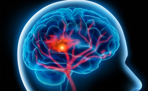
Cerebral congophilic or amyloid angiopathy (CAA) is a clinicopathological entity that has been recognised since the early part of the 20th century.1 It is now considered a common cause of primary non-traumatic brain haemorrhage and traditionally it was described in elderly patients who were thought to be normotensive. Because of its frequent association with Alzheimer’s disease (AD), CAA has become a primary focus of scientific inquiry. The spectrum of intracerebral haemorrhage (ICH) that may occur in CAA includes: cerebral lobar haemorrhage in which several lobes on both sides of the brain may be involved over time; on rare occasions, haemorrhage in deep brain central grey nuclei, the corpus callosum and cerebellum, locations more typically involved when there is hypertensive ICH; the rare purely subarachnoid and subdural haemorrhages, as meningeal vessels may be heavily involved by CAA; and miliary and petechial or cerebral microbleeds.1,2 In addition, there may be scattered associated microinfarcts, an inflammatory type of CAA, leukoencephalopathy, AD-associated changes of the brain and superficial siderosis (SS).3 In this brief article we provide an update on advances in our understanding of CAA-associated ICH. We will focus on the following topics: neuropathology and mechanism of CAA-related haemorrhage; epidemiology, including genetic and other possible risk factors; clinical presentation; diagnosis, including newer imaging modalities; and prospects for prevention and treatment.
Neuropathology and Mechanism of Cerebral Amyloid Angiopathy-associated Brain Haemorrhage
Amyloid-β (AB) deposition in the vascular media and adventitia provides a critical link whereby ICH may occur. AB is generated by proteolytic cleavage of the amyloid precursor proteins β-secretase and γ-secretase to yield a family of AB peptides with 40- and 42-amino acid species (AB40 and AB42, respectively).4 These peptides may undergo degradation by other proteolytic enzymes (e.g., neprilysin, insulin-degrading enzyme), remain in solution and enter the plasma or efflux across the blood–brain barrier, or polymerise to form soluble oligomers or insoluble amyloid fibrils as senile plaques in the brain parenchyma or as deposition in the vascular media and adventitia, as occurs in CAA. AB deposition occurs in the vascular media and adventitia of small arteries of the leptomeninges and cerebral cortex, with heavy involvement of the occipital regions, whereas white-matter brain vessels are much less frequently affected.4 The predominant AB species in CAA is the relatively more soluble AB40. AB is thought to be generated by neurons and possibly in the liver. Microscopically, CAA is characterised by acellular thickening of the walls of small and medium-sized arteries, including arterioles, but less often veins, by an amorphous, intensely eosinophilic material.1 Microvascular amyloid may be identified by periodic acid–Schiff, toluidine blue, crystal violet, thioflavin S or T (fluorescence under ultraviolet light) or Congo red stain under polarised light (yellow-green birefringence). Furthermore, affected vascular channels may show a ‘double-barrel’ lumen, fibrinoid degeneration or necrosis (a hypertension-related change), segmental dilatation with microaneurysm formation and ‘glomerular’ formations of microvessels and obliterative fibrous intimal changes.
The latter changes are characteristic in hypertension and may or may not be associated with the genesis of CAA.1 CAA fibrils replace smooth muscle cells and cause separation of the internal elastic membrane and the external basement membrane, and there is smooth muscle degeneration and capillary occlusion. The latter findings may be linked to cerebral blood flow dysregulation in CAA. Animal and human studies support the concept of vascular dysfunction in CAA, as does the occurrence of associated brain microinfarcts, white-matter lesions and clinical evidence of cognitive impairment in this condition.4–7 Therefore, hypoperfusion and impaired vascular autoregulation in CAA may be responsible for cerebral microinfarcts and white-matter lesions.6,8
Role of Cerebral Amyloid Angiopathy in Cerebral Microbleeds
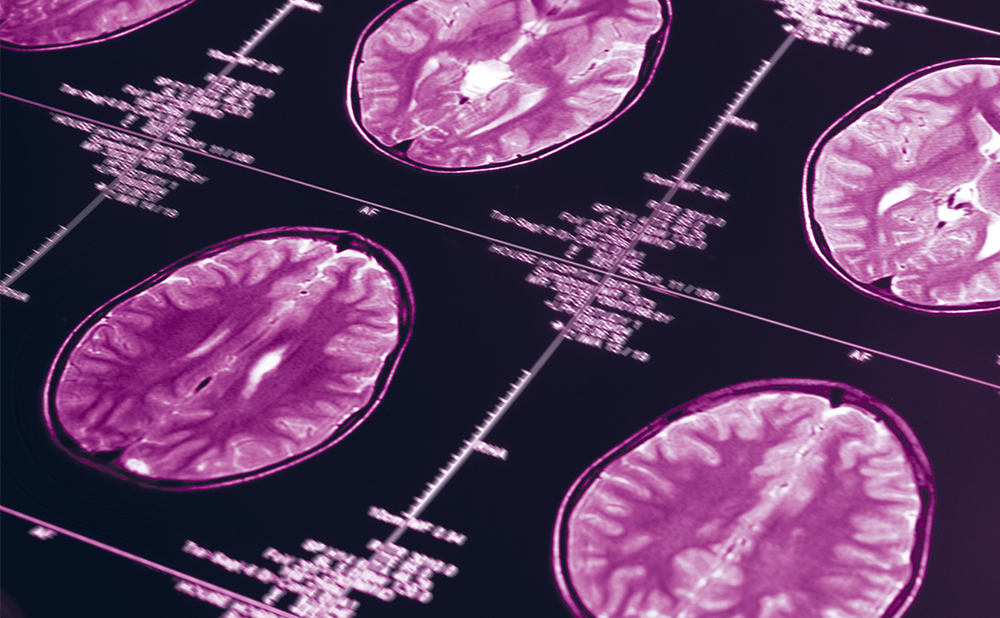
Cerebral microbleeds may be defined on gradient-echo (GRE) or T2*-weighted magnetic resonance imaging (MRI) sequences (or other appropriate magnetic MRI sequences) as rounded foci measuring <5 mm in size that appear as hypointense areas that are distinct from vascular flow voids, leptomeningeal haemosiderosis or non-subcortical mineralisation.9,10 The appearance of a cerebral microbleed on a GRE sequence is believed to be larger than the actual brain tissue lesion due to a ‘blooming effect’ of the MRI signal.2 The reduction of MRI signal is caused by haemosiderin, a blood breakdown product. Haemosiderin is sequestered by macrophages where it remains for years. Cerebral microbleeds located in the cortex are thought to be caused by CAA, whereas those located subcortically are believed to have a hypertensive aetiology.2 Clinically, cerebral microbleeds may be associated with cognitive impairment, higher stroke risk, lower cerebrospinal fluid (CSF) amyloid levels and higher mortality.11,12 What is the mechanism linking CAA to cerebral microbleeds? In CAA, lobar microbleeds are believed to be caused by vessel fragility that leads to vessel rupture mediated by deposition of amyloid within the media and adventitia of small to medium-sized cerebral arteries.13 According to the Boston criteria, the presence of multiple lobar macro- and microbleeds is substantially specific for severe CAA among elderly persons with no other obvious cause for ICH.14 In CAA, both lobar macro- and microbleeds may show a posterior cortical preference in relation to location.13 ICH, therefore, may cluster in the temporal and occipital lobes in these patients and be associated with disease progression and recurrent brain bleeds. Furthermore, cerebral microbleeds generally outnumber lobar macrobleeds, and new microbleeds may be predicted by large numbers of microbleeds at baseline and APOE e2 or e4 genotype.15,16 It has been argued that CAA-related microbleeds and macrobleeds may represent distinct entities, with increased vessel wall thickness predisposing to the occurrence of microbleeds compared with macrobleeds.17
Epidemiology
Sporadic CAA is a common pathological finding in the elderly, which is detected in approximately 10–40 % of brains ≥65 years of age.18 CAA is even more prevalent in elderly subjects with concomitant AD, and has been reported to be present in 80 % of these individuals.18 One study estimated the prevalence of severe CAA as present in 21 % of autopsied brains from individuals 85–86 years of age.19 Spontaneous ICH in the elderly is attributed to CAA in 10–34 % of cases.18,20 The prevalence of cerebral microbleeds in the general population doubles from 20 to 40 % with an increase in age from 60 to 80 years or older, and they are also seen more frequently in people with hypertension.21 In the Rotterdam Scan Study, the presence of microbleeds at baseline was associated with a five-fold risk of developing new microbleeds in a three-year interval compared with subjects without microbleeds at baseline (odds ratio 5.38, 95 % confidence interval [CI] 3.34–8.67).22 CAA-related ICH and hypertensive-related ICH may exist simultaneously in nearly 25 % of individuals with ICH.23 Lobar microbleeds are associated with recurrent ICH and CAA disease progression.15 Subjects with CAA-related microbleeds have a greater than sevenfold risk of death from stroke compared with subjects without microbleeds after adjusting for other risk factors (hazard ratio 7.20, 95 % CI 1.44–36.10, p=0.02).24 The number of amyloid-burdened vessels is increased in carriers of the APOE e4 genotype and the severe vasculopathic changes seen in CAA-related ICH are increased in carriers of the APOE e2 genotype.25 In a genetic association study, carriers of the APOE e2 genotype with lobar ICH had larger ICH volumes, increased mortality and poorer functional outcomes than non-carriers with lobar ICH.26 This association was not found with variant APOE genotypes and deep ICH.26 Hereditary CAA typically presents as an autosomal dominant disease in selected families and is extremely rare.27 APOE genotype does not appear to play a significant role in hereditary CAA, and is associated with an earlier age of onset, severe neurological dysfunction and death.27,28
Presentation
Sporadic CAA can be asymptomatic in the general elderly population, and there is no pathognomonic clinical presentation of CAA-related ICH.29 Depending on haematoma size and location, lobar ICH can present with decreased level of consciousness, headaches, seizures or focal neurological deficits.30 White-matter lesions or leukoencephalopathy are common in subjects aged ≥55 with lobar ICH and are associated with cognitive dysfunction.31 CAA-related microbleeds are associated with an increased risk of cognitive decline and functional dependence.15 CAA and concomitant AD pathology are associated with more severe cognitive impairment than CAA or AD occurring independently.32
One cohort study of 404 patients demonstrated that when controlling for AD pathology, moderate to severe CAA is associated with impairment in the specific cognitive domains of perceptual speed and episodic memory, but not semantic memory or working memory.33 Cerebral microinfarcts are common in severe CAA and may contribute to vascular cognitive impairment seen in these patients.34 A small case-controlled study of 78 subjects with CAA found that 15 % of subjects had diffusion-weighted imaging (DWI)-hyperintense lesions consistent with subacute cerebral infarctions associated with an increased number of haemorrhagic lesions.35 CAA can also lead to ischaemia that presents with focal neurological deficits typically seen with symptomatic cerebral infarctions.20 CAA is the most common cause of non-traumatic non-aneurysmal convexal subarachnoid haemorrhage (SAH) in the elderly.36 SAH at the convexity more commonly presents with transient sensory and/or motor deficits and seizures rather than a headache, which is the typical presentation for aneurysmal or traumatic SAH.36 There have also been case reports of patients with CAA-related inflammation presenting with progressive cognitive decline, headaches and seizures.37 The characteristic MRI in CAA-related inflammation shows asymmetric T2 hyperintensities consistent with vasogenic oedema, and the severity of the presenting symptoms is correlated with the T2 hyperintensity volume.37
Diagnosis
A definitive diagnosis of CAA requires brain biopsy or necropsy for histological examination of affected brain tissue. The Boston criteria were created in part to standardise the diagnosis of CAA during life and include the following diagnoses: definitive CAA, probable CAA with supporting pathology, probable CAA and possible CAA.14 The histological diagnosis requires special amyloid stains as detailed above. The neuropathological severity of CAA can be determined by one of two grading systems proposed by Olichney et al. and Vonsattel et al.38,39 The Boston criteria use the Vonsattel approach that grades CAA severity from mild to severe based on the involvement of pathological changes in the blood vessels.39 SS has also been reported in patients with CAA. In a small case-controlled study of 38 patients with histologically confirmed CAA, the authors reported that SS was present in 60.5 % of patients with CAA, but was not found in any of the control patients.40 Inclusion of SS into the Boston criteria increased the sensitivity from 89.5 to 94.7 %; however, this was not statistically significant and did not change the specificity.40 Although an MRI of the brain is able to detect CAA-related ICH in living subjects, MRI is inadequate to capture the neuropathological changes associated with CAA. A newer diagnostic imaging modality is positron emission tomography (PET) with Pittsburgh compound B (PiB), which binds to AB deposits.41
Studies suggest that PiB-PET can distinctively detect CAA neuropathology and prolonged retention of PiB may be indicative of an increased haemorrhage risk associated with recombinant tissue-type plasminogen activator.42 CSF AB proteins may act as sensitive biomarkers in the diagnostic work-up of dementia. CSF AB42 proteins are decreased in AD compared with controls (p<0.001).43 Further studies have revealed that CSF AB40 proteins are significantly decreased in CAA compared with controls or AD (p<0.01 versus controls or AD) and CSF AB42 proteins are also decreased, although less significantly, in CAA compared with controls or AD (p<0.001 versus controls and p<0.05 versus AD).44
Prospects for Prevention and Treatment
Microbleeds and white-matter lesions may act as surrogate markers of CAA severity.45 MRI modalities such as diffusion tensor imaging may be able to detect small-vessel damage more readily and act as a tool for CAA prevention.46 In multivariate analyses, aspirin use after ICH, including both micro- and macrobleeds, was associated with increased recurrence of CAA-related lobar ICH. The microbleed burden could have significant implications for anti-platelet treatment in patients with CAA.47 A subanalysis of the Perindopril protection against recurrent stroke study (PROGRESS) suggests that blood pressure control reduces the risk of CAA-related ICH and blood pressure lowering could be preventive against multiple causes of ICH.48 Animal studies in mice suggest that AB immunotherapy could theoretically slow or stop the development of CAA; however, it remains unclear if these findings are applicable in humans.49 Finally, corticosteroid or immunosuppressant treatment may be appropriate interventions for patients with symptoms and imaging consistent with CAA-related inflammation.50
Conclusion
CAA is a common cause of primary spontaneous ICH in the elderly and incidence increases with age. CAA is often associated with AD and cognitive impairment. Sporadic CAA is more common in carriers of the APOE e2 and e4 genotypes. The anatomical location and clinical presentation of CAA-related ICH are variable depending on the neuropathological involvement and severity. MRI advances have provided powerful tools in the diagnosis of CAA, but brain biopsy or necropsy is necessary for a definitive diagnosis of CAA. Despite significant progress in our understanding of CAA, further research is needed to determine prospective prevention and treatment strategies to reduce the incidence of CAA-related ICH in the ageing population.


