It cannot be seen, cannot be felt, cannot be heard, cannot be smelt.
It lies behind stars and under hills, and empty holes it fills.
It comes first and follows after, ends life, kills laughter.
The Hobbit. JRR Tolkien
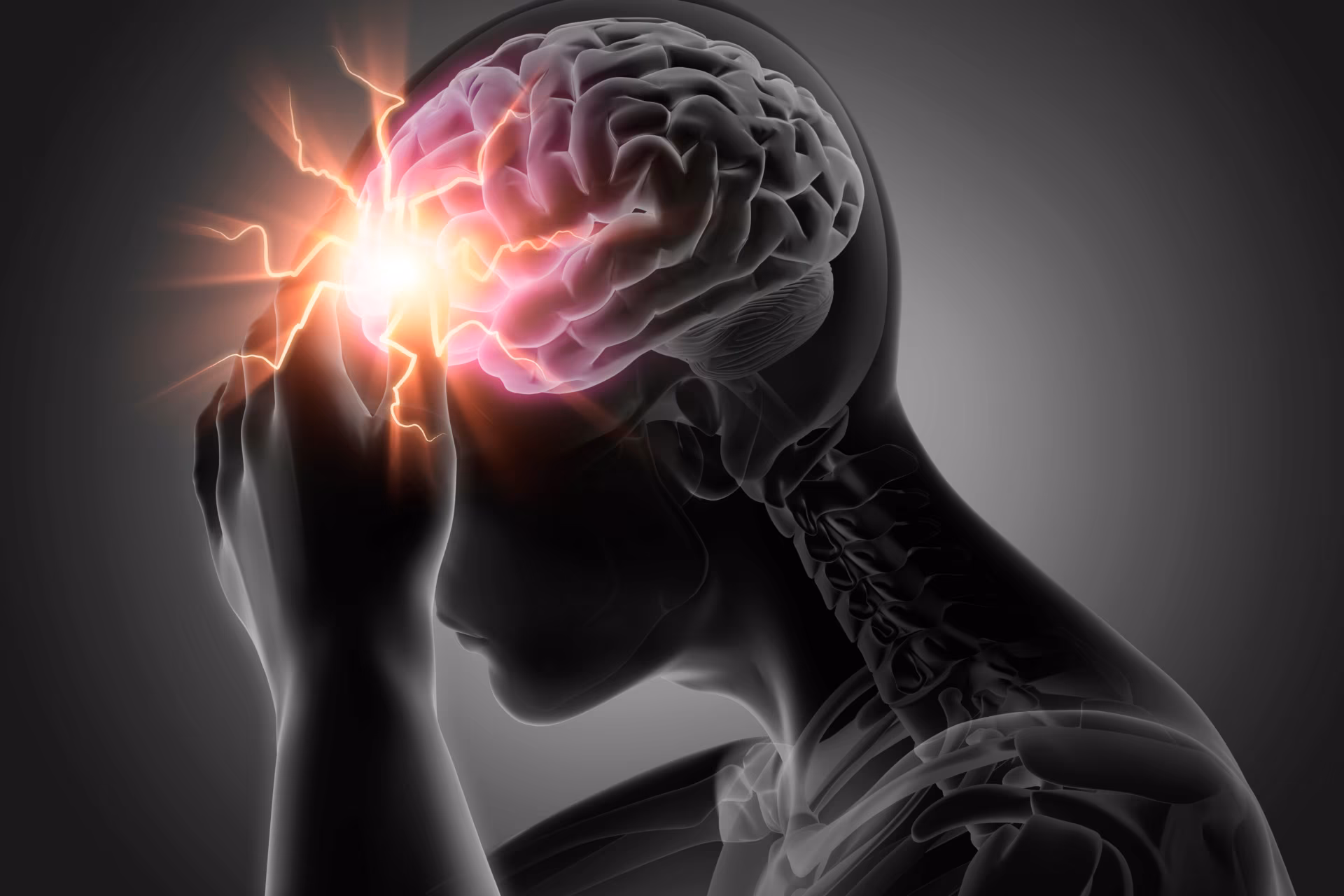
‘Darkness’ was the answer to Gollum’s riddle to get out of the tunnel in The Hobbit.1 Darkness is the only choice for many people with migraine. The reason for this preference for dimmed environments is multifactorial, and the pathophysiology behind it, intricate and fascinating.
The word ‘photophobia’ dates from 1799, and the original Greek terminology -phobia would indeed imply fear or aversion to light.2 Photophobia is described as one of the classic associated symptoms of migraine and is important in the diagnostic criteria for migraine, especially in patients without nausea.3 Symptoms of photophobia can be described by the patient in a variety of ways. Careful attention may be required to understand this symptom completely. This can be challenging if the interpretation of the photophobia symptoms, by the patient and/or the physician, is ambiguous.
Photophobia is defined as “an aversion to, or avoidance of (bright) light, especially as the result of discomfort caused by ocular disorders and certain neurological diseases” (Oxford English dictionary; www.oed.com). Lebensohn commented in 1951 that the term was applied indistinctly for different sensations following exposure to light: uncomfortable visual perception and exacerbation of pain.4 ‘Glare’ and ‘dazzle’ are other terms frequently used, which could have different meanings involving loss of visual acuity, adaptation problems or discomfort following intraocular light scatter,5,6 and should not be confused with photophobia symptoms. Therefore, at least three different characterizations are available, which could, indeed, be translated into three different symptoms that may be related to three different pathophysiological processes.
In this review, we divide photophobia into particular clinical manifestations. Photic sensitivity will be used to describe any bothersome sensation produced by light that does not augment head pain, photic allodynia will refer to the increase in headache intensity, and photo-oculodynia will be used to describe the pain perceived in the ocular region on exposure to light.
Epidemiology
Photophobia is a symptom reported in several neurological conditions, including primary and secondary headaches, meningitis and neurodegenerative conditions, among others.7 In migraine, this symptom might be underrepresented due to reporting bias and subjectivity of the sensation,8 which cannot be corroborated by a witness as much as other associated symptoms, such as vomiting.9 The prevalence of photophobia increased from 51.5% to 82.5% following the use of a specific questionnaire.10
More than 80% of adults with migraine have photophobia associated with the attacks, and in more than half, the photophobia is described as moderate or severe.11 Indeed, photophobia is reported to be twice as likely to occur as nausea or phonophobia, and to be the most bothersome symptom in a migraine attack.12 In paediatric populations, photophobia is also reported in as much as 90% of patients.13 Photophobia is not uniquely associated with the migraine episode, as the threshold for interictal photophobia can be lower in migraine patients,14–16 especially in chronic cases.14 Photophobia is reported in up to 70% of participants when nitroglycerin is used as a migraine trigger,17 and the threshold might be lower in patients with migraine with aura.18
Photophobia can be described as bilateral or unilateral, sometimes ipsilateral to the headache side, although lateralization should suggest a differential diagnosis with other primary headache disorders. More than one-third of a cohort of patients with short-lasting unilateral neuralgiform headache with conjunctival injection and tearing or cranial autonomic features reported photophobia; half of the patients, however, had a migrainous biology.19 The proportion of patients with photophobia in hemicrania continua is also high, at between 55% and 74%.20,21 Half of these photophobia patients experienced unilateral symptoms and up to 66% had migrainous biology;20 the majority reported symptoms ipsilateral to the pain.20,21 Patients with cluster headache manifest more photophobia than controls during the bout, but not outside of it,22 and almost a quarter describe light as a headache trigger.23 Episodic cluster headache might be the primary headache with the higher proportion of lateralized photophobia, up to 80% compared with 48% in chronic cluster headache type.21
Photophobia burden and assessment
Photophobia may also serve as a marker of disability and comorbidities associated with migraine, especially psychiatric comorbidities. Participants with episodic migraine had higher sensitivity than healthy controls in the Photosensitivity Assessment Questionnaire,24 and migraineurs reported a higher discomfort than healthy controls during daily tasks, such as performing social activities, driving, or looking at a bright screen, independently of the chronicity or presence of aura.25 Although causality was not investigated, the presence of interictal photic sensitivity showed higher depression scores on the Beck Depression Inventory, in patients with episodic migraine compared with patients with migraine without interictal photic sensitivity and healthy controls.26 The Beck Anxiety Inventory also revealed differences, with a higher mean in the interictal photophobia group compared with patients without interictal photic sensitivity and controls.26 In addition, photophobia may impact sleep quality. In the same study, up to three-quarters of patients with interictal photic sensitivity reported insomnia or hypersomnia, in contrast to 44% of the controls and 37% of the episodic migraineurs without interictal photic sensitivity. Ictal photophobia has also been positively correlated with age, and interictal photophobia with age and depression, anxiety and stress in patients with episodic migraine assessed by the Depression, Anxiety and Stress Scale.27
Several questionnaires have been developed with the aim of detecting or assessing the intensity of photophobia. The 8-item Korean Photophobia Questionnaire (KUMC-8), focuses on symptoms during the attack, including photophobia behavioural responses, photic allodynia and photo-oculodynia, and also includes a question regarding interictal photophobia.10 The Leiden Visual Sensitivity Scale consists of nine questions about photophobia, but also other visual stimuli, including sensitivity to patterns, palinopsia and impairment of visual acuity.28 The adapted questionnaire by Llop et al.26 takes questions from the KUMC-8, and a previous questionnaire used for blepharospasm,29 and quantifies intensity of photophobia and the outcome in terms of disability on daily activities such as shopping or trips to the cinema.26
The impact of photophobia on quality of life can be assessed using several questionnaires, including the Utah Photophobia Symptom Impact Scale, which does not assess photic allodynia or photo-oculodynia.30
Alternatively, in the clinical setting, where time can be limited, the use of closed-ended questions such as “During a headache, would you prefer to be in bright sunlight or in a dark room?” could increase sensitivity for photophobia.8 For the detection of photic allodynia, some form of the question “If you are in a dark room and someone switches on the lights, would your headache intensity increase from 6 to 8/10?” has proven useful for us.
Photophobia in migraine
Several studies have been conducted in the past four decades, with similar methodology involving exposure of subjects to increasing illumination intensity. Each study, however, used different environmental conditions and lighting intensities. In the 1980s, a study found higher rating of photophobia, ranked 0–40, described as glare and light-induced pain at different intensities, in patients with headaches classified as migraine, compared with subjects with tension or non-migrainous headaches.15 Glare and light-induced pain were higher in headache-free participants than controls, and had a tendency to increase slightly during an episode of headache. However, the control group was defined as having either fewer than 12 headaches or fewer than two severe headaches per year.15 Light-induced pain was doubled in subjects who had a prophylactic, undisclosed, medication.15
In 1997, studies using progressive light intensity showed that patients with migraine manifested more photophobia, described as a lower threshold for discomfort and pain during a migraine attack, and also during the interictal period, compared with controls.16,31 It is unclear whether the disturbance described referred exclusively to the eyes (photo-oculodynia). Pain at the maximal stimulation of 23000 lux was reported in 2/67 controls, compared with 58/67 patients with migraine, and migraine patients also had a lower threshold when attack free.16 Photic sensitivity was bilateral even if the headache was unilateral,16 and the side ipsilateral to the headache tended to be more sensitive than the contralateral side.15,16 Participants reporting discomfort also had a lower threshold for pain.16 Monocular and binocular photic sensitivities were also correlated, and the second eye tested was more sensitive than the first, suggesting a higher connection involving the second- or third-order neurones.16 Furthermore, a threshold decrease was observed with successive measurements, supporting a central sensitization mechanism. Headache intensity and photic sensitivity threshold were not correlated. However, when tested using a questionnaire, those self-reporting interictal photic sensitivity had lower pain thresholds.16 No differences were found according to presence of nausea or uni/bilaterality of the pain.16
In the same study, 19 patients were tested during a migraine episode. Headache was compared before and after the light exposure using a visual analogue scale, with a median increase of 18%.16 Pain and discomfort threshold were lower than outside the attack.16 Photic sensitivity threshold in migraine without aura or migraine with aura was not significantly different, but patients with migraine with aura showed lower discomfort thresholds correlated with age;16 similar results were inferred in a study using unspecified type of migraine.31 More photophobia was reported during winter months, implying possibly a long-term adaptation to the darker season.16
Visual field defects have been analysed using multifocal pupillographic objective perimetry. Patients who had experienced a migraine attack in the 2 weeks preceding an assessment using yellow stimuli delivered to different locations in the visual field, presented consistent defects on the inferotemporal regions, whereas no defects were consistent following the blue protocol.32 This could imply either a superonasal retinal impairment or a defect at a cortical level.32
A disabling, long-standing photophobia may lead the patient to pursue certain lighting preferences and behavioural patterns that are reported spontaneously during the anamnesis.33 A study in the mid-1990s found it more likely that patients with migraine chose “reddish” lights to be more uncomfortable.34 Patients with migraine recognized lower and higher wavelengths to be more uncomfortable,35 and this also extrapolated to white, unfiltered light,35 with medium wavelengths better tolerated.35
Sensitivity to flickering, reflected lights and patterns were increased in women with migraine.36 Additionally, sensitivity to other visual phenomena has also been detected among subjects with higher frequency of headaches (not classified as migraine), who reported the perception of more illusions during the observation of patterns,37 as well as in migraineurs after exposure to striped patterns.38 A recent review described the pathophysiology of aversion to patterns, flicker and colours in migraine.39
There are conflicting results involving photosensitivity related to the colour of the iris.16
Along with photic sensitivity, cutaneous allodynia is a frequent associated symptom in migraine patients40–42 that has been attributed to central sensitization.43–45 This symptom can be higher in patients with photic sensitivity46–48 and photic allodynia,49 especially in patients with chronic forms of migraine.50
Pathophysiology
A certain degree of photic sensitivity is essential for retinal protection in situations of maximal light exposure.51 However, a disruption in the pathway receiving and processing light may imbalance this threshold, and, consequently, alterations in light intensity could enhance the perception of dural nociceptive inputs to the trigeminocervical complex and thalamus, with a subsequent increase in the recognition of pain.52
Seventy years ago, Lebensohn disclosed that photophobia is but a symptom, and that treatment is best accomplished by the cure of the underlying disorder, which he believed to be dependent on four factors, including vasodilation, oculomotor function and sensation.4 The pathways for photophobia have been hypothesized to consist of a sophisticated network involving, at least, eye structures, nerves with motor, sensory and autonomic function, and brainstem, diencephalon and cortex.
Pupil
It is not infrequent that a change in pupillary diameter is reported motu proprio by the most observant migraineurs during clinical anamnesis. Pupillary assessment was, therefore, a logical way forward in the investigation of the pathophysiology of photophobia.
Mean pupil diameter was smaller on infrared photographs in patients examined during a migraine attack compared with that of controls; outside an attack, mean pupil diameter was similar between the two groups.15 However, by using pupillography, the pupils of subjects with migraine were 0.4 mm smaller in comparison with controls and tension headache subjects,53 at several light intensities. Pupil diameter decreased in subsequent measurements in participants with and without headache, and some asymmetry was seen during the interictal period in those with frequent migraine episodes or with other headache types without their typical associated migraine symptoms. Independently of the type of headache, participants also had less and slower dilatation compared with controls on the side ipsilateral to the headache during the attack and in the interictal phase, in a darkened environment.53,54
A dysregulation of the central monoamine pathway was consequently proposed, and therefore the sensitivity to mydriatic or miotic changes in the iris sphincter has been assessed in several studies. The instillation of the unspecific adrenergic agonist adrenaline caused hyperreactive, bilateral mydriasis in patients with “classic” or “common” migraine, which, after 15 minutes, was significant in the side ipsilateral to the headache.55 An increased mydriatic response was induced in adults after the ingestion of a selective alpha-adrenergic agonist, phenylephrine56 or following the local instillation at 1%, with responses occurring promptly as well as at 1 and 2 weeks after the attack, compared with controls; responses were especially pronounced in patients with pain described as pulsatile from the onset and with associated cranial autonomic symptoms.57 Phenylephrine also caused mydriasis after 90 minutes in a paediatric population with “classic migraine”, a finding that was similar in those with tension headache, “common migraine” or in healthy controls.56 Guanethidine, a blocker of the post-ganglionic adrenergic nerves, caused a more prolonged ipsilateral miotic response in migraine patients when instilled unilaterally, without contralateral change in diameter.58 Interestingly, males without migraine showed a substantial and early mydriatic response, which was milder and earlier in all females independently of the group, whereas this response was absent in males with migraine.58 Decreased mydriatic responses were found following the local instillation of fenfluramine, a serotonin-releasing agent in paediatric59 and adult58 migraine patients compared with healthy controls. In patients with cluster headache, the pupil ipsilateral to the cluster headache was less dilated than the contralateral side at 30 minutes following the instillation of the catecholamine-releasing agent, tyramine,58 whereas this response was bilateral or slightly anisocoric in migraine patients.57 This miotic response may also be mediated by neurokinins.60
This possible sympathetic hypofunction has been investigated more recently. Patients with migraine had a slightly smaller pupil diameter than controls, and reduced velocity and amplitude of contraction within 2 days of an attack.61 Quantitative pupillary light reflexes showed reduced parasympathetic constriction and sympathetic re-dilation in migraine patients with severe symptoms,14 and the reduced diameter change was lower if the patient had a lower interictal light threshold.14 Infrared measurements after administration of the alpha-2 adrenergic agonist apraclonidine demonstrated a longer latency of the light reflex in migraine patients.62 No significant differences with the control group were found interictally or during the migraine episode.62
Vertical distance between eyelids was greater on the symptomatic side.54 When applying cold pressure test as a stressor, the majority of migraineurs had a reduced pupillary response.63
Pupil diameter in subjects with episodic migraine was measured following yellow and blue stimuli, and were larger following the yellow stimuli, both in migraineurs and controls.32 This study did not identify any changes in pupillary responses that could potentially predict the next attack.32
Furthermore, systemic catecholamine alterations have been observed after photic stimulation, with an increase in urinary excreted epinephrine and a decrease in the excretion of norepinephrine, compared with controls.64 Similar responses were obtained after exercise,65 which may account for movement sensitivity and hypersensitization.
Retina
Retinal photoreceptors include rods, cones66 and retinal ganglion cells with different types of photopigments.67 The opsins are pigments that couple to a G-protein and activate them in a light-dependent manner, with more than a thousand described to date.68 Melanopsin-expressing intrinsically photosensitive retinal ganglion cells account for less than 3% of retinal ganglion cells69,70 and possibly have a role in the mammalian circadian rhythm in the suprachiasmatic nucleus.69–71 These constitute an important element of the so-called non-image-forming visual system or non-visual phototransduction.
In mammals, intrinsically photosensitive retinal ganglion cells have broad connections with several anatomical structures, including the olivary pretectal nucleus, broadly known for its function in the pupillary reflex, intergeniculate leaflet70 and the dorsolateral geniculate nucleus, from where thalamocortical projections originate,72 superior colliculus, medial amygdala or periaqueductal grey.73 A high proportion of these neuronal projections are crossed.73 These cells may also be involved in photic allodynia,74 as described in the diencephalon section (see below). Axonal connections also spread to structures related to autonomic function, as shown in preclinical studies, which may account for sympathetic and parasympathetic responses described in humans. These include the superior salivatory nucleus, located in the pontine tegmentum in the brainstem, or the sympathetic intermediolateral nucleus in the spinal cord.75
Mutations in the melanopsin gene may also be related to affective conditions. Participants presenting the homozygous genotype for the missense variant P10L were more than five times more likely to be in the group with seasonal affective disorder than in the control group.76 Melanopsin has been extensively studied in recent years, but it is not the only opsin involved in the phototransduction pathways. Others include neuropsin,77 which can be implicated in the prevention of myopia,78 or encephalopsin, which is highly expressed in the cortex and cerebellum,79 can be activated with transcranial illumination and might regulate monoamine concentration.80
Cranial nerves: The optic nerve and trigeminal pathways
In a cohort of 19 completely blind patients (13/19 with no light perception) with painful eyes due to several comorbidities, eye enucleation produced relief of prominent ipsilateral or contralateral photophobia.81
Luminance changes stimulating peripheral and parafoveal areas of the retina caused increased visually evoked potential (VEP) amplitudes, whereas VEP was not increased with stimuli transmitted through the fovea.82 Sensitization might not be dependent exclusively on the optic nerve, as seen in animals with increased blink reflex in response to bright light following optic nerve section.83
Preclinical studies in adult rats showed that neurones in the trigeminal nucleus caudalis related to nociceptive responses can be activated by light stimuli.84 However, this is not the case in neonatal mice, a finding that has been attributed to the immaturity of the eye vasculature at that age.74 These neurones can also be inhibited by injecting local anaesthetic into the vitreous and trigeminal root ganglion, superior salivatory and olivary nuclei, but not with topical treatment at the ocular surface.85 Curiously, similar responses were obtained with intravitreal alpha adrenergics, implying an intraocular mechanism.85 In rabbits, mechanical stimulation on the trigeminal nerve produced miosis and the intraocular release of substance P86 and calcitonin gene-related peptide (CGRP).87 The latter has also been shown to be involved in central sensitization generated through mechanical stimuli.88 Furthermore, other peptides such as pituitary adenylate cyclase-activating polypeptide could be involved in light-aversive behaviours triggered in mice after nitroglycerin injection.89
In humans, the involvement of the trigemino-cervical complex would justify the decrease in pain perception thresholds in subjects with migraine. Along with several associated symptoms, severe dazzle was reported in a patient with a demyelinating lesion involving, among others, the nucleus of the trigeminal nerve.90 The authors postulated that these disturbances may be caused by any lesions in the central course of the trigeminal tract.90
In particular, photophobia arising from corneal irritation can activate similar regions involved in the trigeminocervical pathway.91 This involved areas innervated by trigeminal branches V1-V3 and also cervical regions innervated by the occipital nerve after photic stimulation.92 Reciprocally, the reduction of the photosensitivity threshold following application of a cold stimulus above the glabella was significant in subjects with migraine with and without aura, compared with controls who reported fewer than 12 headaches per year.93
Brainstem
The mid brain, and especially the pretectal area of the olivary nucleus is involved in light-evoked pupillary and blink reflexes,94 and might be involved in regulating rapid eye movement sleep responses to photic stimulation,95,96 chronic pain syndromes and descending pain control mechanisms.97,98
A significant increase in regional cerebral blood flow was observed in brainstem structures on positron emission tomography (PET) scan during spontaneous migraine attacks, especially the locus coeruleus and dorsal raphe nuclei.99 This activation persisted after relief of headache and photophobia symptoms following subcutaneous administration of sumatriptan, and may represent the duration of migraine attack without sumatriptan treatment as symptoms can recur after the short effect of sumatriptan wears off.99 The locus coeruleus may play a role, as another region activated in the premonitory phase100 and with a modulatory effect on the excitability of the cortex,52 and trigemino-cervical complex.101 The mid pons has also been found more activated towards the next migraine attack102 and the dorsal pons, in the premonitory phase100, with activation of the latter being ipsilateral to the side of the migraine.103
In patients with migraine subjected to visual stimulation, functional magnetic resonance imaging with blood-oxygen-level-dependent imaging (MRI-BOLD) revealed that activation of brainstem structures preceded the onset of activation of the occipital cortex.104 These structures included mesencephalic areas such as the red nucleus and substantia nigra.104
Diencephalon and limbic system
In the early 1960s, Huber proposed that the mesencephalon and diencephalon may regulate retrogradely either the response in the synapsis in the “external geniculate body” or the “state of adaptation” of retinal nerves.105 Severe photic sensitivity has been described in patients with damage in the meso-diencephalic area.106
The thalamic nuclei, especially the posterior and lateral posterior areas have been related to extra-cranial allodynia and photic sensitivity in preclinical studies107,108 and also in human studies using MRI-BOLD.108 These thalamic areas have also been mapped retrogradely in rats, by injection of a tracer into the ventromedial and ventral tuberomammillary nuclei of the hypothalamus and the reticular thalamic nucleus, but also extra-diencephalic areas including the cortex, diagonal brand of Broca, medial lemniscus, superior colliculus, periaqueductal grey, locus coeruleus or the spinal cord and trigeminal nuclei.107
Activation of the posterior thalamic area, also activated by nociceptive stimuli, was seen in mice following light stimuli.74 This area also had a lower baseline neuronal activity in melanopsin-knockout mice.74 This supports the convergence of sensory information that may be involved in photic allodynia. However, the activation was also present in melanopsin-knockout mice.74 Indeed, the relationship between these cells and photic allodynia might not be uniquely dependent on the melanopsin pathway, as seen in studies using mice lacking intrinsically photosensitive retinal ganglion cells, which showed light aversion behaviours following treatment with opioids.109 Using functional MRI, the pulvinar nuclei were activated in a patient with long-standing isolated photo-oculodynia, following checkerboard visual stimuli.33
The retinohypothalamic tract is responsible for the regulation of circadian rhythm and light-induced production of melatonin.110,111 The suprachiasmatic71 and preoptic112 nuclei in particular may play a key role. Different types of visual information are encoded in these areas, which connect with the periventricular areas of the hypothalamus and ventral thalamus, exhibiting different responses in cell firing for changes in steady state illumination or light transitions.113
The exposure to light can cause a normal neuroendocrine response consisting of suppression of plasma melatonin concentrations, in a percentage of blind patients lacking a conscious perception to light, who presented severe retinal disorders or no evident pupillary reflexes.114 This suppression can be stronger in subjects exposed to short wavelength light.115 The hypothalamus was found to be more active in the 24 hours preceding a migraine attack in a patient with photic sensitivity, and this was coupled with the activation of the spinal trigeminal nuclei.102 This activation accompanies premonitory-like symptoms, including photophobia, and can also be ipsilateral to the side of pain.100
The limbic system, and particularly, the amygdala, is believed to be involved in the emotional response to pain inputs116–118 and modulation of long-term peripheral hypersensitivity.119 The limbic system is densely innervated by neurons containing CGRP, which spread projections to the thalamus, hypothalamus, striatum, cerebellar peduncles and prefrontal cortex.120,121 The neuronal plasticity in this region may also be mediated by CGRP.122 Certain regions in the amygdala can be activated by light, and have a similar activation by nociceptive stimuli caused with supraorbital injection of formalin. These responses were absent in melanopsin-knockout mice.74 In humans, the amygdala and insular areas have a higher activity during spontaneous migraine attacks,123 and a dysfunction in the neurolimbic pain network, including connections between the abovementioned regions and the thalamus, has been demonstrated interictally in comparison with other disorders involving chronic pain.124
Cortex
Patients with migraine may have luminescence-induced cortical hyperexcitability or lack of habituation that might be absent in healthy control subjects. The right insula may play a key role in the perception of visual inputs125 and the posterior insula may be involved in the altered habituation response to non-nociceptive stimulus in migraineurs.126 Certain cortical regions could be thickened in patients with interictal photophobia, including left perirolandic and supramarginal areas, as well as right posteromedial areas involving the isthmus cingulate, and the occipital pericalcarine region near the cuneus area and lingual gyrus.24
Up to one-third of patients in a paediatric cohort with disturbance of the occipital lobes or the posterior visual pathways had mild, persistent photophobia.127 The generation of phosphenes was elicited by transcranial magnetic stimulation in 13/15 patients with migraine during the interictal period, compared with 2/8 controls, and eight migraine patients developed a headache, whereas no headache was reported in the control group.128
VEP amplitudes were also increased in migraine patients, compared with controls,82 and increased amplitudes have been seen in healthy subjects when stimulated with red light, which did not occur in migraineurs.129 There may be potentiation of response in migraineurs, instead of the habituation found in controls, following repeated stimuli during the interictal period,130,131 which can be reversed with selective serotonin reuptake inhibitors132 and worsens with hyperventilation.133 Other studies, however, showed no differences in habituation134 but suggested a possible dysfunction at a precortical level.135
In a study of migraineurs in 2010 using H2O PET during the interictal phase to measure regional cerebral blood flow, light activated areas of the visual cortex including the cuneus, lingual gyrus, posterior cingulate cortex.136 The activation of different cortical areas was potentiated by trigeminal pain; in non-migraineurs, however, this occurred exclusively when applying concomitant trigeminal pain.136 In another study by the same group, continuous luminous stimulation was applied at low, progressive intensity, over the whole visual field, during spontaneous attacks of migraine in eight subjects.137 This caused photic sensitivity and photic allodynia, exclusively during the migraine attack, with very different thresholds among patients. The stimulation provoked activation of the primary visual cortex on the left cuneus during the attack and after headache relief, and on the right lingual gyrus after headache relief. There was no significant activation during the interictal period.137 The lingual gyrus has also been identified as a possible key structure of the visual network, with an altered connectivity function in patients with migraine with visual aura138 and an alteration in patients presenting visual snow syndrome.139
When migraine attacks are triggered in patients with episodic migraine, activation of the frontotemporal cortex ipsilateral to the headache and the bilateral occipital cortex can be seen in a cohort, with 3/8 presenting photophobia.100
In another study following a patient with spontaneous episodic migraine for 1 month, the activation of areas of the visual cortex, including Brodmann areas 17 and 18, was significant during the last 24 hours before the migraine attack, followed by ictal deactivation.102 Following attack termination, the visual cortex had a stronger response to painful stimuli than during the migraine attack.102
The cortex of patients with migraine with aura might be more active than in migraine without aura, as seen on electroencephalography and with repetitive photic stimulation.140 Similarly, patients with migraine with aura may have altered metabolism, with a reduction of N-acetylaspartate and an increase in lactate.141 Photic sensitivity could be negatively correlated with photic driving amplitude, which was increased before the attack in migraine without aura.142
Management rationale
Given that sensitivity to light is a symptom, rather than a diagnosis, the clinician should focus on the management of the cause, in this case, migraine. In this section, we have focused on the evidence that supports the effects of acute and preventive migraine medications and other treatments on photophobia.
Photophobia can be provoked with nitroglycerin in migraineurs, which was not the case when using placebo,9 and may be enhanced by treatment overuse.109
Patients with photic sensitivity may have a better response to triptans if taken early.143 Beta-blockers including propranolol and metoprolol can modify the velocity of VEP in the occipital cortex, independently of the therapeutic effect; this effect was not observed with calcium channel blockers.82 Similar results have been seen with other electrophysiology studies.144 In addition, propranolol may play a role in the dysregulation of catecholamines, as seen by the modification in the urinary excretion of epinephrine and norepinephrine,145 which might be induced by light in patients with migraine.64 A block in the photic-derived hyperexcretion of epinephrine without changing the excretion of norepinephrine was seen in eight patients with migraine following treatment with flunarizine 5 mg for 10 days. The tricyclic antidepressant amitriptyline 36 mg for 10 days in 20 patients with migraine had similar effects.146 Depression-related photic sensitivity responded to antidepressant therapy in a small study that used tricyclic medication, namely doxepin,147 which has also been used in migraine.148 These patients also had a lower threshold to blue light, which was reversible with the tricyclic medication.147
Botulinum toxin has been effective in isolated photo-oculodynia secondary to ocular pathologies.149 These patients also responded to blocks with lidocaine150 and FL-41 tinted lenses,33 although some of them presented nausea, which may be interpreted as an associated symptom of migraine.
As with triptans, the presence of photophobia can be a clinical predictor for response to CGRP antibodies.151 In common with CGRP pathway antibodies, CGRP receptor antagonists, including olcegepant,152 telcagepant,153 atogepant154 and ubrogepant,155 can reduce photophobia. Genetic modifications of receptor activity-modifying protein 1, implicated in the CGRP pathway as a subunit of the receptor, are capable of generating mice sensitive to light, and this hypersensitive state can be reversed with CGRP antagonists.156,157
Green light has been reported to have a potential soothing effect on headache intensity, compared with blue, white, amber and red light,158 and also in headache frequency,159 and this effect might be mediated by opioids.160 Spectacles filtering wavelengths of 480 and 620 nm diminished migraine disability,161 and rose-coloured glasses reduced migraine frequency in a paediatric group.162 Patients who chose their more comfortable colour found a marginal benefit, and the majority were in blue tones.163 A recent study found no differences in migraine generation following stimulation with a yellow or blue protocol.32 Unfortunately, none of the trials report experiences with the colour pink, an extra-spectral shade with calming properties,164 frequently chosen as part of the attire and other items carried by patients with chronic migraine.
Conclusion
Our knowledge of the pathophysiology of photophobia has advanced considerably in the past decades, contributing to our understanding of migraine, and vice versa. Nevertheless, a detailed characterization of the exact symptoms described by a patient with light sensitivity, including photic sensitivity, photic allodynia and photic ocullodynia, is crucial, and may help us define different migraine phenotypes that potentially require distinct treatment strategies. Research into the mechanisms of habituation in healthy subjects and the systems that control soothing effects are needed. Illuminating the elaborate pathways of photophobia will continue to elucidate migraine mechanisms, and eventually bring the migraine patient out of the shadows.