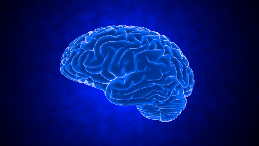
With the ever-increasing availability and clinical use of brain magnetic resonance imaging (MRI), the frequency of white matter hyperintensities (WMH) has become more readily apparent. As a heavy burden of WMH has been shown to be an important risk factor for incident stroke as well as vascular cognitive impairment,1 it is important to understand the pathophysiology, aetiology and clinical implications of WMH volume. Evidence suggests that hypertension is the strongest modifiable risk factor for WMH2–8 and raises the possibility of both prevention and treatment strategies, but clinical trials are lacking and further studies are needed.
Neuroimaging
WMH are demonstrated on T2-weighted MRI sequences. These damaged regions are evidenced on the T2 sequence as high signal intensity, which appear bright.9 They are considered white matter lesions if hyperintense on T2-weighted, fluid-attenuated inversion recovery and proton density images, without prominent hypointensity on T1-weighted images.1 They indicate areas of increased brain water content in white matter tissue, but the degree of damage to axons and supporting cells is not clear and remains an area under study (see Pathology section below).9 Recent evidence using diffusion tensor imaging has suggested a linear relationship between blood pressure levels and the microstructural integrity of both normal-appearing white matter and white matter hyperintensities. In hypertensive patients, the microstructural integrity of the cerebral white matter is significantly more affected than in normotensive patients.10
Pathology of White Matter Hyperintensities
The most common areas affected by WMH are in the deep white matter of the cerebral hemispheres, especially in the distribution of end-arteries and arterioles that supply a border-zone territory with minimal irrigation by collateral vessels. In areas identified as hyperintensities on MRI, pathological studies have reported myelin pallor, loss of tissue density, myelin and axonal loss, and gliosis.11,12 These lesions are hypothesised to be caused by chronic hypoperfusion of the white matter due to low-grade vascular insufficiency resulting in ischaemia,12 and are often associated with structural changes of the vessels, including hyalinisation, tortuosity, elongation and narrowing with arteriosclerosis and lipohyalinosis.12,13 Many of these changes have been associated with hypertension.14
To view the full article in PDF or eBook formats, please click on the icons above.