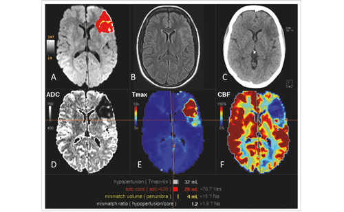
Lacunar ischaemic infarctions, white matter lesions (WML) (hyperintensities) or leukoaraiosis and cerebral microbleeds constitute the spectrum of ischaemic cerebral small-vessel disease (SVD) documented on magnetic resonance imaging (MRI) studies. History of ischaemic SVD is frequently present in patients with cognitive impairment of vascular origin.1–4 Cerebral lacunes are small infarctions of less than 20 mm in diameter localised in the vascular territory of penetrating arterioles, and represent the most-frequent manifestation of cerebral SVD (see Figure 1).5,6
Clinically, ischaemic lacunar stroke presents five well-recognised features, including pure motor hemiparesis,7 pure sensory syndrome,5,8 sensorimotor syndrome,5,9 dysarthria clumsy-hand10 and ataxic hemiparesis.5,11 Atypical lacunar syndrome is occasionally observed.12 Hypertension and diabetes are well-known cardiovascular risk factors for lacunar stroke.2,5 Other presenting forms of cerebral infarction of the lacunar type are silent lacunar infarcts and transient cerebral ischaemia.2
Deep or penetrating arterioles 100–400 μm in diameter that originate directly from a large cerebral artery are typically involved in the pathogenetic mechanism of lacunar infarct.13 These arterioles, without terminal anastomoses or collateral branches, provide blood supply to the deepest and nearest territories to the middle cerebral hemispheres and the brainstem. Lacunes are frequently found at the level of the lenticular branches of the anterior and middle cerebral arteries, the thalamoperforating and thalamogeniculated branches of the posterior cerebral artery and the paramedian branches of the basilar artery.
Symptomatic lacunar infarctions are commonly related to microatheromatosis or branch atheromatous disease.13 Patients with hypertension frequently present multiple asymptomatic lacunar infarctions usually due to lipohyalinosis. In different series, silent lacunes have been reported to occur in 52 % and 77 % of cases.5,13
Also, silent lacunes have been detected by MRI in about 30 % of patients with first-ever lacunar stroke.1,14 Contributing factors to vascular cognitive impairment in patients with lacunar infarction are summarised in Table 1. This review is focused on the clinical evidence and mechanisms underlying the relationship between cognitive impairment and lacunar stroke. The description of neuropsychological consequences of haemorrhagic lacunar stroke15 or the role of ischaemic cerebral SVD to the development of Alzheimer disease16 is not discussed in detail, although this remains an intriguing area of research and overlaps with the topics covered in this review.
Neuropsychological Deficits
Symptomatic Lacunar Infarctions
The clinical relevance of symptomatic lacunes as a causative factor of motor or sensory deficits has been extensively researched, but the degree of which cerebral symptomatic or silent lacunes may affect cognitive function remains unclear, except for subcortical dementia associated with multiple lacunes – the état lacunaire of Pierre Marie – which has been recognised for over a century, and perivascular leukoaraiosis associated with clinically multiple lacunar infarcts (Binswanger disease).2,17 Some classic studies have shown that during the symptomatic acute phase of lacunar stroke, neuropsychological deficits are usually absent. By contrast, neuropsychological abnormalities including verbal fluency, dysmnesia and abulia or spatial heminegligence, atypical aphasia and alteration of cognitive performance have been reported in patients with single lacunar infarctions in the dorsomedial and anterior thalamic region.4,18–20
Recently, it has been observed that ischaemic lacunar infarction is an important predictor of post-stroke cognitive decline and vascular dementia. In a previous study of 40 patients with lacunar infarction and studied 1 month after stroke, mild neuropsychological disturbances were found in 57.5 % of patients, being especially common among patients with pure motor hemiparesis and atypical lacunar infarction. Also the mean score of the Mini-Mental State Examination (MMSE) was 28.4.21
The cognitive profile generally considered to be associated with ischaemic cerebral SVD involves preserved memory with impairment in attention and executive functioning.17,18 Moreover, dysfunction of the frontal system is relatively frequent in cases of multiple subcortical lacunar infarcts.22 Vascular dementia has been reported in 11 % of patients after 2 to 3 years of the initial lacunar infarct and in 15 % of patients after 9 years.14,17 Symptomatic lacunar infarctions are particularly relevant as a predisposing factor for progression to subcortical dementia of the vascular type, and this risk increases with recurrent lacunar events and the presence of concurrent white matter disease.
In a study of 122 patients with recurrent lacunar stroke, cognition was studied in a subset of 59 patients, cognitive impairment, defined as an MMSE score <24, was detected in 16 % of patients with first lacunar infarction recurrence and in 40 % of those with multiple lacunar infarction recurrences.23 Adequate management of lacunar stroke patients is the first step to prevent vascular dementia associated with stroke recurrence.
In summary, recurrent ischaemic lacunar stroke, silent progression of SVD and perivascular leukoaraiosis in cases of clinically symptomatic multiple lacunar infarctions are associated with an increased risk of cognitive impairment.24
Silent (Asymptomatic) Lacunar Infarctions
Silent (or asymptomatic) lacunar infarctions are very common. In subjects older than 65 years, 20–28 % of lacunar infarctions are incidentally discovered on MRI studies. It has been shown that silent lacunar infarctions are risk factors for cognitive impairment and recurrent lacunar stroke.14,25 Between 10 % and 50 % of patients with a history of asymptomatic lacunar infarcts present new silent lacunar infarctions on the MRI examination at 3 years, which confirms asymptomatic progression of lacunar disease.26 On the other hand, 40 % of patients with lacunar stroke present progression of leukoaraiosis. The risk of vascular recurrence and cognitive impairment is particularly important in elderly healthy individuals with silent ischaemic cerebral SVD.25–27
The Leukoaraiosis and Disability Study (LADIS)28 investigated whether incident lacunes on MRI determined cognitive changes in elderly subjects with WML. The main finding was that a longitudinal increase in new lacunes on MRI was significantly associated with steeper longitudinal decline in specific cognitive domains, independently of the baseline WML, volume, number of lacunes and progression of WML. In particular, incident lacunes had a significant individual contribution to the deterioration of mental and motor speed and executive functions, while global cognitive function and memory functions remained unaffected. Asymptomatic progression of small-artery disease was a characteristic feature. The individual contribution of silent lacunes on cognition was modest, but the asymptomatic lacunes found on MRI should not be considered a benign vascular condition, but rather a potentially severe disease as a risk indicating progressive cognitive decline requiring adequate and rigorous therapeutic control and medical follow up.28
However, this clinical and neuropsychological aspect is still poorly studied. Long-term follow-up studies with large samples should be carried out to assess the outcome of patients with silent lacunes and to determine clinical and neuroimaging predictors of cognitive decline.
White Matter Hyperintensities
White matter hyperintensities (WMH) (also known as white matter changes or leukoaraiosis) are frequently documented in computed tomography (CT) and MRI studies of patients with lacunar stroke, elderly subjects with cerebrovascular risk factors and in cognitively impaired subjects. WMHs are believed to be caused by incomplete white matter infarction associated with SVD24,29 and a reflect of haemodynamic ischaemia of the white matter secondary to atherosclerotic thickening of the perforating arteries supplying the white matter.30 Some authors have suggested that patients with lacunar infarcts have more severe WML than patients with ischaemic infarcts of the non-lacunar subtype. Baseline WMH volume has been shown to be the strongest predictor of future cognitive decline in ischaemic cerebral SVD.28
Microbleeds
Cerebral microbleeds are small dot-like hypointense lesions corresponding with hemosiderin deposits in the cerebral microvascular perivascular spaces. These lesions are commonly observed in patients with lacunar ischaemic stroke and hypertension.31,32 Echo-planar gradient-echo T2-weighted MRI has high sensitivity for detecting cerebral microbleeds, which are most frequently observed in the basal ganglia (68 % of cases), although they are also common in the frontal, parieto-occipital and temporal regions.32 The occurrence and the number of cerebral microbleeds are associated with the degree of cerebral white matter abnormalities as well as with lacunar stroke and hypertension. On the other hand, cerebral microbleeds have been related to executive dysfunction and cognitive impairment. The results of a recent systematic review suggest that rather than being clinically silent, cerebral microbleeds might be a factor inducing cognitive function decline and the assessment of cerebral microbleeds should be included in the evaluation of patients with vascular cognitive dysfunction.31,32
Mild Vascular Cognitive Impairment
In a recent quantitative systematic review (12 cross-sectional studies) of domain-specific cognitive impairment associated with symptomatic lacunar infarcts, Edwards et al.33 reported impairment in the domains of attention/working memory and executive functioning as well as impairment in other additional domains, including memory, language and visuospatial abilities, thus extending previous characterisations of subcortical vascular cognitive impairment.
In a study of our group, more than half of patients with first-ever lacunar infarction showed cognitive impairment of the executive functions and fulfil criteria for mild cognitive impairment of vascular origin.34 In these patients voxel-based morphometry was used for assessing grey matter atrophy.35 It was observed that grey matter shrinkages mainly affected the following cerebral regions: bilaterally the temporal lobes, parietal and frontal regions and the left cerebellum as well as bilaterally the parahippocampal gyro and the right hippocampus (the latter tworegions emerged when region of interest analysis was restricted to these specific regions) (see Figure 2). These findings may be interpreted as corroborating those of the functional neuroimaging literature in which the remote effects of subcortical damage beyond the immediate area of infarction were described.34
Within the Secondary Manifestations of ARTerial disease-Magnetic Resonance (SMART-MR) study,36 a prospective cohort on MRI changes in patients with symptomatic atherosclerotic disease, 565 patients without large infarcts had vascular screening and 1.5 T MRI at baseline and after a mean follow-up of 3.9 years. The presence and progression of periventricular WML and lacunar infarcts were associated with greater progression of brain atrophy, independent of vascular risk factors.
In a longitudinal follow-up study, Nikunan et al.37 showed that the rate of atrophy in patients with SVD, presenting with lacunar stroke and leukoaraiosis, is approximately 1 % per annum and twofold that found in age-matched control subjects.
In another recent study, Duering and colleagues38 reported compelling proof-of-principle evidence that small subcortical infarcts in patients with hereditary stroke (cerebral autosomal dominant arteriopathy with subcortical infarcts and leukoencephalopathy [CADASIL]) have remote consequences on grey matter volume. Using MRI scans acquired before and after an incident subcortical infarct, it was shown that the appearance of a new subcortical infarct was associated with cortical thinning in connected brain regions. The authors hypothesised that the new infarct would cause focal cortical thinning in connected brain regions. Indeed, the remote ‘action at a distance’ may be the key to solving the riddle of why small subcortical infarcts have such large consequences on cognition.39
Brain atrophy can be the end-stage of multiple pathological processes and is a well-recognised feature of many neurological diseases, including Alzheimer disease and multiple sclerosis. Ischaemic cerebral SVD should be associated with greater progression of brain atrophy.
Subcortical Vascular Dementia
In clinical studies, the proportion of dementia caused by SVD ranges between 36 % and 67 %.17 Dementia is a leading cause of dependency after stroke and has devastating impact on post-stroke quality of life. Subcortical vascular dementia related to ischaemic lacunar stroke probably results from the interruption of prefrontal-subcortical loops by the lacunar infarcts and WML. Interruption of the dorsolateral prefrontal-subcortical loop results in executive dysfunction.39 Brain atrophy and other neurodegenerative changes have been documented in patients with subcortical vascular dementia and cognitive impairment, suggesting that the pathogenesis of the cognitive deficits is variable and may reflect a combination of degenerative and ischaemic causes.17,34,40,41 However, relatively few studies have investigated longitudinal changes in cognitive function following lacunar stroke. Long-term follow-up studies (more than 3 years) with large samples should be carried out to assess the outcome of patients with mild cognitive impairment of the vascular type and to determine clinical and neuroimaging predictors of vascular subcortical dementia.
Strategic infarct dementia is unusual and is caused by isolated infarcts in regions that are important for cognition of the brain, such as the thalamus, hippocampus, caudate or genu of the internal capsule. It is characterised by the abrupt onset of cognitive or behavioural changes, which vary depending of the infarct location.42
In a recent clinical study, Duering et al.43 applied a voxel-based lesionsymptom mapping approach to data from 215 patients with CADASIL, and identified strategic locations for subcortical ischaemic lesions, which affect processing speed, as a major cognitive aspect of vascular cognitive impairment. Significant clusters were found for both lacunar lesions and WMH and identify the anterior thalamic radiation and the forceps minor as major anatomical structures.
The mid- and long-term prognosis of lacunar ischaemic stroke patients with subcortical vascular dementia is generally poor because of the high frequency of adverse outcomes, such as cognitive and functional deterioration, nursing home placement and death.14
Recent Advances and Therapeutic Strategies
The Secondary Prevention of Small Subcortical Stroke (SPS3)44 phase III study is the first double-blind, multicentre stroke prevention trial focusing specifically on a large group of patients (n=3,020) with recent symptomatic, confirmed MRI lacunar infarcts. Its aims to define the optimal interventions (antiplatelet therapy of either enteric-coated aspirin 325 mg/day or aspirin plus clopidogrel 75 mg/day) to target levels of systolic blood pressure (either intensive <130 mm Hg or usual 130–149 mm Hg) to prevent stroke recurrence, cognitive decline and major vascular events in this population. The secondary prevention of lacunar stroke as detected by the use of MRI has not previously been the focus of a randomised trial. After a mean follow-up of 3.4 years, the first published data showed that adding clopidogrel to aspirin did not reduce recurrent stroke and increased bleeding in lacunar stroke.45
All English-speaking participants in SPS3 underwent neuropsychological testing at baseline (n=1,636).46 A total of 47 % of the cohort met psychometric criteria for mild cognitive impairment with amanestic deficits as prevalent as non-amnestic.46 Younger age, male gender, less education, post-stroke disability and impaired activities of daily living were independently associated with mild cognitive impairment. The observation of impaired processing speed, motor dexterity and executive function was consistent with the impairment pattern hypothesised for subcortical ischaemic disease.46 On the other hand, the observation of episodic memory deficits was a novel aspect of the study given that incident lacunes have been reported to impact processing speed and motor control but not memory.46 The authors emphasised that mild cognitive impairment found in half of the participants is an important clinical sequela of lacunar stroke, which in turn is more prevalent than physical disability defined by m-Rankin score ≥2 (33 %) and present in 41 % of patients with no significant disability (m-Rankin score 0-1’ Barthel index score 100). It is speculated that the cognitive effects of lacunar stroke are at least as important as disability related to motor or sensory deficits, and can occur in their absence.
Regardless of the paucity of data for cognitive end-points, adherence to current guidelines for primary stroke prevention with smoking cessation, Mediterranean-type diet, physical activity, weight control and control of cardiovascular risk factors, mainly hypertension and diabetes, as well as secondary stroke prevention with antiplatelet therapy, are first-line pharmacological strategies in the management of patients with ischaemic lacunar stroke subtype.1–3,47–49
Recently, cognitive stimulation has been advocated as one of the tasks of neuropsychological rehabilitation with the aim of improving cerebral neuroplasticity based on a set of stimuli for an integral improvement of cognitive, emotional, physical and inter-relationship skills with a final benefit of a better quality of life. Cognitive stimulation may act on some of the three processes involved in neurorepair, such as angiogenesis, neurogenesis and synaptic plasticity.50 Biomaterials for promoting brain protection, repair and regeneration are new hot targets in lacunar ischaemic stroke patients.
Concluding Remarks
The cognitive effects of acute ischaemic lacunar stroke have been identified in half of the patients and are at least as important as disability related to motor or sensory deficits, and occur in their absence. The classic cognitive profile involves preserved memory with impairment in attention and executive functioning. However, in a recent systematic review, impaired cognition appears less selective than previously thought, involving all major cognitive domains and episodic memory impairment may appear as an equally neuropsychological central feature in acute lacunar stroke.
Recurrent lacunar ischaemic stroke is one of the major factors involved in producing vascular subcortical dementia. Brain atrophy can be the end-stage of multiple pathological processes and is a well-recognised feature of many neurological diseases, including Alzheimer disease and multiple sclerosis. Atrophy is another feature recently associated with cerebral small vessel diseases.
Some aspects of cognitive impairment related to lacunar infarction are not definitively established. Further research in the following fields include: a) the investigation of relevant genetic forms and suitable animal models would be valuable for exploring the pathogenesis as well as the prevention of the lacunar stroke causes of cognitive impairment; b) diffusion tensor imaging MRI and cerebral microbleeds in relation to prognosis of lacunar stroke; c) assessment of the role of cerebral volume in cognitive impairment suffered by patients with lacunar infarction. The vascular basis of loss of neuronal and dendrosynaptic integrity in relevant cortical lobes also needs further investigation, and d) the inclusion of cognition as an outcome in future prospective observational studies and clinical trials is urgently required. Ischaemic lacunar stroke offers a target for beneficial interventions to preserve cognition in older people.


