Stroke is one of the leading causes of death and disability in developed and less-developed countries.1,2 Effective therapeutic approaches to brain protection and recovery in stroke remain elusive, due to an incomplete understanding of the endogenous neurobiological processes involved in repair and a consequent inability to adequately stimulate them.3 Many trials of neuroprotective therapies for ischaemic stroke over the past 2 decades have failed, largely due to a failure to recognise the complexity of stroke and the repair processes involved in recovery.4–7
Various factors alter stroke severity, rehabilitation and outcomes. These include co-morbidities, such as diabetes and cardiovascular disease, and other factors, such as stress.8–12 Imaging techniques such as positron emission tomography (PET) and animal model studies are increasing knowledge of pathophysiology and the neurorehabilitation process.13,14 These studies also indicate targets for new drugs and alternative roles for existing therapies, particularly those that enhance brain repair processes. Furthermore, non-invasive brain stimulation (NIBS) can stimulate recovery at specific brain regions.15 In addition, emerging biomarkers have the potential to identify risks in stroke and inform treatment strategies.16 Adequate stroke rehabilitation services at treatment centres are critical in improving outcome but the these are often under-resourced.17 Better outcomes in stroke can also be achieved using current best practice, the application of which can be assisted by meta-analyses of multiple clinical trials.
This article presents important information on current experimental models and clinical approaches to recovery and rehabilitation after stroke, based on presentations given at the 3rd International Salzburg Conference on Neurorecovery (ISCN) in September 2014.
Non-invasive Brain Stimulation in Rehabilitation after Stroke
NIBS techniques that represent a promising new area of therapy for post-stroke recovery and affect the specific and localised interactions between the brain hemispheres that determine recovery of a patient, were reported by Professor Wolf-Dieter Heiss from Cologne, Germany. He stressed that these methods are derived from two main principles: transcranial direct current stimulation (tDCS) or repetitive transcranial magnetic stimulation (rTMS).15 rTMS allows for very precise, focal and noninvasive electrical manipulation of nervous tissue, including the cerebral cortex, spinal roots and cranial as well as peripheral nerves.18 The effects of NIBS, the interaction in functional networks and their reorganisation in recovery after focal brain damage can be visualised using functional imaging PET and functional magnetic resonance imaging (fMRI).13
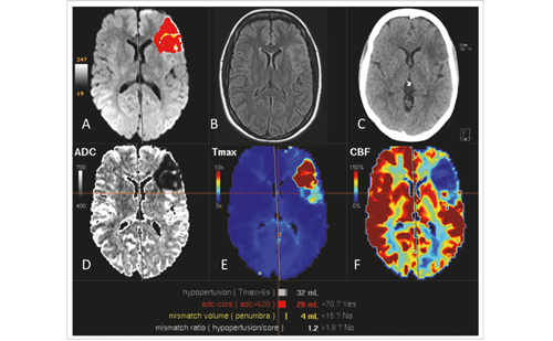
Within the lesional area in ischaemic stroke there is the core zone that is, for the most part, irreversibly damaged but this is surrounded by the penumbra or peri-infarct, which is ischaemic but is still potentially viable and can be recovered if reperfused rapidly.19 Interventions enabling reperfusion can limit the spread of damage and protect neurological function. In some instances the unaffected hemisphere inhibits the recovery of ipsilateral functional networks but this effect of transcallosal inhibition can be reduced by NIBS methods, such as contralateral inhibitory rTMS.20
Motor recovery stimulation following stroke has been mainly performed using an rTMS approach. Studies have employed diverse time windows for initiating treatment, ranging from 5 days to 7 years post-stroke.21 In a review of 22 studies including 307 patients, motor functions improved after NIBS, clinical effects were highly variable depending on patient characteristics but all the improvements were maintained until the end of the observation periods.22,23 In addition, early stimulation following stroke was more beneficial than late stimulation.
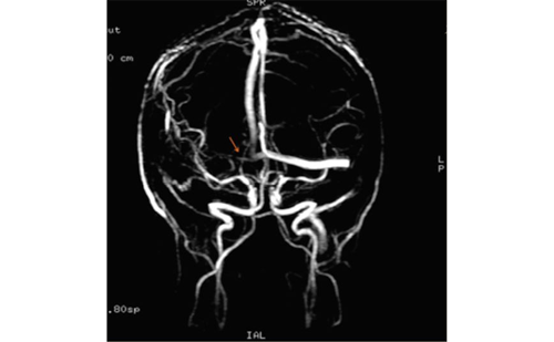
Aphasia affects more than a third of all stroke victims and is one of the most disabling functional defects after ischaemic stroke. It improves during the first 4 weeks in one-third of patients, increasing to approximately half after 6 months. Speech and language therapy (SLT) is the only effective treatment to date, but this is usually limited in duration and intensity and should be provided early.24 NIBS is an important approach to address aphasia: rTMS modulates cortical excitability, has been shown to suppress naming function and can facilitate language recovery in aphasic patients (see Figure 1).20
NIBS acts on specific networks involved in the pathophysiology of language processing and promotes adaptive cortical reorganisation after stroke. Rehabilitation of post-stroke aphasia involves two strategies: recruitment of perilesional cortical regions in the dominant (left) hemisphere and development of language ability in the non-dominant (right) hemisphere. The majority of NIBS trials in post-stroke aphasia aimed to reinforce the activity of brain regions in the left hemisphere due to more effective recovery in strokes involving this side.25 This can be achieved using an excitatory NIBS protocol to reactivate the lesioned area or an inhibitory NIBS protocol to reduce activities in the contralesional homologous area.
Professor Heiss concluded that further developments are needed to firmly establish NIBS protocols in the treatment of stroke. These include larger-scale brain-stimulation studies for post-stroke recovery, as well as early intervention, ideally within the first 2 weeks post-stroke in stroke units rather than rehabilitation centres. Furthermore, NIBS should always be combined with rehabilitation and pharmacological therapy. Future studies should directly compare tDCS and rTMS or excitatory versus inhibitory protocols. The trials should also further explore advantages of contralateral inhibitory protocols that include better localised stimulation site but have no direct infarct influence and no risk of seizures.
Co-morbidity and Stroke Outcomes
Stroke patients have many co-morbidities, which can influence prognosis8 and should be taken into account during evaluation and treatment, as emphasised by Professor Jaakko Tuomilehto from Helsinki, Finland, and Krems, Austria. Appropriate measures must be undertaken to properly evaluate co-morbidity bias in the clinical trials. A recent systematic literature review of 26 studies (acute myocardial infarction n=8; heart failure n=11; stroke n=7) that measured co-morbidity in cardiovascular research concluded that “…measurement of co-morbidity remains limited to a list of conditions without stated rationale or standards, increasing the likelihood that the true impact is underestimated”.26
The importance of co-morbidity was emphasised in an analysis of a cohort of 451 patients with ischaemic stroke, which revealed an interesting correlation between stroke outcomes and periventricular white matter disease (PVWMD).27 Severe PVWMD was significantly associated with poor functional outcome at 3 months, independent of other factors, such as diabetes and age. Outcome at 3 months was also a significant predictor of long-term mortality and functional recovery.
The investigation of patients with ischaemic stroke in neurologic rehabilitation (INSIGHT) registry was the first to provide large-scale data on patients with acute stroke (<3 months) who survived the initial phaseof high risk and were undergoing neurological in-patient rehabilitation (n=1,167).28 Analysis of registry data demonstrated a significant association between microalbuminuria (MAU) and polyvascular disease and supported previous findings that MAU predicts cardio-/cerebrovascular events in patients recovering from ischaemic stroke. MAU could therefore be used as a biomarker during neurological in-patient rehabilitation, identifying patients with increased risk of recurrent events.
Acute ischaemic stroke (AIS) and diabetes appear to be correlated in both children and adults. This was emphasised in a study in Finland in which the cumulative incidence of stroke in childhood onset type 1 diabetes patients with nephropathy was 20 % at age >40.29 In the Nationwide Inpatient Sample, hospital admissions in the US for AIS between 1996 and 2007 decreased but there was a steep rise in the proportion with comorbid diabetes (from one in five to almost one in three).12
Finally, Professor Tuomilehto concluded that these examples and others suggest that co-morbidities should be taken into account in the evaluation and treatment of stroke patients.
Organisation of Stroke Rehabilitation Services
Professor Michael Brainin reported that the use of health care resources for stroke rehabilitation has been greatly influenced by local availability but is often under-resourced.17 This was emphasised by the GAIN trial that assessed 1,422 patients (from 19 countries), which found that any link between resources used and outcomes is unclear and showed a threefold variation in the average number of days in hospital/institutional care (20 to 60 days).30 Additionally, there was no relationship between health care resource use and survival or activities of daily living (ADL) at 3 months.
In Austria and Germany, a phase system has been developed that involves institutional rehabilitation linked directly with acute stroke care.31 This system is based on a continuous flow of competences from acute treatment (Phase A) to outpatient and community rehabilitation (Phase E) and has been used to justify the establishment of large rehabilitation clinics. In some Western countries, stroke units are now considered essential platforms on which other important therapeutic means are delivered and around which the major progress in stroke management has been made in recent years. In other Western countries, however, outpatient rehabilitation centres play major roles. The Cochrane Outpatient Service Trialists systematic review showed that therapy for patients with stroke who live at home could prevent deterioration in ADLs (absolute reduction in risk of deterioration was seven per 100 patients) but concluded that the best delivery model could not be identified.32
Early mobilisation post-stroke has been regarded by many physicians as an important element of rehabilitation services. This is supported by animal models of stroke recovery and currently available clinical data that suggest that spontaneous biological recovery occurs very early and lasts probably up to 3 months post-stroke. Findings of the recently completed AVERT study, however, appear to be contrary to this principle (see Figure 2).33,34 This study followed-up patients who received very early mobilisation/higher treatment dose, within 24 hours of their stroke (n=1,054) compared with patients who received ‘usual care’ (n=1,050) consisting of an early, lower dose out-of-bed activity regimen. Three months after stroke, fewer patients in the very early mobilisation group had favourable outcomes than those in the ‘usual care’ group (46 % versus 50 %; p=0.005). These results suggest that ‘usual care’ is preferable to very early mobilisation/higher dose intervention, but clinical recommendations on this require more investigation and analysis.
Major efforts are in progress to classify and unify stroke rehabilitation services worldwide and build on the 2004 WHO International Classification of Functioning, Disability and Health for Stroke, which defines the spectrum of post-stroke disabilities.35 Additionally, the Global Stroke Community Advisory Panel recently developed guidelines that identified promising areas of stroke rehabilitation:36–38
- Drug therapy for motor recovery;
- Early mobilisation;
- Body-weight support treadmill training;
- Robotics;
- Virtual reality;
- Transcranial magnetic stimulation; and
- Hypothermia.
Organisation of rehabilitation after stroke must be redefined by the need to effectively manage processes of recovery in the brain together with the need to avoid complications. Commencing rehabilitation earlier is under investigation (AVERT trial) but longer rehabilitation is needed due to the dynamics of chronic stroke.
Principles of Stroke Recovery
In neurorehabilitation, Professor Dafin Muresanu from Cluj, Romania, began by stating that there is a wide gap between the results of randomised control clinical trials (RCTs) and real-world benefits of treatments. In the last 30 years, few RCTs in brain protection and recovery have shown positive results. The unclear neurobiological concepts that generated inconsistent pharmacological strategies (suppressive/stimulating strategies) using inappropriate molecules for various therapies have been blamed for these shortcomings. How theory and basic research can improve this situation is a major question that has to be answered before stroke therapy can progress.
To reduce the gap between theory and clinical practice, there are three major factors to address:
- Elementary rules must be derived from basic science.
- Rules must be applied to develop therapeutic procedures.
- Therapeutic approaches must be assessed in sophisticated RCTs.
An acute brain lesion is always followed by an endogenous continuous brain defence response consisting of two overlapping main sequences: 1) an immediate and short-lasting one that reduces brain damage and impairments (NEUROPROTECTION) and 2) a later but long-lasting one in which brain damage is repaired (NEUROREPAIR). The latter process consists of neurotrophicity, neuroplasticity and neurogenesis and leads to NEURORECOVERY and reducing disability.39,40
The major advantage of neurorestorative treatments is that they are not restricted to the first few hours or minutes post-stroke, where there is a race against the accelerating process of cell death.41 Neurorestorative treatments can be performed with an extended therapeutic window lasting days if not weeks post-stroke. Many patients exhibit improved neurological function post-stroke, yet current treatment does not enhance the intrinsic restorative mechanisms driving this recovery.
Recovery post-injury is a multi-layered process involving a dynamic network level in which the brain can be considered to function as a small-world network.42,43 The practical implication of this theory is that the brain can efficiently share information across brain regions and can be more resilient to network disruptions/lesions. This understanding of brain function should move the emphasis of treatment and restorative trials towards modulating the level of activity in pre-existing networks.44
In the treatment of stroke, chemical drugs can be monomodal in that they target only one neurobiological process (e.g. neuroprotection or neuroplasticity) and have failed in clinical trials. By contrast, multimodal drugs have the capacity to simultaneously regulate two or more endogenous neurobiological processes and more closely address the complex reality of the stroke recovery processes. Among the few existing drugs of this class is the neurotrophic peptide preparation Cerebrolysin®, which is in clinical use and two others in clinical development.
Pharmacological support of neuroprotection and neurorehabilitation is a valid concept but a correct neurobiological concept should be observed and a correct strategy should also be implemented, integrating acute and long-term pharmacological treatment. Only drugs that work within this strategy should be used (multimodal drugs and pleiotropic metabolic modulators) and should involve appropriate dosage and longterm administration strategies.
Meta-analysis and Outcomes
Professor Jan Merholz from Germany summarised his experience of systematic reviews and meta-analyses as important elements of the decision-making process in clinical practice. The Cochrane Library has become a major source of high-quality, independent evidence in such decision-making and the Cochrane methodology is now a benchmark for critical reviews of various medical treatments, including those of stroke.45 The process of clinical research and evidence-based practice consists of the following activities and stages:
- Primary research studies (clinical trials);
- Interpretation of research (e.g. Cochrane reviews other systematic reviews and meta-analyses);
- Decision support (assisted by clinical guidelines and treatment policy); and
- Health care decisions (involving patients, clinicians, MS nurses, pharmacists, managers and health care organisations)
An example review examined the effects of electromechanical and robotic-assisted gait training devices for improving walking after stroke and an assessment of the acceptability and safety of this therapy.46 The authors searched the medical, sports and engineering databases as well as conference proceedings, trials and research registers and identified further published, unpublished and ongoing RCTs. Primary outcomes were the proportion of participants walking independently at follow-up; secondary outcomes were walking speed and walking capacity.
The meta-analysis found clear benefit of assisted gait training and found that one in five walking disabilities after stroke might be avoidable using the assessed therapy. The secondary endpoints were not significantly positive, but the acceptability of the therapy was favourable. The results also show that non-ambulatory patients, especially in the acute/subacute phase after stroke, who receive electromechanical-assisted gait training in combination with physiotherapy are more likely to achieve independent walking than patients receiving gait training without these devices.
There is excellent evidence for stroke rehabilitation. Cochrane systematic reviews and meta-analyses enhance decision-making in clinical practice. These set a high standard for evidence-based clinical data evaluation and help inform clinical guidelines. The importance of evaluating currently available treatments in a rigorous way is underlined by the complexity and difficulty of conducting large-scale RCTs in neurology.
Drug Enhancers
Professor Alla Guekht from Moscow outlined that there are three main groups of neurological drugs that enhance brain processes: the noradrenergic agonists and levodopa, the selective serotonin reuptake inhibitors (SSRIs) and the trophic factors. Among the noradrenergic agonists, dexamphetamine induces physiological and/or structural changes in the brain that may be relevant to recovery (sprouting and synaptogenesis) and can facilitate long-term potentiation. The combination of dexamphetamine and physiotherapy appears to have potential as a stroke treatment. Animal models of stroke have been used extensively to assess the effects of amphetamine on recovery processes. These have mostly delivered positive results but there was a failure to translate them into human clinical trials.47,48
A recent Cochrane meta-analysis assessed SSRI use for stroke recovery, and included 52 (4,059 participants) of the 56 completed trials f SSRI versus control.49 This revealed statistically significant benefits of SSRIs on both the relative risk for reducing dependency at the end of treatment (0.81) and for disability score (standardised mean differences = 0.91). SSRIs produced statistically significant benefits in terms of neurological deficit, depression and anxiety. SSRIs also appeared to improve dependence, disability, neurological impairment, anxiety and depression after stroke, but methodology was inconsistent and had limitations in many of the trials.
There are various potential targets for stroke treatment. Recently, a polymorphism in brain-derived neurotrophic factor (Val66Met) was found to have direct impact on motor skill acquisition in both experimental models and in humans.50 There is also growing evidence that nerve growth factor is of potential clinical use in variety of disorders (see Figure 3).51 Additionally, a recently published Cochrane systematic review/metaanalysis of RCTs investigating the efficacy and safety of Cerebrolysin® in the treatment of vascular dementia suggests clinical benefit.52 The recently finalised trial: Cerebrolysin and Recovery in Stroke (CARS), a randomised, placebo-controlled, double-blind, multicentre study, will shed more light on potential clinical benefits of this neurotrophic therapy in stroke patients undergoing rehabilitation. Various other ongoing clinical trials are investigating treatments for stroke including: memantine, lithium carbonate, galantamine, Actovegin® (calf blood extract), cholinergic drugs (donepezil), anti-inflammatory agents and antiepileptic drugs.
The common underlying therapeutic assumption for all drug enhancers is support for processes of recovery post-stroke. Cerebrolysin® appears to support physical rehabilitation in the early acute phase as well as in the early post-acute period. Moreover, the recently published Cochrane systematic review/meta-analysis52 points to its efficacy in long-term treatment of cognitive impairment and dementia post-stroke.
PET Studies for the Evaluation of Dynamics in Stroke Pathophysiology
In ischaemic stroke research, the use of PET has enabled the progressionof irreversible damage, the core of ischaemia into functionally impaired area, the penumbra, to be followed in experimental models as well as demonstrating the potential for recovery of these areas with reperfusion within the time window.13 Professor Wofl-Dieter Heiss from Cologne, Germany, stated that this imaging technique prompted the use of thrombolysis and other reperfusion therapies in the treatment of ischaemic stroke.
In an experimental model in cats using PET imaging, cerebral artery (MCA) occlusion resulted in severe ischaemia in 55–75 % of the hemisphere after 3 hours occlusion. Hyperperfusion after reopening of the MCA turned into hypoperfusion leading to severe global ischaemia with transtentorial herniation.14 In a clinical study, patients with MCA occlusion, PET measurements within 24 hours after stroke showed larger volumes of ischaemic core (mean, 144.5 versus 62.2 cm3) and larger volumes of irreversible neuronal damage (157.9 versus 47.0 cm3) in patients with a malignant course (i.e. oedema formation with midline shift) than in patients with a benign course. PET allowed prediction of malignant MCA infarction within the time window suggested for hemicraniectomy.53
These examples and others underline the role PET has played for translational research in stroke in the last 30 years. Its impact might even be increased by the advent of combined MR/PET equipment (see Figure 4) and the introduction of more sophisticated molecular tracers into clinical application.
Insight into the Variability of Outcomes After Stroke Therapy in the Diabetic Brain
The failure of clinical trials in stroke can be largely attributed to afailure to recognise the complexity of stroke in the context of comorbidities and other individual factors such as genetic variability.
Professor Michael Chopp from Detroit, US, argued that in the case of diabetes, some experimental models indicate major physiopathological changes that can influence both the impact of stroke and the outcomes of stroke treatments. In rat models, following stroke, type 2 diabetes mellitus (T2DM) exacerbates neurological deficits, including cognitive deficits, fibrin deposition and loss of axons, oligodendrocytes in the hippocampus and reduces spine density and dendritic arborisation.54
In mouse experimental models, T2DM worsens the impact of stroke by increasing neurovascular and myelinated axonal damage, and increasing inflammatory response. T2DM also increases angiopoietin 2, decreases angiopoietin 1 in the ischaemic brain leading to worsened functional outcome compared with non-T2DM mice.55–57
The treatment of experimental diabetic rats with thrombolytic factors (e.g. with tissue plasminogen activator [tPA]), failed to improve functional outcome after stroke in type 1 (T1) DM rats (see Figure 5). tPA, however, significantly increased blood–brain barrier (BBB) leakage, brain haemorrhage and inflammatory response.58
The efficacy of treatment of stroke with cell-based therapies in diabetic animals is strongly time-window dependent, with early treatment on day 1 showing no beneficial effects, in contrast to functional benefit with delayed treatment at day 3.59 However, potential adverse effects of cell-based treatment in diabetic animals under these conditions can be profound. Data show, in both T1DM and T2DM diabetic animals receiving day 3 treatment of stroke, that there is strong evidence of arteriosclerotic-like vascular lesions. This alteration in vascular structure may increase the risk of secondary stroke and atherosclerosis. Further studies on the mechanism of accelerating atherosclerotic-like changes in DM are therefore required.
The neurovascular status of the brain and the response to stroke and therapeutic intervention differ significantly between the diabetic and non-diabetic animals. This knowledge must be taken into account when designing new clinical trials in stroke therapy.
Strategies for Neurorecovery
Professor Ludwig Aigner from Salzburg focused on the failure of clinical trials in stroke neurorecovery. These can be attributed at least in part to the fact that they were focused solely on neuroprotection, which is only a part of the complex story of rejuvenation of the brain postinjury. The recovery from damage is based on natural, spontaneous processes leading to repair, regeneration and restoration of neurological structures and functions. These processes can be stimulated by drugs such as leukotriene receptors antagonists (like montelukast, anasthma treatment agent) that reduce neuroinflammation, stimulate neurogenesis and improve functional outcomes.60
Drugs used formerly in the context of neuroprotection might be tried in neurorestorative strategies within re-evaluated and modified protocols. Cerebrolysin® is such a drug currently undergoing a clinical developmentprogramme aimed at establishing neurotrophic and multimodal therapeutic standards in stroke and other neurological disorders.61–64
b>Epidemiology of Vascular Biomarkers
Personalised medicine strongly relies on the availability of reliable biomarkers, enabling prediction of disease risk or progression. Professor Stefan Kiechl from Innsbruck, Austria, observed that biomarker research in stroke focuses on the ascertainment of stroke or transient ischaemic attack (TIA) status, classification of stroke subtypes, estimation of prognosis after stroke (including complications) and prediction of stroke and cardiovascular disease (CVD) risk (first-ever strokes and recurrent events).
Promising areas in the development of stroke biomarkers include: emerging risk factors, lipid markers (Lp(a), Lp-PLA2, lipidome, apoC3), calcification markers, markers of plaque instability, ultrasound biomarkers and circulating microRNAs (miRNAs) and the microbiome and gut metabolome.16,65–67 The development and validation of biomarkers facilitate personalised medicine with the goal of targeted prevention, and provide new insights into pathophysiological mechanisms and disease pathways implicit for designing new treatment concepts.
Among the emerging risk factors of cerebrovascular disease are inflammation and anaemia. These could guide the use of anti-inflammatory agents and blood donation, which are promising new areas of treatment. All the newly developed biomarkers can be used as a means for multistage risk scores assessment and for prevention of cerebrovascular events through personalised medicine.
An important initiative in stroke biomarker development is VASCage67 a new project conducted at Medical University in Innsbruck. This project focuses on ageing processes within vasculature and to utilise current knowledge of biomarkers for advancing into the emerging area of personalised medicine.
Stress and Stroke – A Biomedical Condition
Finally, Professor Natan Bornstein from Tel Aviv, Israel, pointed out that stroke is a stressful, life-threatening experience. In addition to the damage to the brain, the patient’s sense of wholeness and safety can be shattered, leaving a lasting sense of vulnerability. Exposure to physical or psychological threat activates the hypothalamic–pituitary– adrenocortical (HPA) axis.10,11,69,70 Hippocampal atrophy has been described in various neuropsychiatric disorders that are associated with HPA axis dysregulation, such as depression, post-traumatic stress disorder (PTSD) and Alzheimer’s disease.71 Studies of adults with PTSD demonstrate significantly reduced hippocampal volumes compared with controls.72–74
The Tel Aviv Acute Stroke cohort (TABASCO)75 is an ongoing, prospective cohort study that plans to recruit approximately 1,000 consecutive firstever mild–moderate, non-demented stroke patients (see Figure 6).75 It aims to evaluate the association between pre-defined demographic, psychological, inflammatory, biochemical, neuroimaging and genetic markers (during the acute phase) and long-term outcome, including subsequent cognitive deterioration, vascular events (including recurrent strokes), falls, affect changes, functional everyday difficulties and mortality. Chronic brain exposure to physical or psychological threats could lead to HPA axis over-activation leading to neuronal damage with resulting hippocampal atrophy and cognitive impairment.
Preliminary findings of this study show that admission bedtime saliva cortisol levels are inversely correlated with total hippocampal volume. These levels are related to a reduction in hippocampal volume 24 months later and are also associated with worse executive functioning scores. Patients with increased saliva cortisol levels also show inferior cognitive scores at baseline and 6 and 24 months later.
Discussion and Conclusion
The failure of many candidate treatments for stroke in clinical trials over previous decades is most likely a result of a lack of knowledge regarding the pathogenesis of the disease and repair processes. The presentations summarised above indicate that stimulating recovery from stroke is highly dependent on multiple parameters that may be evaluated in the clinic and the laboratory, including: age, gender, co-morbidities, type and location of stroke and individual genetic and epigenetic factors. Previous investigations largely failed to account for these factors and thereby limited their potential to stimulate or recognise neurological repair and improve patient outcomes. Many of these parameters, however, can be used to provide a more tailored treatment of stroke to enhance neurological recovery.
Understanding of the pathophysiology of post-stroke repair is improving; it is recognised that monomodal drugs have little effect but a multimodal approach with multiple modes of action may be more effective. This approach could lead to neurorestorative treatments. In the search for effective treatments, a variety of potential drugs that enhance brainrepair processes are under investigation. These include the SSRIs, which show significant benefits in many studies when used to treat stroke. Another example of such repurposing of a treatment is the use of Cerebrolysin® in patients undergoing rehabilitation after stroke. Various other drugs are being evaluated for use post-stroke and may eventually increase the limited options available to physicians treating stroke.
The prospect of reliable biomarkers in stroke for future use in the clinic to routinely predict risk and indicate treatment response is becoming more likely. Initiatives such as VASCage will assess biomarkers that are currently available and could expand the possibility of personalised medicine in patients post-stroke. A further pivotal initiative is the ongoing TABASCO study, which has already found strong associations between stress, as determined by cortisol levels, and patient condition and outcomes in stroke. Monitoring stress could potentially identify patients at risk of PTSD and other conditions and thereby improve post-stroke therapy. Meta-analyses of multiple trials in stroke have identified strong evidence in support of stroke rehabilitation, leading to improved patient care. Assisted gait training, for example, was found to substantially reduce walking difficulties post-stroke.
The provision of stroke rehabilitation services is highly variable in different world regions and this has a direct effect on patient outcomes. It is likely that the soon-to-be-completed AVERT trial will indicatethe importance of commencing rehabilitation measures very soon after stroke and may encourage health authorities to adopt an early intervention approach in all patients. In addition, the ongoing CARS trial may provide valuable data in support of Cerebrolysin® therapy during stroke rehabilitation. An alternative treatment approach may be to target intercellular communication via exosome and miRNA expression, which regulate various responses in organs.
In addition to drug therapies, NIBS also shows promise, especially when administered soon after a stroke. This technique has demonstrated lasting improvements in aphasia and motor functions. NIBS can be targeted at specific regions of the brain where damage has occurred. This approach, however, requires further evaluation in larger populations and should be used in conjunction with rehabilitation and drug treatments.
Knowledge of stroke rehabilitation, pathophysiology and clinical interventions to stimulate it is expanding on many fronts. In recent decades, attempts to restore or improve function post-stroke were beset by failure. Recent developments using existing technologies, however, should improve outcomes. Application of these developments in the clinic through wider understanding and awareness of current treatments and the translation of experimental findings to more effective therapies should improve rehabilitation in the post-stroke patient.


