The classic definition of transient ischemic attacks (TIAs) based on focal neurological deficits most likely due to cerebrovascular diseases with full recovery within 24 hours dates back to the 1960s.1 With the advent of advanced neuroimaging techniques (computed tomography [CT] and magnetic resonance imaging [MRI]), new insights into pathobiology and prognosis of cerebrovascular events, as well as the approval of recombinant tissue plasminogen activator (rtPA) treatment and the increasing emergency management in stroke units, this definition has become outdated and suggestions for its change have been frequently made.2 Although many patients with acute ischemic stroke have repeatedly been demonstrated to benefit from this advanced concept of ‘time is brain’ and ‘stroke is an emergency’, those patients with rapid, spontaneous recovery after onset of symptoms (within less than three hours) have still been investigated less sufficiently: among the extremes are misdiagnosis of ‘TIA mimics’ resulting in no treatment at all or premature thrombolysis as well as underestimation of the high risk for suffering a full stroke within 10–14 days if adequate diagnosis and prevention is omitted or patients do not seek medical advice in due time.
With this in mind and considering the fact that TIAs are well recognized risk factors for stroke (mean annual stroke risk after TIAs has been found to be up to 15%),3 TIAs stopped to be considered harmless long ago. Currently, different strategies have been inaugurated for the management immediately after onset. While many prefer admitting these patients to a stroke unit for full work-up within 72 hours, others propose 24-hour open ‘TIA clinics’ or even a quick work-up in a specialized outpatient department.4 Predictors such as the ABCD2 score5 or fluctuations of symptoms6 characterizing the individual risk for stroke are clinical or imaging-related (CT, MRI).7 Good TIA management requires a practical definition and confident diagnosis, based on good and reliable diagnostic tools, separation from TIA mimics, a valid prognosis and stroke risk assessment to identify potential sources of stroke and risk factors and a strategy for treatment and prevention.
Definition and Diagnosis of Transient Ischemic Attack
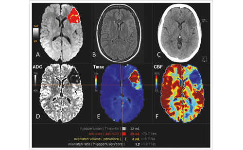
The original TIA definition as “a cerebral dysfunction of ischemic nature lasting no longer than 24 hours with a tendency to recur” was based on pure clinical findings and was formulated in a time period in which neuroimaging was rudimental and acute stroke treatment missing. Despite neuropathological evidence already available at that time,8 people thought that TIAs did not cause permanent brain damage. A new definition of TIA and even the abandoning of this term have been proposed during the last decades. Many experts support the idea of changing the time limit to <1–2 hours with practical consequences in the acute situation, when the decision whether the patient should be thrombolysed or not has to be taken.2,9,10 As a consequence, TIAs have been proposed to represent “brief episodes of neurological dysfunction caused by focal brain or retinal ischemia, with clinical symptoms typically lasting less than an hour and without evidence of acute infarction.”2,93,11,12 In this context, neuroimaging, and especially MRI, plays an important role as it is able to detect acute ischemic lesions in diffusion-weighted imaging (DWI) early. Studies have shown that up to half of the patients diagnosed clinically with TIA had DWI lesions in the MRI.7,13–15 Rovira et al. even found ischemic DWI lesions in two-thirds of TIA patients in the acute phase.16
Transient Ischemic Attack Mimics
Diagnosis of TIA can be challenging, not only in the acute phase. The wide range of manifestations and clinical symptoms, as well as the frequent lack of valid information given by the patient when the episode is over, make a large number of differential diagnoses possible, especially in the absence of DWI lesions and/or under time pressure. Migraine attacks, focal epileptic seizures, metabolic disorders such as hypoglycaemia, dystonias, sepsis (especially in older patients), or less valid signs or symptoms associated with ischemia in the posterior circulation such as vertigo, diplopia, ataxia, vomiting, etc. are only some of the many differential diagnoses often referred to as TIA mimics. Some studies have shown these TIA mimics to be present in up to 60% of patients presenting with transient focal neurological deficits in emergency units.17
This dilemma has prompted many attempts to determine patterns that reliably distinguish TIA from TIA mimics. Prabhakaran et al.17 found three clinical characteristics for TIA mimics in a population of 100 patients: prior history of unexplained transient neurological symptoms, presence of non-specific symptoms and gradual symptom onset. Hand et al.18
reported 336 patients with possible stroke or TIA and suggested eight items predicting the correct diagnosis of a TIA: cognitive impairment, abnormal signs in other systems, exact time of onset, definite focal symptoms, abnormal vascular findings, presence of neurological signs, lateralization of signs of the left or right hemisphere, and the ability to determine a clinical stroke subclassification. Albucher et al.19 provided useful clinical practice guidelines for the diagnosis of TIA, making bedside clinical evaluation of possible or probable TIA easier. Factors suggestive of TIA or TIA mimics are summarized in Figure 1.
Prognosis and Risk Assessment
The main reason why TIAs are considered to be an emergency condition that requires immediate attention is the high (especially short-term) risk for permanent disability from any subsequent stroke.20
Short-term Stroke Risk
A systematic review of 18 different cohorts with a total of 10,126 patients examined in the last few years found the short-term stroke risk to be 3.1% at two days and 5.2% at one week.21 Another meta-analysis of 11 studies on the stroke risk after TIA revealed pooled risks of 8% at 30 and 9.2% at 90 days.22 According to a recent study, even in a stroke unit and despite sufficient acute treatment, worsening of neurological condition took place after a mean duration of about three days in 4.5% of patients with TIA or minor stroke.23 Although there were considerable differences of stroke risk between the cohorts, obviously depending on the quality of stroke management offered, TIAs definitely represent a substantial risk factor for stroke. One should consider that the early risk for stroke following TIAs is higher than the risk for myocardial infarction (MI) in patients with acute chest pain 12 and can be even higher (up to 25%) in patients with an underlying carotid stenosis, atrial fibrillation, and high ABCD2 scores.24–27
Long-term Stroke Risk
In community-based studies, the average annual risk for stroke after TIAs was between 2.4 and 6.7%, in a follow-up ranging from three to nine years. Four major hospital-based studies showed similar results, with an annual stroke rate of between 2.2 and 5% over four to five years.28 The Life Long After Cerebral Ischemia (LiLAC) study followed 2,473 patients, who were recruited after the period of highest stroke risk.29 At 10 years the risk for a major vascular event was 44.1%. Risk Assessment after Transient Ischemic Attacks
Although an estimate of the general risks of stroke after TIAs is straightforward and well evidenced, affected patients are heterogeneous and present with different manifestations (symptoms, duration, frequency) and diverse risk factor profiles.30 Most of these patients will not suffer an early stroke after a TIA, but it is a great challenge for physicians seeing these patients in the acute phase to distinguish those with high from those with low stroke risk. Tools for risk assessment are useful in order to achieve a fast, adequate and cost-effective management of TIA patients. Table 1 summarizes the most important factors associated with high stroke risk after TIAs.
Clinical Features and Scores
Many factors were found to independently predict high stroke risk after TIAs. Of these, age (>60 years), blood pressure, unilateral weakness, speech disturbance, duration of symptoms, and diabetes were found to be significantly associated with early stroke risk in many studies, so they were used to compile the ABCD and later the ABCD2 scores.25,26 Both scores have been validated in many studies. They also have a good and statistically significant predictive value to detect ‘true’ TIAs as opposed to mimics.5,31 However, results are less valid for a short-term individual estimate and a recent study showed that the ABCD2 score was less prognostic, but rather predicted a worse severity of recurrent ischemic events.30 Furthermore, recent studies have cast doubts on the validity of the ABCD2 score at least in some settings.27,31 Nevertheless, this score has been adopted by several guidelines to characterize a standardised profile of patients once admitted to a stroke center.32 An important and presumably the best individual clinical feature that helps predict early stroke after a TIA are symptom progression or fluctuation within a short time window (<12 hours). Patients with such ‘unstable’ TIAs have repeatedly been shown to be at highest risk for subsequent stroke.6,33,34
Neuroimaging
Computed tomography (CT) was shown to be of prognostic value. Detection of probably previous, silent infarction in TIA patients is associated with increased stroke risk.35 MRI–DWI (in contrast to CT) is able to detect even small areas of ischemia very early including vascular territories of the posterior circulation and to deliver much more additional information (microbleeds, white matter, ischemic lesions, cerebral vessels, cerebral perfusion deficits). It is of greater usefulness in compliant patients without contraindications and should be first choice in early TIA work-up.36–38
Etiology and Vascular Territory
The etiology of TIAs is also thought to play an important role in the early risk for stroke. This assumption originates from the fact that patients with an associated active large artery disease (e.g. symptomatic carotid stenosis) have almost double the risk for early recurrent stroke compared with patients with lacunar stroke.3,20,39
Strategy for Treatment and Prevention
Since TIAs are not a benign entity, all efforts should be made to quickly and efficiently manage patients after onset and despite full recovery from signs or symptoms. Increased public awareness is mandatory to admit patients in an emergency department or stroke unit and to gain better support for an update of healthcare facilities all over Europe and beyond for full work-up and best stroke prevention. Figure 2 summarizes the recommended management in a flowchart.Acute Management, Examinations, Work-up, and Thrombolysis
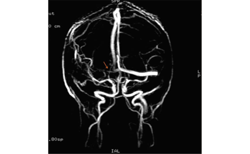
Patients with suspected acute TIAs (within 24–48 hours) should be treated as an emergency to expedite assessment and possibly allow for immediate prevention or even thrombolytic treatment in case of rapidly recurring symptoms. Stroke units are probably better than TIA clinics or other outpatient facilities to guarantee expert recognition of repeat symptoms during the short time window of highest stroke risk.40,41 First, the diagnosis of TIA should be verified taking a detailed history (including vascular risk factors, risk indicators—recent strokes and/or MI—and current medication) and neurological medical assessment. If a TIA seems possible or even probable, immediate brain imaging should take place. MRI should be the first choice, including DWI to look for acute ischemic lesions, perfusion weighted imaging (PWI), T2* weighted images, and MR-angiography. MRI not only reveals acute ischemic lesions (which would change the diagnosis from TIA to stroke according to the new definition), but also demonstrates the burden of previous cerebral ischemia and allows more insight into the vascular state (including possible perfusion deficits).
Patients at highest risk are those with TIA within the past 48 hours, ABCD2 scores >3, symptom fluctuations on presentation, ipsilateral carotid stenosis or potentially active known embolic sources (e.g. atrial fibrillation) after recent changes or withdrawal of antithrombotic medication, after surgery or interventional treatment, etc. They should be admitted into a specialized stroke center to allow for a quick and comprehensive work-up. In case of repeat or permanent symptom fluctuations, immediate thrombolysis is recommended.6,34 An initiation of antiplatelet therapy (if not contraindicated) is generally recommended because of its rapid action and its known efficacy in secondary prevention.19 Patients who had TIAs days before presentation should also receive a full work-up in a specialized stroke setting to conduct the necessary examinations quickly.4
Most guidelines agree that initial examinations (within 24 hours) should include extracranial and transcranial Doppler/duplex imaging, electrocardiography (ECG), laboratory tests, and continuous 24-hour monitoring of clinical, ECG, blood pressure, respiration, fever, and other parameters (oxygen, blood sugar, electrolytes, inflammation parameters).19,41 If a cardioembolic mechanism is suspected or no distinct etiology has been found, transthoracic and/or transesophageal echocardiography for endocarditis, atrial thrombus or for right to left shunting is recommended. This is particularly recommended in (but not limited to) patients below 55 years of age.42
Stroke Prevention
Several different treatments have been shown to independently improve long-term outcome and stroke prevention. Antiplatelet therapy should be immediately used in patients found to have non-cardioembolic TIAs. In most cases, aspirin is sufficient alone or sometimes in combination with dipyridamole.43,44 In cardioembolic TIAs due to atrial fibrillation, oral anticoagulation should be initiated45 either after early low-molecular-weight heparin administration or (in the near future) with new fast-acting anticoagulants (oral direct Xa-inhibitors).46,47 Statins are also effective in reducing recurrent stroke risk, as shown by the Stroke Prevention by Aggressive Reduction in Cholesterol Levels (SPARCL) trial, and should be administered immediately after TIAs.48,49 The Perindopril Protection Against Recurrent Stroke Study (PROGRESS) could show that a blood-pressure-lowering regimen with perindopril and indapamide lead to a risk reduction over a follow-up period of four years.50 In the case of high-grade carotid stenosis identified as the cause of TIA, early carotid endarterectomy is of benefit if performed shortly after the TIA and there is a favorable benefit–risk ratio.51,52 It is very important to stress that early identification, work-up and treatment of TIAs play an essential role in the effective stroke risk reduction. This we
l-known insight was evidence-based by studies that examined the impact of urgent TIA management and start of secondary prevention. In the EXPRESS study, it was demonstrated that the fast assessment of TIA patients and the commencement of suitable preventive treatment in UK reduced the risk for early stroke by about 80% after TIA or minor stroke compared with current standards.53
Conclusion
TIAs carry a high risk for early stroke and also influence long-term prognosis. While it can be challenging to recognise a TIA correctly and assess the individual risk for each patient, clinical features, risk scores, brain imaging, and other investigations or parameters can help not only to make the correct diagnosis, but also estimate the stroke risk properly. Urgent assessment in a specialized stroke unit or dedicated emergency unit with special expertise and initiation if suitable preventive treatment such as antiplatelet agents, anticoagulation, statins, antihypertensive drugs, or even early carotid endarterectomy, can greatly affect outcome and reduce the risk for a permanent stroke.


