Stroke is the second most common cause of death and a major cause of disability worldwide. Because of the ageing population, the burden of stroke is likely to increase during the next 20 years, especially in developing countries.1 The majority (85 %) of strokes are ischaemic: patients present with asymptomatic bruits, transient ischaemic attacks (TIA) or manifest neurological symptoms. A TIA is a transient neurological deficit lasting from a few seconds to a few hours. Reversible ischaemic neurological deficit (RIND) is often included within the category of stroke and is a neurological deficit that lasts longer than 24 hours but less than 3 days and results in complete recovery. Prolonged reversible ischaemic neurological deficits (PRIND) may last for up to 7 days.
Non-ischaemic or haemorrhagic stroke is associated with higher mortality rates than ischaemic stroke.2 Patients present with intracerebral or subarachnoid haemorrhage, causes of which include hypertension, intracranial aneurysms, arteriovenous malformations (AVMs), dural arteriovenous fistulas (DAVF), or cerebral amyloid angiopathy The first stage in the evaluation of patients with acute stroke is to elucidate the nature and aetiology of stroke (haemorrhage or infarction), identify infarcted and threatened tissue and visualise thrombi (see Table 1). Neurovascular imaging techniques can assess these parameters within minutes of the patient arriving at the hospital and allow accurate diagnosis, prompt initiation of appropriate treatment, characterisation of disease progression and monitoring of the response to interventions.
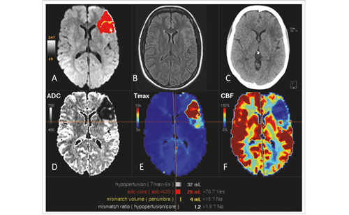
Computed tomography (CT) has traditionally been the mainstay of imaging patients with acute stroke but its sensitivity for early infarction is less than ideal and it cannot accurately define the infarct core. It is also subject to substantial inter-rater variability in interpretation and magnetic resonance imaging (MRI) has demonstrated superiority for detection of acute intracranial haemorrhage.4,5 Traditionally, to obtain vascular and perfusion information with CT, multiple injections have been required although newer techniques have enabled the simultaneous acquisition of CT-perfusion and CT-angiography.6 MRI techniques are becoming increasingly used in the diagnosis of stroke and enable identification of the infarct core and the relationship to the typically larger volume of ischaemic tissue (penumbra) (see Figure 1).
Magnetic Resonance Imaging of Stroke
A number of MRI techniques are used in stroke diagnosis. These are defined in Table 2. The imaging regimen in acute stroke includes diffusion weighted imaging (DWI), T2 and fluid attenuated inversion recovery (FLAIR) weighted imaging, gradient recalled echo imaging (GRE) or susceptibilityweighted imaging (SWI) and magnetic resonance angiography (MRA), followed by physiological assessment with perfusion weighted imaging (PWI).7 Stroke evaluation protocols should include a combination of dWI and PWI, because together they define the location and extent of ischaemia and infarction within minutes of onset. In addition, when performed in series, they can provide information about the pattern of evolution of the ischaemic lesion and treatment monitoring.8
Perfusion imaging can play an important role in identifying haemodynamic insufficiency and in grading its severity, and can be readily integrated into neuroimaging protocols.9Contrast-based PWI, also known as dynamic susceptibility contrast magnetic resonance perfusion (DSC-MRP) is based on a magnetic susceptibility contrast phenomenon involving T2 and T2*-effects of intravenous (iv) bolus-injected contrast agents. Images are acquired by serial imaging of the whole brain as a bolus of gadolinium-based contrast agent (GBCA) passes through the tissue capillary bed. From these images it is possible to calculate functional parameters such as relative cerebral blood flow (RCBF), relative cerebral blood volume (RCBV) and transit parameters including the mean transit time (MTT), time-to-maximum (Tmax) and time-to-peak (TTP) maps.
Complete interruption of blood flow in acute stroke results in irreversible injury within minutes.10 In many cases, however, vascular occlusion by atherosclerotic plaques develops over time, which allows the development of a collateral blood supply to the affected tissues and this can sustain brain tissue for hours after the occlusion of major arteries to the brain. Cerebral tissue that is viable but at risk for infarction, the ischaemic penumbra, can be saved if appropriate intervention is promptly initiated.8 The viability of this region may extend to 48 hours after the onset of stroke. Even in acute stroke, PWI can capture and characterise blood flow changes in the critical hours after symptom onset, allowing the opportunity assess the ischaemic penumbra and to intervene. determining the volume of the ischaemic penumbra helps to identify patients who would benefit from thrombolytic therapy or conventional treatments, such as carotid endarterectomy or blood pressure elevation.
The diffusion-perfusion mismatch, i.e. the difference in size between lesions captured by dWI and PWI, may also be used to assess the ischaemic penumbra and is a strong predictor of lesion volume growth. It can measure the tissue at risk and has been increasingly used in the evaluation of hyperacute and acute stroke.11,12 Two clinical trials involving acute ischaemic stroke patients, (dEFUSE (n=74) and EPITHET (n=101), have established that a mismatch between PWI and DWI imaging volumes may be used to select patients for reperfusion treatment (see Figure 2).13,14,15
Infarct core is assessed by DWI, which has been found to have substantially better sensitivity and accuracy than CT in the assessment of hyperacute ischaemia.4,16 It is widely accepted that lesions with diffusion slowing represent tissues prone to infarction. After acute stroke, damage to the blood–brain barrier occurs17 and allows leakage of contrast agent into cells from the extracellular space. However, increased use of DWI in acute stroke has revealed that lesions that initially show diffusion slowing may undergo diffusion normalisation. It has therefore been concluded that acute slowing of diffusion is not necessarily an indicator of infarct.18
There is a need for a consensus on which perfusion measurement and processing methods should be routinely used in clinical practice.19,20 DSC-MRP in combination with dWI is most commonly used in the evaluation of acute stroke and TIAs.21,22,23
The Use of Contrast-enhanced Magnetic Resonance Angiography in Imaging of the Neurovasculature
Ischaemic Stroke
An important component of the diagnosis of acute stroke, TIA, or suspected cerebrovascular disease is the imaging of the extracranial and intracranial vasculature. Several different MRA techniques may be used for imaging cerebral vessels, including 3d TOF and contrast-enhanced MRA (CE-MRA).24 CE-MRA is increasingly specified in standard protocols for the diagnosis of acute stroke. In a study (n=66) comparing TOF-MRA and CE-MRA, the latter enabled superior visualisation of vessels distal to the occlusion. Furthermore CE-MRA allowed visualisation of extracranial vessels and faster image acquisition.25 CE-MRA can also be used to diagnose intravascular occlusion due to a thrombus or atherosclerotic plaque and for evaluating the carotid bifurcation in patients with acute stroke.26 The technique can be performed rapidly alongside PWI in acute stroke and demonstrate arterial segments with low flow and avoid overestimation of vascular obstruction.27
Vascular Malformations
Several different forms of vascular malformations exist, including capillary telangiectasias, developmental venous anomalies, cavernous malformations, arteriovenous malformations and arteriovenous fistulae. Some are benign and require no treatment. However, other malformations such as arteriovenous malformation (AVM) and arteriovenous fistulas (AVF) may result in haemorrhagic stroke. CE-MRA has been increasingly used for the diagnosis and characterisation of these malformations and gives the detailed images of their location, feeding vessels and drainage patterns.
Three-dimensional time-of-flight (TOF) CE-MRA has become an established non-invasive imaging tool in the evaluation of atherosclerotic pathology of carotid arteries28,29,30 and provides better image quality, greater diagnostic confidence and more inter-observer agreement compared with 2d ToF MRA.31 It has also proven a useful supplement to digital subtraction angiography (DSA). The technique shows high sensitivity and specificity at detecting disease not only in the carotid vessels, but also in the vertebrobasilar circulation, and has the potential to provide a comprehensive and non-invasive evaluation of the head and neck arteries in a single study.32
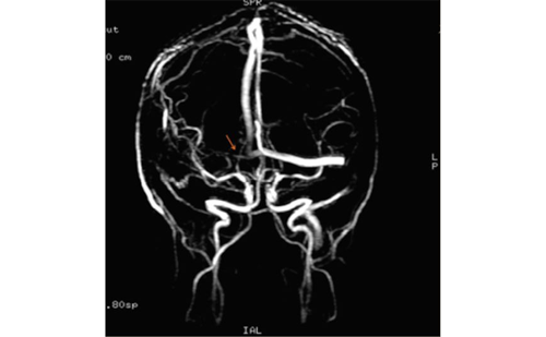
Cerebral arteriovenous malformations (AVMs) are a common cause of non-ischaemic stroke and are a rare developmental abnormality of the intracranial vasculature involving an abnormal tangle of arteries and veins with no capillaries in between. This causes shunting of pressurised blood from arteries directly into veins, exposing the veins to high pressure DSA has been the standard method of diagnosis for AVMs; however, CE-MRA has high sensitivity and specificity in the assessment and grading of AVMs.33,34 Two recent studies comparing DSA and 4D dynamic MR Angiography (4D-MRA) have confirmed that the latter is a promising technique in the diagnosis and follow-up of cerebral AVMs, providing functional information that has until recently been gained only with DSA.35,36 In both studies, classification by 4D-MRA and DSA matched in all patients. An example of the visualisation of AVM using CE-MRA is given in Figure 3.
CE-MRA is useful in the evaluation of intracranial aneurysms treated with detachable coils. However, aneurysm recanalisation may occur because of coil compaction or regrowth of a residual neck and therefore follow-up imaging of these structures is important. Such imaging has historically involved repeated intra-arterial cerebral catheter angiography, an invasive procedure with associated risks. MRA provides a noninvasive alternative with less discomfort and morbidity for patients. A retrospective study (n=232), in which patients underwent CE-MRA and DSA for depiction of aneurysmal remnants of coiled cerebral aneurysms, showed that CE-MRA was at least equivalent to DSA. In addition, contrast filling within the coil mass was more clearly seen with CE-MRA than with DSA.37 A recent study has also shown that CE-MRA is an accurate technique for follow-up of aneurysms following stent-assisted coiling with excellent depiction of remnants in spite of the presence of a stent.38 An example of the use of CE-MRA in a patient with turbulent flow basilar aneurysm is given in Figure 4. The contrast-enhanced study allowed a better assessment of the flow inside the aneurysm and a better appreciation of the thrombosed parts. Furthermore, a better visualisation of smaller vessel segments can be achieved. This allows a substantially better detection of potentially present additional vascular malformations.
An arteriovenous fistula (AVF) also is rare and is very similar to AVM. An AVF is an abnormal connection between arteries and veins. The most common types are a dural arteriovenous fistula (DAVF) and a carotidcavernous fistula (CCF). A DAVF is an abnormal connection between arteries and veins in the dura. It is characterised by a direct connection between the arteries and the vein) without any vessels between. CEMRA has proven an effective technique in the screening and surveillance of DAVF in specific clinical situations, providing a degree of resolution that was previously impossible.39 In 93 % of DAVF cases, separate readers of the scans were unanimous and correct in their independent interpretation of time-resolved MRA scans at 3 T, correctly identifying or excluding all fistulas and accurately classifying them.
Use of Gadolinium-based Contrast Agents in Neurovascular Imaging
The first GBCA became commercially available in 1988 and many GBCAs are now approved for use in neurovascular MRI; a summary of commercially available agents is given in Table 3. Typically, gadolinium (GD) agents are extracellular, non-tissue-specific, non-protein-binding, water-soluble compounds. The GD3+ ion has strong magnetic properties, which causes a T1-relaxation shortening of protons and the production of a high signal in T1-weighted MR sequences.40 The structure of GBCAs is either a linear or macrocyclic chelate and they are available as ionic or non-ionic preparations. The molecular structure and ionicity determines the stability of GBCAs.41 Linear chelates are flexible open chains that do not offer a strong binding to GD3+ ions. Macrocyclic chelates, however, bind strongly to GD3+ ions as they comprise rigid rings which cage the gadolinium atom, and therefore significantly reduce the level of free GD3+ions after application. Non-ionic linear molecules are also less stable compared with ionic ones: the binding between GD3+ ions and negatively charged carboxyl groups is stronger than that with amides or alcohol in the non-ionic preparations.42
The ability of GBCAs to bind GD3+ ions has important safety implications. Some GBCAs have been associated with adverse effects resulting from the release of GD3+ ions in vivo. These include an increased risk of nephrogenic systemic fibrosis (NSF), a rare disorder that principally affects the skin but may affect other organs, in patients with renal insufficiency.43–48 Agents carrying higher risk for NSF are contraindicated for use in patients with acute kidney injury or chronic severe kidney disease. These agents include gadodiamide (omniscan®), gadopentetate dimeglumine (Magnevist®) and gadoversetamide (optiMARK®) (see Table 3). Agents in the lower-risk category must still carry warning labels, but they do not require contraindications. These include gadoteridol (ProHance®), gadoterate (dotarem®) and gadobutrol (Gadovist®).49
Gadobutrol was approved initially for contrast enhancement in cranial and spinal MRI by the European Medicines Agency (EMA) in 2000 and has since then found wide acceptance in applications in various body regions. Gadobutrol is a new-generation, macrocyclic, non-ionic Gd chelate that is highly hydrophilic with negligible protein binding and good clinical tolerance.50,51 Furthermore, gadobutrol has a number of properties that allow superior image quality in MRI. The relaxivity of gadobutrol is higher than that of other non-protein-binding agents, leading to the highest available T1-shortening per volume and better image contrast than other agents.52,53 The T1 relaxivity of gadobutrol in plasma is significantly higher that of gadopentetate dimeglumine (5.2 versus 4.1 at 1.5 T and 5.0 versus 3.7 at 3 T).53 Gadobutrol is available in a highly concentrated (1.0 mol/L) formulation and requires half the injection volume compared with other low molecular weight contrast agents, the formulations of which are less concentrated (0.5 mol/L). When injected in an iv bolus, gadobutrol produces a significantly smaller bolus width at half maximum signal intensity decrease, a smaller mean peak time, a higher contrast and contrast-to-noise ratio (CNR) between grey and white matter.54 This is advantageous for PWI as image quality is dependent on the geometry of the contrast bolus, which in turn is optimised by an increased concentration of the injected contrast solution in brain tissues.9,55 Gadobutrol leads to shortening of the relaxation times even in low concentrations. In healthy volunteers, 1.0 mol/L gadobutrol administered at a dose of 0.3 mmol/kg body weight (BW) yielded MR perfusion images that were superior to those obtained with the 0.5 mol/L formulation.54 The standard dose currently used is 0.1 mmol/kg bw.
Gadobutrol is well tolerated and has a good safety profile. In a review of 14,299 patients receiving iv injection of gadobutrol in routine clinical radiology practices, the percentage of patients reporting at least one adverse drug reaction (AdR) was low (0.55 %). Two (0.01 %) serious AdRs were reported. The most frequently reported AdR was nausea, which occurred in 0.25 % of patients. The observed occurrence of AdRs was similar to published safety data of other GD-based contrast agents.56
Clinical Studies of Gadobutrol in Neurovascular Imaging
Gadobutrol in Magnetic Resonance Imaging
The first study of gadobutrol in stroke evaluated its efficacy and safety in the assessment of cerebral haemodynamics by DSC-MRP. The multicentre double-blinded study involved patients (n=89) with carotid artery stenosis or cerebral infarcts and used dosages from 0.1 to 0.5 mmol/kg bw. The dose of 0.3 mmol/kg bw was found to be diagnostically adequate. gadobutrol was safe and well-tolerated.57
A randomised intraindividual study (n=12) found that gadobutrol was equivalent to gadobenate dimeglumine for cerebral PWI at 1.5T . The susceptibility effect, described by percentage of signal loss was similar for both agents (gadobutrol 29.4 % versus gadobenate dimeglumine 28.3 %). Both agents allowed the calculation of high quality perfusion maps at a dosage of 0.1 mmol/kg bw.58 A further study (n=12) compared 0.1 and 0.2 mmol/kg bw doses of gadobenate dimeglumine and gadobutrol for cerebral PWI at 1.5 T. A single dose (0.1 mm/kg bw) of both agents was sufficient to achieve high-quality, diagnostically valid perfusion maps at 1.5 T and there was no significant benefit for one agent over the other for quantitative or qualitative determinations. The susceptibility effect was 29.4 % for gadobutrol versus 28.3 % for gadobenate dimeglumine. double doses of the two agents produced better overall image quality but no clinical benefit over the single-doses.59
In a study of cerebral perfusion parameters obtained by dSC-MRP, sixteen healthy volunteers underwent three separate dSC-MRP examinations at 3 T receiving single-dose (0.1 mmol/kg bw) gadobutrol, double-dose gadobutrol and single-dose gadobenate dimeglumine. The study found that a single gadobutrol or gadobenate dimeglumine dose of 0.1 mmol/kg bw is sufficient for DSC-MRP. The T2* relaxation effects of the two agents were almost identical.60,61 An ongoing prospective, single centre observational study (n=1,200) employing gadobutrol as contrast agent in acute stroke patients aims to describe the incidence of mismatch between dWI lesion and PWI deficit volumes and the predictive value of PWI for final lesion size and functional outcome depending on delay of imaging and vascular recanalisation.62
Gadobutrol in Contrast-enhanced Magnetic Resonance Angiography
Gadobutrol offers advantages over other contrast media in cerebrovascular applications of CE-MRA. When gadobutrol is given at 1 mol/L, enhanced MRA demonstrates significantly higher (up to 70 %) signal-to-noise ratio (SNR) and CNR values compared with 0.5 mol/L gadobutrol- and gadopentetate dimeglumine-enhanced images, respectively.63 CE-MRA of the neck vessels with gadobutrol allows a tighter contrast bolus than other GBCAs and can more accurately estimate the extent of vessel stenosis.64
Until recently, the diagnosis of intracranial atherosclerosis was difficult owing to lack of adequate imaging techniques. Conventional MRA techniques are only able to visualise intracranial wall thickening when it causes narrowing of the lumen and may therefore underestimate the presence of intracranial wall pathology. Moreover, the intracranial arteries, which have a diameter less than 1 mm in distal regions, have proved difficult to visualise by conventional methods. A high-resolution method of intracranial vessel wall imaging at 7.0-T MRI using 3d turbo spin-echo (TSE) sequence has recently been developed using gadobutrol as the contrast agent.65 This technique allows high image resolution and sufficient sensitivity to allow identification of vessel wall of the narrow arteries of the circle of Willis and can detect lesions and healthy intracranial vessel wall. A combination of MRA and duplex sonography using gadobutrol as the contrast agent was found to be effective for the accurate grading of stenoses in the assessment of stenoses of the internal carotid artery.66
A recent study evaluated the feasibility of low-dose, 3d time-resolved CE-MRA (TR-CE-MRA) in the assessment of the supra-aortic vessel and compared the results with high-resolution contrast-enhanced MRA. Acquisition of low-dose TR-CE-MRA by administration of gadobutrol was found to be feasible and the two techniques gave consistent results in the haemodynamic grading of stenosis. In addition, TR-CE-MRA rapidly provided haemodynamic information, enabling improved evaluation of atherosclerosis of the supraaortic arteries.67
In a comparison of gadopentetate dimeglumine and gadobutrol in the evaluation of 56 patients with cerebral AVMs using 4d MRA, vessel sharpness and diameter did not differ substantially between the two groups. However, vessel-to-background contrast was significantly higher in the gadobutrol group (p<0.005).35
Summary and Conclusion
Neuroimaging in acute stroke is essential for establishment of an accurate diagnosis and monitoring of the response to interventions. Magnetic resonance techniques including MRI and MRA have higher accuracy, fewer safety risks and provide a greater range of diagnostic information than CT and can provide a definitive diagnosis within minutes of a patient’s arrival to the hospital. The utility of MR imaging in acute stroke management lies not only in its capability to detect early ischaemic lesions with high sensitivity, but also in its capacity to reveal specific features of cerebrovascular pathology. The role of MRI in acute stroke management is an area of active research. Results from MRI-based clinical trials are helping to refine the mismatch concept and penumbral imaging is a promising technique that will enable the identification of individuals who might benefit from thrombolytic treatment beyond the current therapeutic window. These advances may result in improvements in patient outcomes and cost-effectiveness.
Contrast agents have greatly enhanced the utility of MR techniques. Gadobutrol is the only 1 M contrast medium currently available and has an excellent safety profile. Its concentrated formulation and high relaxivity, result in superior bolus characteristics and enhancement allowing improved diagnostic efficacy in PWI and CE-MRA. The use of new high-relaxivity contrast agents is critical when defining more appropriate and effective therapeutic management approaches.


