There are no official guidelines on how to manage stroke-related seizures. In a prospective multicentre study, the incidence of seizures in relation to stroke was 8.9%, with a frequency of 10.6 and 8.6% in haemorrhagic and ischaemic stroke, respectively. In subarachnoid haemorrhage the incidence is 8.5%. Due to the fact that infarcts are significantly more frequent than haemorrhages, seizures are mainly related to occlusive vascular disease of the brain.1 The general view is to consider stroke-related seizures as harmless complications in the course of a prolonged vascular disease involving the heart and brain.
Seizures can be classified as those of early and those of late onset in a paradigm comparable to post-traumatic epilepsy, with an arbitrary dividing point of two weeks after the event. Most early-onset seizures occur during the first day after the stroke. Late-onset seizures occur three times more often than early-onset ones. A first late-onset epileptic event is most likely to take place between six months and two years after the stroke. However, up to 28% of patients develop their first seizure several years later.
Simple partial seizures, with or without secondary generalisation, account for about 50% of total seizures, while complex partial spells, with or without secondary generalisation, and primary generalised tonic–clonic insults account for approximately 25% each. Status epilepticus occurs in 12% of stroke patients, but the recurrence rate after an initial status epilepticus is not higher than after a single seizure.2 Inhibitory seizures, mimicking transient ischaemic attacks, are observed in 7.1% of cases.
Risk Factors
Early-onset seizures in ischaemic stroke are mainly found in patients with large cortical lesions, decreased consciousness and additional haemodynamic and metabolic – mainly renal – disturbances. In haemorrhagic stroke, the products of blood metabolism, such as haemosiderin, may cause a focal cerebral irritation, leading to seizures.
In the Seizures After Stroke Study (SASS), the main predictors for late-onset seizures after an ischaemic stroke were the severity of the initial neurological deficit and the presence of a large cortical infarct.1 In our series we found that the only clinical predictor of late-onset seizures was the initial presentation of partial anterior circulation syndrome due to a territorial infarct. Patients with total anterior circulation syndrome had less chance of developing epileptic spells, not only due to their shorter life expectancy but also due to the fact that the large infarcts are sharply demarcated in these patients. Large cortical infarcts with irregular borders located in the temporal–parietal regions represent an increased risk of developing seizures. Epileptic spells can also occur after a ‘silent’ cerebral infarct triggered by alcohol or drug abuse. Finally, a late-onset seizure can be due to recurrent infarction, mainly of cardioembolic origin. Lacunar infarcts and ischaemic white-matter lesions are probably not a direct cause of seizures and epilepsy, and there is no evidence that post-stroke epilepsy occurs more frequently in patients with cardioembolic, as opposed to thromboembolic, infarcts. Arterial hypertension, coronary and valve disorders, diabetes and hypercholesterolaemia are not risk factors for seizures in stroke patients. Only chronic obstructive pulmonary disease (COPD) is more frequently found in patients with stroke-related seizures. Lobar haematomas are frequently accompanied by early-onset fits. Surgery for an intra-cerebral haemorrhage is an additional risk for the development of late-onset seizures.2
Diagnostic Procedures
Every patient who suffers a convulsive seizure episode following a previous stroke has to be investigated for the possibility of a new cerebrovascular accident. Furthermore, patients with delayed transient worsening of neurological deficits after an ischaemic stroke have to be investigated for the possibility of inhibitory seizures. An electroencephalogram (EEG) should be performed as soon as possible after the ictal event. Periodic lateralised epileptiform discharges (PLEDs) are observed in 25% of patients with early-onset seizure, but in only 1% of those with late-onset seizures. However, the combined frequency of PLEDs, intermittent rhythmic delta activities (IRDAs) and diffuse slowing on EEG is 26.5% in stroke patients with late-onset seizures compared with 6.2% in those without seizures.
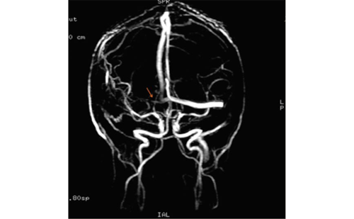
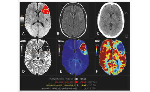
A computed tomography (CT) scan of the brain is helpful for three reasons. First of all, it can sometimes demonstrate a new infarct in patients who have already had one. Second, a previous silent infarct can be demonstrated as the cause of the epileptic spell in 10% of patients with a history of vascular risk factors but not of stroke. Finally, patients with a capricious distribution of cortical infarct with apparent areas of preserved cerebral tissue within the infarct region are more prone to develop seizures and epilepsy than those with a sharply demarcated infarct zone. Magnetic resonance imaging (MRI) of the brain is more sensitive than CT and is useful in ruling out conditions other than cerebrovascular accident that may have been the cause of a seizure. Diffusion-weighted imaging (DWI) can also demonstrate a new infarct to be the cause of an epileptic spell, or can reveal additional secondary damage due to the seizure itself. Patients who develop seizures after a stroke should also have an extensive blood examination in order to exclude or demonstrate possible metabolic or toxic provoking factors. After the first epileptic fit, stroke patients should again have an exhaustive cardiovascular examination, including a 24-hour electrocardiogram (ECG), cerebrovascular ultrasound of carotid arteries and transthoracic echography or transoesophagial Doppler of the heart in order to investigate for possible new sources of emboli.2
Outcome
Seizures related to stroke are harmful for patients. The outcome for patients with early-onset seizures is poor with a high in-hospital mortality rate. On the other hand, patients with early-onset seizures have a recurrence rate of just 16%. Patients with late-onset seizures have a recurrence rate of more than 50%. In patients who have their first seizure between six months and two years after stroke, recurrence rate reaches 62%, while for very-late-onset seizures it is 47%. Recurrence of late-onset seizures or post-stroke epilepsy increases the disability of stroke patients and promotes the occurrence of vascular cognitive impairment.2
Treatment
Most anti-epileptic drugs (AEDs) impair cognition in elderly patients. This side effect has been reduced with the new generation of AEDs, such as lamotrigine, gabapentin and levetiracetam.3 However, these are more costly than phenytoin, valproate sodium and carbamazepine.
The choice of AED should be guided by the individual characteristics of each patient, including concurrent medications and medical co-morbidities. First-generation AEDs should be the first choice, although they undergo significant hepatic metabolism, and phenytoin and vaproate sodium are highly protein-bound. For example, the recognised interaction of warfarin with phenytoin makes it difficult to maintain consistent therapeutic ranges of both agents in patients with atrial fibrillation. No controlled trials have been conducted so far to assess the efficacy of specific agents in stroke-related seizures.4 In early-onset seizures and status epilepticus, intravenous benzodiazepines are the first choice, eventually followed by phenytoin sodium or valproate sodium. Due to the low recurrence rate after this type of seizure, maintained AED treatment does not seem necessary.
The question of whether prevention with AEDs should be started in some stroke patients who are particularly at risk of developing late-onset seizures, particularly those with COPD and a large irregular cortical infarct in the temporal–parietal region, remains unanswered. The probable answer is that treatment with AEDs ought not to be started due to the possible side effects of the AED treatment and because eventual worsening of the neurological condition and an increase in vascular cognitive impairment mainly occur after repeated seizures.
The high-risk patients who develop a first epileptic spell between six months and two years after stroke should be treated. Given the typical focal onset of late-onset seizures and the low cost, the first-line option is to use carbamazepine as single therapy. In the case of seizure recurrence under this therapy, treatment is switched to lamotrigine, as a recent multicentre, double-blind, randomised trial demonstrated that this drug was better tolerated and protected elderly patients from seizures for longer intervals than carbamazepine.5
In patients with very-late-onset seizures one could probably wait to start AEDs until after a second seizure, according to the general guidelines on treatment of epilepsy. Patients with a single epileptic event and in whom only old lacunae are found on CT or MRI of the brain should be managed in the same way, particularly since the relationship between seizures and lacunae is uncertain. It is necessary to search for other possible causes of such events, and readjustment of the antithrombotic treatment may be required, as well as better control of the vascular risk factors.
In patients who develop seizures due to recurrent infarction, a cardioembolic source should be suspected and, if proved, anticoagulant treatment should be started or readjusted. In the case of repetitive transient worsening of neurological symptoms after an initial ischaemic stroke, inhibitory seizures should be suspected. Even in the absence of typical EEG findings, a trial with AEDs is worthwhile.2
Conclusions
Seizures related to stroke need special attention and should not be considered as benign complications occurring during the long-standing course of cerebrovascular disease. They should be correctly managed as this could improve the quality of life of stroke patients and avoid further deterioration of their condition. ■


