Sleep-disordered breathing (SDB) refers to momentary, often cyclical, cessations in breathing (apnoeas) or momentary or sustained reductions in the breath amplitude (hypopnoeas), sufficient to cause arousal from sleep and/or significant arterial hypoxemia and hypercapnia. causes of breath cessation during sleep are classified as obstructive (OSA), or central (CSA).1 The observation of both obstructive and central events at different times in the same patient, have brought to consider them as extremes along a continuum of SDB with considerable overlap between the two apnoea types.2 Sleep studies on continuous series of patients have shown that variably ‘mixed’ rather than pure obstructive or central (‘control’) patterns are often recognisable. in some cases, what starts as clearly obstructive disease at the beginning of the night, evolves into predominantly central disease by the end of the recording. This phenomenon is particularly frequent in patients affected by congestive heart failure (CHF).3,4 in most cases the central sleep breath disorder is precipitated by the continuous positive airway pressure (CPAP) treatment of an OSA. 5–7 This pattern of disease has been named ‘complex sleep apnea’ to convey the high likelihood that both obstructive and control factors are involved in its creation.6
Recent data have shown that the prevalence of both obstructive and central sleep disordered breathing in patients with first-ever stroke or transient ischaemic attack (TIA) is higher than expected.8 although CPAP is the ‘gold standard’ treatment for OSA, acute stroke cases affected by obstructive sleep apnoea-hypopnoea (OSAH) are treating with CPAP with controversial results, suggesting that CPAP may not be the appropriate treatment for everyone with OSA after stroke.9 other treatment options of SDB after stroke are scarce, mainly cause of the poor knowledge of the pathophysiology of SDB after stroke, in particular the interaction between the consequences of stroke on the breathing system and the primary disturb itself.
In order to characterise SDB in patients with stable stroke, a prospective observational study is ongoing in Rome. We report the results of the first 55 cases studied.
Methods
Patients
A consecutive series of patients with acute stroke, admitted to the stroke unit of two different Roman university hospitals, were screened. after the exclusion of patients who did not consent to enter the study, elected cases were submitted to both clinical and instrumental diagnostic tests in the acute phase of the illness and after 4 months of stroke onset. Stroke risk profile was assessed and classified according to the italian SPREad guidelines classification.10 Participants were visited firstly at stroke onset and then at 4 months on the day of the diagnostic tests, that included polisomnographyc (PSG) study and brain MRI. The severity of neurological deficit was assessed by means of NIHSS, that was administered by an accredited physician or nurse.11causes of stroke were classified as to TOAST criteria.12 daytime sleepiness was estimated with the Epworth Sleep Scale (ESS) questionnaire.13 The presence of symptoms other than daytime sleepiness has also been investigated and classified so as: choking, gasping, fragmented sleep, unrefreshing sleep, reported daytime sleepiness, inattention.13,14 in order to identify possible predisposing conditions to OSAHS, such as the nasal obstruction and pharyngeal constriction, an otolaryngology (ORI) evaluation has been performed.
Definitions
apnoea was defined as the cessation of airflow for at least 10 seconds. hypopnoea was defined a clear amplitude reduction of a validated measure of breathing during sleep (but less than a 50 % reduction from baseline) that is associated with an oxygen desaturation of >3 % or an arousal. apnoea-hypopnoea index (AHI) was the sum of all apnoeas and hypopnoeas occurring per hour of analysis time. SDB, either central or obstructive, was diagnosed in presence of an AHI value ≥5.15 Three different degrees of severity were outlined: mild SDB; AHI value ≥5 ≤15; moderate SDB; AHI value >15 ≤30; Severe SB: AHI value > 30.16 Each event was considered central if during apnoea/hypopnoea there were no efforts to breathe (central sleep apnoea/hypopnoea [CSAH]); it was instead considered obstructive if there were efforts to breathe (OSAH). a diagnosis of OSAH syndromes (OSAHS) was set in cases in whom at least 50 % of the total respiratory events showed a clear decrease (>50 %) from baseline in the amplitude of a valid measure of breathing during sleep lasting at least 10 seconds. a CSAH syndromes (CSAHS) was instead diagnosed when at least 50 % of the total respiratory events were central in origin. central periodic breathing (CPB) was defined as ≥3 cycles of regular crescendo-decrescendo breathing associated with reduction of ≥50 % in nasal airflow and respiratory effort lasting ≥10 seconds. a cycle was defined from the end of one apnoea/hypopnoea to the end of the next.17– 19arousals were defined as “an abrupt change from a ‘deeper’” stage of non-REM (NREM) sleep to a “lighter” stage, or from REM sleep toward wakefulness, with the possibility of awakening as the final outcome”.20 arrhythmias were defined as any disturbances of the normal rhythmic beating of the heart.
Sleep Studies and Polysomography
The ambulatory polysomnography (PSG) study was performed by the hand-held 34 channel Morpheus ambulatory recorder by Micromed® Srl ambulatory PSG was preferred to standard laboratory recording in order to allow the patients to sleep in their home setting, without needs of adaptation. The following parameters were recorded: 1) body position; 2) ribcage and abdominal respiratory efforts (dual thoracoabdominal RiP-respiratory inductance Plethysmography-belts by Micromed® Srl accessories EPM 915x a); 3) oro-nasal airflow (thermistor transducer by Micromed® Srl accessories EPMs 1450-S); 4) haemoglobin saturation (finger pulse oximeter sensor); 5) eight electroencephalograph (EEG) channels (frontal, central temporal, occipital); 6) right and left electrooculography; and 7) submental electromyography from surface electrodes; 8) electrocardiogram (EKG). Morpheus was retrieved the following morning the sensors were attached and the data downloaded to the SystemPlus Evolution software, and subsequently analysed by a dedicated software Rembrandt SleepView. Were considered adequate only PSGs with a total recording time >4 hours and a total sleep time (TST) >2 hours, with presence of both NREM and REM sleep episodes. Sleep-stage scoring was done visually according to standard criteria.18
Brain Imaging
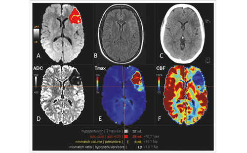
Brain imaging at stroke onset included magnetic resonance image (MRI) T2-fluid attenuated inversion recovery (FLAIR) images and diffusionweighted imaging (DWI) by means of a 1.5 T magnet (Gyroscan NT-15; Philips, Eindhoven, The Netherlands). MRI images were analysed by a neuroradiologist blinded as respect to the clinical profile of cases.
Statistical Analysis
Statistical analysis was performed with SPSS version 18.0 for Windows. The following PSG parameters entered the analyses: age, sex, time from stroke to PSG evaluation (days); Nih-SS at entry and at discharge from stroke unit; body mass index (BMi), ESS, TST; TSP (TSP) time; sleep efficiency index; arousal/h sleep; number and type of apnoeas and hypopnoeas; oxygen saturation; arrhythmias/h of sleep; total number of central apnoeashypopnoeas per hour of sleep; total number of obstructive apnoeashypopnoeas per hour of sleep; and oxygen desaturation index (ODI). Patient‘s demographic data, risk factors for stroke, and sleep study data are expressed as mean and Sd or percentages. continuous data were analysed using analysis of variance (ANOVA). on the bases of PSG study, three groups were identified: patients with normal sleep study, cases with OSAHS and cases with CSAHS. comparisons were made between cases (either OSAH or CSAH) and controls (normal cases). Statistical comparisons between groups were made with the use of χ2 tests and via Fisher’s exact test depending on which was more appropriate. correlation analyses were done with nonparametric tests (Spearman’s rho). Statistical significance was set at p<0.05. Results
Fifty-seven patients accepted to enter the study. at the time of PSG, two cases had deceased, therefore 55 PSG studies were performed. Fifty-two patients had a complete ischaemic stroke and three were TIAs. The 54.6 % of cases were embolic in nature, the 29.1 % were lacunar, in the 12.7 % of cases the cause of stroke/TIA was undetermined and in 3,6 % a large artery cause was revealed. None of included cases had a haemorrhagic stroke. Patients mean age was 63±13. Forty cases were males (71.4 %).The Nih-SS at entry and at discharge were respectively 4±3 and 2±2. The PSG study was performed 126±40 days after stroke onset. Forty-two cases (76.4 %) had a SDB (i.e. AHI ≥5). OSAH were the most frequent breathing abnormality (n=2,823 total events), representing the 69 % of all events (n=4,082). Twenty-eight out of the 42 cases with an AHI ≥5 (66.6 %) had an OSAH (i.e. a >50 % decrease from baseline in the amplitude of a valid measure of breathing during sleep that lasted at least 10 seconds in more than the 50 % of the total events) and 12 cases (28.6 %) had a CSAH (i.e. showed no efforts to breathe in more than the 50 % of all abnormal respiratory events). None of CSAH cases presented central periodic breathings during sleep (CPBS). The BMi was 27±5 kg/m2. Thirteen patients had a BMi >30 of them were among the subgroup of cases with OSAH, among cases affected by CSAH and patients in the subgroup of normal cases at PSG. The mean AHI was 12 (SD 10 range 1 to 38; median 10). Twenty-seven cases had a mild (AHI value ≥5 ≤15), 12 had a moderate (AHI value >15 ≤30) and three cases a severe SDB (AHI value >30) (see Table 1).
The TST was 354±105; the TSP was 430.8±108; the SEI was 78±22. The ORI evaluation was performed in 32 cases and was abnormal in 15 of them. all 15 cases had an obstruction of the upper airways and in eight of them it was combined with an increase in pharyngeal tissue. Nine patients with positive ORI evaluation had an OSAH at the PSG and three was affected by CSAHS. all these three cases were affected by an obstruction of the upper airways combined with an increase in pharyngeal tissue; in two of them BMi was higher than 30 (see Table 1).
At the comparison analysis, patients affected by CSAH were significantly older than controls (p=0.043), as well as than those affected by OSAH (p=0.052). Patients with OSAH have been submitted to PSG significantly later than normal cases (p=0.028). increase in pharyngeal tissue resulted to be significantly more frequent in cases affected by CSAH as respect to controls (p=0.040) (see Table 1). a significant difference between patients affected by OSAH and by CSAH was found in the number of arrhythmias/h of sleep (p=0.016) (see Table 2)
Inverse correlation in patients with OSAH, with a significance respectively of p=0.019 and p=0.006, not in the group of CSAH events (respectively p=0.144). TST was directly correlated to the SEI in the group of cases with OSAH and with CSAH (respectively p=0.002 and p=0.033). In the group of cases with OSAH, TST was significantly directly related with the total number of obstructive apnoeas-hypopnoeas (p=0.029). The correlation was not demonstrated in CSAH cases. In cases with CSAH, a direct association was found only between the TST and the SEI (p=0.033). A direct correlation was found between the number of arousals/h sleep and the number of arrhythmias/h of sleep. The correlation was not found in the group of cases with SAH. The sleep efficiency index was inversely related to number of arrhythmias/h of sleep in the group of cases with OSAH and with CSAH (respectively p=0.042 and p=0.034). In the group of cases with OSAH, arrhythmias/h of sleep were found to be directly related to the number of arousals/h sleep (p=0.009) and inversely related to the SEI (p=0.042). In the group of cases with CSAH, arrhythmias/h of sleep were found to be directed related to TST and to TSP, inversely to the SEI (p=0.034) and the total number of central apnoeas/hypopnoeas (p=0.048). In the group of cases with OSAH, ODI was found to be directly related to the total number of obstructive events (p=0.000). In the group of cases with CSAH, ODI was found to be directly related to the number of days from stroke onset to PSG (p=0.041).*
As to the location of brain lesion, the following sites were documented: eight in the frontal/precentral gyrus infarction, six in the insula, five in the brainstem, three in the thalamus/hypothalamus and two in the deep middle cerebral artery territory. No significant association was found at the Spearman rho between predominantly central apnoeas and the location of the brain lesion. Breathing parameters and stroke classification as to the TOAST classification is reported in Table 3.
Discussion
in our series of cases, nearly the 74 % of patients had an abnormal breathing during sleep; the 52 % of them were obstructive, and the 22 % were central events, all percentages were very close to what was reported in a similar population of patients.8 as well, the clinical and instrumental characteristics of cases with prevalently obstructive respiratory events during sleep are similar to the clinical features reported in of previous studies.8,21–23 The longer the time interval from stroke to PSG, the shorter both the TSP and the TST. Patients with predominantly CSAH, were more frequently arrhythmic and more frequently affected by increase in pharyngeal tissue and referred significantly more frequently inattention or unrefreshing sleep. The hypothesis we postulate is that, in a subgroup of patients with stroke, the effect of stroke itself on the collapsibility of the airways moves to worsen patients’ pre-existing condition, causing obstructive events that alternate with central ones. in some cases the frequency of central events may be higher than the obstructive, giving reasons for classifying them as ‘predominantly central sleep disordered breathings’. instead, at least some of predominantly CSAH cases might become affected by a more severe form of OSAH than before stroke. The hypothesis is supported by several evidences. it is in fact known that stroke can worsen breathing by a number of mechanisms, such as inducing swallowing abnormalities, narrowing of oro- and/or hypo pharynx, or hypomobility of ribcage.24–31 Furthermore, individuals with severe OSAH have higher loop-gain than those with milder OSAH, 32,33 therefore their breathing during sleep can shift from OSAH to CSAH. a condition that has been named complex sleep-apnoea (Complex-SA) and to what very severe OSA may predispose.34
CSAH during the non-rapid eye movement (NREM) sleep, in cases affected by OSAH has been frequently described in patients with concomitant congestive heart failure (CHF). in those cases it has been postulated that an increase in carbon dioxide sensitivity can destabilise breathing during sleep, giving rise to recurrent cycles of central apnoea and hyperventilation during sleep.35–42 This study has several limitations, the most important being the small sample size of mild stroke cases, who were not submitted to neither ORI evaluation nor a sleep study before stroke. Since no relationship was found in this study between the site of brain injury and SDB, we are not able to evaluate its possible role on SDB. in any case, the relationship between breathing and brain are so complex, so close and mutual, which is outside the scope of the present paper.43
Conclusions
our findings suggest that a ventilatory instability is induced by a stroke, that can worsen the breath disturb of cases affected by OSAH since before the cerebrovascular event. in these cases, central apnoea may take precedence over the obstructive one, in a loop gain circuit higher than alternating obstructive and central events (possible complexsleep apnoeas). identification of stroke patients predisposed to this ventilatory instability in the long term of stroke, as well as the eventual predicting factors in the early phase of stroke, has potenTIAl important therapeutic implications, particularly in the selection of cases that benefit most of mechanical therapy. Multicentre studies, capable of enrolling larger cohorts, are needed to confirm this hypothesis.
*Additional data can be provided by the Authors on request


