More than 700,000 acute strokes1 and 300,000 transient ischaemic attacks (TIAs)2,3 occur annually in the US. It is estimated that between 15 and 26% of acute stroke cases have a prior history of TIA.4 TIAs are important because they are associated with high short-term risk of both stroke and cardiac events. In a widely quoted emergency department (ED) study of over 1,700 TIA cases from California, the three-month stroke risk was found to be 10.5%.5 A recent meta-analysis of 11 TIA cohort studies found that the summary estimate for the 90-day stroke risk was 9.2% – very similar to the Californian study.6 This meta-analysis also confirmed that most of this stroke risk occurs in the first few days after the TIA event; the risk of stroke was 3.5% at two days and 8.0% at 30 days.6 Similar findings were found in another recent meta-analysis of 18 cohort studies, which estimated that the seven-day risk of stroke was 5.2%.7 Patients with TIA are also at high risk of other cardiovascular events. In a meta-analysis of 39 cohort studies, the annual risk of myocardial infarction and non-stroke vascular death following TIA was 2.2 and 2.1%, respectively.8 These studies, which serve to illustrate the high risk of cardiovascular events following a TIA, suggest that patients suspected of having a TIA event require an expedited clinical work-up. Historically, TIA has been defined on the basis of focal neurological deficits due to transient and reversible cerebral or retinal hypoperfusion lasting for less than 24 hours.9 However, because the duration of symptoms for most TIAs is much less than 24 hours – typically less than TIA10 – there has been a proposed change in the definition of TIA to include only cases with a symptom duration of less than one hour.11,12 The advent of diffusion-weighted imaging (DWI) technology adds further challenges to the traditional definition of TIA – up to 50% of TIA patients have DWI abnormalities indicating ischaemic changes.13–15 The presence of positive DWI changes in TIA cases has been shown to be associated with longer symptom duration (>60 mins), the presence of speech disturbance, atrial fibrillation and ipsilateral carotid stenosis.13
Accuracy of Transient Ischaemic Attack Diagnosis in the Urgent Setting
The diagnosis of TIA has always been a clinical challenge even for neurologists. Two carefully conducted diagnostic studies undertaken in outpatient or non-acute settings demonstrated that the inter-rater agreement among neurologists for the diagnosis of TIA was actually good (kappa = 0.65–0.77).16,17 In one study of 56 patients, the overall agreement for TIA diagnosis between pairs of neurologists who each interviewed the patients was very high (85%; n=48).16 In the other study evaluating the validity of TIA diagnosis in 72 patients, the overall agreement between neurologists was also very high (88%; n=64).17 However, many cases of TIA present to the ED, where an ED physician rather than a neurologist evaluates them. Achieving optimal diagnostic accuracy for TIA is even more challenging in the ED setting. The reported accuracy of TIA diagnosis among non-neurologists is quite variable, with overall agreement rates varying between 39 and 67%.18–21 In a recent study of 100 hospitalised patients who had a presumptive ED-based diagnosis of TIA, a retrospective chart review by two stroke neurologists found that 60% were misdiagnosed.22 However, such results should not be interpreted as necessarily reflecting the clinical skills of neurologists and ED physicians; rather, these data are a reflection of the fact that more complete and definitive diagnostic information (e.g. brain imaging, carotid imaging) is typically not obtained until after the patient is admitted to the hospital.22 Difficulties in making an accurate diagnosis of TIA in the ED setting arise from several factors. First, time constraints resulting from pre-hospital delays23,24 and rapid triage and assessment requirements make the process especially difficult in a busy ED. Second, differentiating common stroke mimics from TIA can be difficult, particularly for nonstroke physicians.25 Even differentiating TIA from ischaemic stroke can be challenging when reliable information on the exact onset time of symptoms is lacking. Third, terms such as ‘TIA’, ‘TND’ (transient neurological deficits), ‘mini stroke’ and ‘minor stroke’, which may signify different underlying pathology and aetiology, are often liberally applied in the ED setting, and may or may not identify a patient who meets the formal definition of TIA (i.e. transient focal neurological symptoms of <24 hours’ duration). Fourth, the frequent use of terms such as ‘rule-out TIA’, ‘suspect TIA’, ‘possible TIA’ or ‘TIA/stroke’ in the ED setting may reflect either a reluctance on behalf of the ED physician to make a definitive diagnosis based on limited clinical information, and/or the inherent difficulty in ruling out alternative diagnoses in the limited time available.
Finally, and perhaps most importantly, the recent emphasis on the rapid identification and evaluation of TIA cases in the ED setting means that the target population of interest shifts from confirmed TIA cases to ‘suspect TIA’ cases,26 who may or may not have a final diagnosis of TIA. This shift in focus makes the appropriate identification of suspect TIAs, as well as the confirmation of their true TIA status, even more challenging.
The Role of Clinical Prediction Rules in Risk Stratification of Transient Ischaemic Attack Cases (the ABCD2 Score)
Over recent years two clinical prediction rules, the California Rule and the ABCD score, have been developed with the goal of risk-stratifying TIA cases with respect to their short-term risk of stroke.5,27 Recently, these two rules were combined to create a new rule, called the ABCD2 score.28 This rule includes information on the following five factors to create a score scaled from 0 to 7: age >60 years (1 point); blood pressure >140/90mmHg (1 point); clinical features: unilateral weakness (2 points), speech impairment without weakness (1 point); duration: >60 minutes (2 points), 10–59 minutes (1 point); and diabetes (1 point). A subsequent validation study conducted among over 4,800 TIA cases showed that the ABCD2 score was highly predictive of subsequent stroke risk. Twenty-one per cent of these cases were classified as high-risk based on the fact that they had a score of 6 or 7, which was associated with a very high two-day stroke risk of 8.1%. Forty-five per cent of cases were classified as moderate-risk on the basis of a score of 4 or 5, which was associated with a two-day stroke risk of 4.1%. Finally, 34% of cases were classified as low-risk (score ≤3, two-day stroke risk 1.0%). Importantly, the ability of the ABCD2 score to predict the risk of stroke after a TIA is due in part to the fact that it helps to identify patients who are more likely to have had a true TIA.29 In a study conducted using the Californian ED-based TIA cohort,5 questionable cases of TIA underwent an independent review by a neurologist.29 The ABCD2 scores were lower in the 10% of cases that were judged not to have been a true TIA and, importantly, the 90-day stroke risk in these cases was very low (1.4%).29 In summary, the ABCD2 score facilitates the identification of TIA cases in the acute setting, especially those at moderate or high risk of stroke, in whom urgent evaluation and intervention is justified. Conversely, patients with a low ABCD2 score are at low risk, in part because many of them are likely not TIA cases. Low-risk TIA cases may not require urgent evaluation, although such a clinical strategy has yet to be formally tested.
Potential Role of Diffusion-weighted Imaging in Assessing Prognosis of Transient Ischaemic Attack Patients
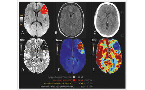
The presence of DWI lesions in patients with TIA can provide useful prognostic insights. As mentioned previously, many TIA patients have DWI abnormalities,13–15 and these changes are associated with more definitive TIA symptoms and vascular risk factors.13 The presence of DWI lesions in TIA patients predicts a higher risk of subsequent stroke,30 as well as other vascular events.31 In a study of 200 TIA patients who underwent brain imaging three or more days after the event, higher scores on the California and ABCD rules5,27 (which indicate higher short-term stroke risk) were associated with positive DWI lesions.32 In another study33 of 180 TIA patients who underwent imaging within 24 hours of symptom onset, 38 (21%) had DWI abnormalities, and among these subjects those patients who were symptomatic (i.e. those who had symptoms at the time of initial evaluation) were much more likely to go on to develop stroke during their hospitalisation compared with those who were asymptomatic. Finally, another study found that, compared with TIA patients with DWI abnormalities, patients who did not have abnormalities were much more likely to have a recurrent TIA event but were less likely to develop stroke.34
Current Clinical Recommendations for the Evaluation and Treatment of Transient Ischaemic Attack in the Acute Setting
In the acute setting, suggested clinical recommendations for the evaluation and treatment of patients with TIA include brain imaging, carotid imaging, cardiac imaging, antiplatelet therapy, anticoagulation therapy and statin therapy.35,36
Brain Imaging
Although TIAs are diagnosed clinically, the use of brain imaging – either computed tomography (CT) or magnetic resonance imaging (MRI) – is prudent to rule out other rare lesions such as subdural haematoma or brain tumour.36
Vascular (Carotid) and Cardiac Imaging
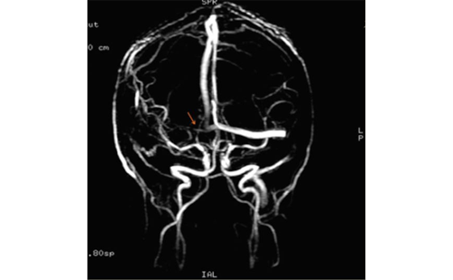
Because carotid stenosis is a major risk factor for stroke recurrence in stroke and TIA patients, carotid imaging (e.g. carotid Doppler) is commonly recommended.35 Also, because endarterectomy for patients with significant symptomatic carotid stenosis is more valuable when performed within two to four weeks of the event,37 it is recommended that carotid imaging be undertaken in a timely fashion.36 MR angiography (MRA) and/or CT angiography (CTA) are recommended if Doppler ultrasonography does not reveal reliable results. However, in cases of discordant results, conventional angiography remains the gold standard for the examination of cerebral vasculature. Finally, transthoracic or transesophageal echocardiogram can also be considered to evaluate for a cardioembolic source of TIA.
Antiplatelet and Anticoagulation Therapy
An antiplatelet agent, such as aspirin 75–325mg, clopidogrel or aspirin plus extended-release dipyridamole, is recommended for all patients with TIA.38 The most recent update of the AHA/ASA stroke prevention guidelines,39 which were published in May 2008, strengthened the recommendation that aspirin (50–325mg/d) monotherapy, the combination of aspirin and extended-release dipyridamole and clopidogrel monotherapy are all acceptable options for initial antiplatelet therapy (Class I, Level of Evidence A). On the basis of the results of the ESPRIT trial, these guidelines also strengthened the recommendation for combination therapy, specifically recommending the combination of aspirin and extended-release dipyridamole over aspirin alone (Class I, Level of Evidence B). No changes were made to the recommendation that clopidogrel may be considered over aspirin alone on the basis of direct-comparison trials (Class IIb, Level of Evidence B), or that the combination therapy of aspirin and clopidogrel is not routinely recommended for ischaemic stroke or TIA patients unless there is a specific indication (Class III). However, these recommendations were made before the release of the results of the PRoFESS (The Prevention Regimen for Effectively Avoiding Second Strokes) randomised trial, which compared the combination of aspirin and extended-release (ER) dipyridamole with clopidogrel. This large, well-executed trial found that the risk of recurrent stroke was almost identical in the two treatment arms (8.8% with combined therapy versus 9.0% with clopidogrel).40 For patients with persistent or paroxysmal atrial fibrillation who have had a cardioembolic TIA, long-term oral anticoagulation (i.e. warfarin) is also recommended. For these patients, a target international normalised ratio (INR) of 2.5 (range 2.0–3.0) is recommended. Aspirin is recommended for patients with contraindications to oral anticoagulation. There is no benefit to combining aspirin and warfarin therapy to prevent stroke.41
Statin Therapy
Lipid-lowering therapy can reduce the risk of stroke by 25%.42 On the basis of the SPARCL trial,43 which demonstrated a 16% reduction in stroke risk with atorvastatin treatment compared with placebo, intensive lipid-lowering statin therapy is recommended for all patients with atherosclerotic ischaemic stroke or TIA.
Carotid Endarterectomy
Carotid endarterectomy is of proven benefit for patients who have had recent (within two to four weeks) hemispheric, non-disabling carotid artery ischaemic events, and who have ipsilateral carotid artery stenosis of 70–99%.44 Endarterectomy may also be beneficial for symptomatic patients with retinal transient ischaemia.
Finally, other risk factors for recurrent cerebrovascular ischaemic events should be treated appropriately. This includes lowering blood pressure and blood cholesterol (with lifestyle modifications and/or drug therapy) in all patients with atherothrombotic TIA.36
The Need for Rapid and Cost-effective Evaluation of Transient Ischaemic Attack Cases in the Acute Setting
As has been discussed above, it is now well recognised that TIAs represent an important clinical opportunity to prevent the short-term risks of stroke and other cardiovascular events.45,46 Current clinical guidelines outline what diagnostic and treatment interventions need to take place;35,47 however, the place and timing of this clinical work-up is the subject of considerable debate.48,49 Currently, there are no definitive recommendations concerning the early evaluation and disposition (i.e. hospitalisation versus outpatient care) of TIA patients.50–52 Although the approach of admitting all TIA patients is recommended by some,53,54 there is no definitive study that demonstrates the benefits of this approach.
Given the absence of clear guidelines, there is considerable variation in current practice;55,56 published hospitalisation rates have varied from as low as 14%5 to more than 50%.57,58 Depending on the availability of diagnostic procedures and the clinical approach taken, it is possible for the TIA work-up to be completed in the ED, in a short-stay (<24 hours) observation unit, as an inpatient or as an outpatient.
Two recent observational studies conducted in Europe have demonstrated the benefit of early assessment and treatment of TIA patients in the outpatient setting. The Early Use of Existing Preventive Strategies for Stroke (EXPRESS) study was conducted in England and examined the effect of rapid outpatient assessment and treatment of TIA and minor stroke cases referred by primary care physicians.59 In the first phase of this quasi-experimental study, 310 patients were referred to an outpatient TIA clinic. However, the clinic recommended but did not initiate any new treatments, and so the median time from referral to first prescription was 20 days. In the second phase of the study, 281 patients were referred to the TIA clinic, which now initiated treatment immediately – the median time to first prescription was reduced to one day instead of 20 days. The 90-day risk of stroke was 80% lower during the second phase of the study (2.1%) compared with the first phase (10.3%). The second study, called SOS-TIA,60 was conducted between 2003 and 2005 in Paris, France, and evaluated the impact of a 24-hour outpatient TIA clinic. A cohort of 1,085 TIA patients were referred to the clinic, where they underwent rapid evaluation including neurological, arterial and cardiac imaging. For these cases the 90-day stroke risk was only 1.2% (95% confidence interval [CI] 0.7–2.1%), compared with the predicted risk of 6% calculated using the ABCD2 rule.60 These studies serve to illustrate the importance of completing the clinical evaluation in a timely manner, and also that this assessment can be achieved in an outpatient rather than a hospital environment.
Although hospitalisation may make sense for some TIA cases, especially those at high risk, it is clear that many TIA cases are at low risk and could be appropriately managed in an outpatient setting. Risk stratification using the ABCD2 score can help to identify which patients should be hospitalised. Patients with an ABCD2 score of ≥4 (i.e. moderate to high risk) stand to gain the most from urgent evaluation and treatment, which, in the US at least, can be reliably provided only in the inpatient or observation unit setting; in contrast, low-risk patients (i.e. ABCD2 score ≤3) could be appropriately evaluated on an outpatient basis.61 However, objective data on the effectiveness, safety, costs and cost-effectiveness of such a clinical strategy are needed before it can be formally recommended. ■
This work was supported by Boehringer Ingelheim GmbH. The authors had full control over content and did not receive editorial assistance from outside parties.


