Transient ischaemic attack (TIA) is common, with approximately 200,000–500,000 reported to medical attention in the US each year.1 The risk of TIA rises steeply with age, with the majority of all events occurring in people over 70 years of age.2 In contrast to major stroke, the incidence of TIA is not declining and an increase in overall rates is expected over the next two to four decades as a result of the ageing of the population.3 Doctors from a wide range of specialities (primary care, neurology, emergency medicine, geriatrics, ophthalmology) are likely to encounter patients with suspected TIA, among whom some will have confirmed TIA with a high risk of stroke or serious non-cerebrovascular pathology; obtaining an accurate differential diagnosis and estimating risk for individual patients is therefore important for many clinicians.
Over the last decade there have been considerable advances in the understanding of the pathophysiology, prognosis and treatment of TIA and stroke, leading to changes in the proposed definitions and approach to management. This article will discuss the definition of TIA and stroke, how to formulate a differential diagnosis in a patient with suspected TIA and how to predict risk in individuals with a confirmed TIA.
Definitions of Transient Ischaemic Attack and Stroke
Obtaining a differential diagnosis in a condition depends on its definition, and in the case of TIA this has been hotly debated in recent years. The previous distinction between TIA and stroke was established over 30 years ago,4,5 and used time-based criteria. TIA was defined as “an acute loss of focal brain or monocular function with symptoms lasting less than 24 hours, of presumed vascular cause”, while a stroke caused symptoms that lasted longer than 24 hours (or led to death). A new classification has been proposed that distinguishes between TIA and stroke on the basis of the presence or absence of brain infarction on imaging, regardless of symptom duration.6 It is argued that this ‘tissue-based’ distinction is more consistent with current knowledge of pathophysiology and prognosis.
One of the strengths of the old, time-based definition was that it carried a clear differential diagnosis that was clinically helpful when evaluating a patient with suspected TIA, presenting with transient and focal neurological symptoms. For the purposes of this article, I will therefore discuss the differential diagnosis in relation to the old time-based definition, without reference to imaging findings.
Differential Diagnosis
TIA is one of several causes of ‘transient focal neurological attacks’ (alternative causes are often termed ‘mimics’). There is no test to confirm a TIA and the gold standard method of diagnosis remains a thorough clinical assessment as soon as possible after the event by an experienced stroke physician. The advent of new imaging techniques, particularly diffusion-weighted (DWI) magnetic resonance imaging (MRI), has allowed the diagnosis to be made or excluded with more certainty in some patients.
Several tools have been developed to aid diagnosis in different clinical settings, but these have focused more on stroke than on TIA. The Recognition Of Stroke In the Emergency Room (ROSIER)7 tool was developed for use by paramedic and emergency department (ED) staff for the rapid distinction between stroke and mimics prior to referral for specialist assessment. A further, more complex scoring system8 has been developed for use at the bedside, again to distinguish between stroke and its mimics, in the hands of non-specialists.
More recently, the distinction between TIA and its mimics has been studied and tools have been proposed.9,10 Such tools are developed by studying a cohort of patients with suspected TIA, of whom some have an eventual TIA diagnosis and others have a mimic diagnosis; predictive features of each diagnosis are identified and combined in a scoring system. The drawback of such an approach is that TIA is a heterogeneous condition with a wide range of possible presentations so that only a very complex tool would have the necessary sensitivity, at the expense of its specificity. They therefore do not present an alternative to expert assessment, but may be useful in primary or emergency care for use by the non-specialist. In general, a diagnosis of TIA is supported by a sudden onset of definite, focal symptoms attributable to a specific vascular territory, while a diagnosis of a mimic is supported by other features, such as non-sudden onset, seizure activity, pre-syncope or syncope.
Some conditions are particularly frequently misdiagnosed as TIA (see Table 1), but features in the history are often helpful in distinguishing TIA from mimics (see Table 2).
Migraine with Aura
Typical migraine presenting with aura and headache, with or without nausea or vomiting, does not present a diagnostic challenge. However, sometimes a migraine aura can develop in an individual without previous migraine and without subsequent headache. In this situation, the slow intensification and then fading of symptoms over time, often with gradual spread from one domain to another (for instance vision to speech), is suggestive of migraine as opposed to TIA.11
Epilepsy
Partial seizures and post-ictal paralysis are often mistaken for TIA. Todd’s paresis is a focal neurological deficit that can follow up to 10% of seizures, most commonly grand mal seizures, and typically causes a unilateral motor weakness but can also cause diplopia or speech disturbance. The cause of Todd’s paresis is unknown, but ‘exhaustion’ of the primary motor cortex or inactivation of motor fibres by N-methyl-D-aspartate (NMDA) receptors have been postulated. Like a TIA, Todd’s paresis can last for several hours and differentiation depends mainly on establishing the presence of seizure activity at onset.12 Partial sensory seizures tend to cause positive symptoms such as tingling, and symptoms ‘march’ across a hand or foot and up the limb in around a minute, and may eventually be accompanied by focal motor seizures or secondary generalisation.
Intracranial Structural Lesions
Occasionally, but importantly, intracranial structural lesions such as subdural haematoma or tumour may cause transient neurological deficit, although the mechanism is unclear. Compression of an intracranial artery, sudden expansion caused by in situ haemorrhage or oedema, or focal seizures are all possible mechanisms for transient symptoms in otherwise ‘chronic’ conditions. Additional features in the history such as headache or nausea, systemic symptoms and stuttering or gradual onset are suggestive of non-TIA diagnoses. Imaging with non-contrast computed tomography (CT) lacks sensitivity for space-occupying lesions, and MRI is superior.
Transient Global Amnesia
Transient global amnesia (TGA) presents with a characteristic syndrome of sudden-onset, severe, anterograde amnesia, often accompanied by retrograde amnesia. Attacks last several hours, during which the patient appears bewildered and typically repetitively asks the same or related questions, and after which the patient makes a full recovery but has no memory of the attack.13,14 The aetiology of TGA is unclear, but mechanisms including temporary metabolic abnormality in the medial temporal lobes, venous hypertension, focal ischaemia and seizure activity have been proposed.15
Vestibular Dysfunction
The acute onset of vertigo is common and presents a diagnostic challenge, especially in elderly patients with pre-existing risk factors for vascular disease. ‘True vertigo’, or the false illusion of movement of the patient relative to the surroundings, should be distinguished from other, less specific symptoms of ‘unsteadiness’ or ‘light-headedness’.
The differential diagnosis of true vertigo is divided into peripheral causes, including benign positional vertigo, vestibular neuritis and Meniere’s disease, and central causes, one of which is TIA affecting the brainstem. Generally, peripheral causes of vertigo are more common than central causes. Features in the history suggestive of TIA mimics include recurrent stereotypical episodes, presence of provoking factors (head movement), other symptoms of middle ear disease (tinnitus, hearing loss) and absence of other focal brainstem symptoms (visual or speech disturbance, weakness or numbness). Features on examination that are thought to identify a central cause of vertigo include nystagmus that is not suppressed by visual fixation, a normal head thrust test and other features of posterior circulation ischaemia including dysphagia, dysarthria, limb or facial weakness, gaze palsies or upgoing plantar responses.
Delirium or Toxic Confusional State
Delirium, toxic confusional state, metabolic encephalopathy or acute confusional state are terms that are used interchangeably and often loosely to describe a syndrome of acutely disordered cognition, sometimes associated with a reduced level of consciousness and abnormal attention. The syndrome is very common, especially in the elderly and in patients with dementia, and presentations vary widely in terms of both speed of onset and severity.16 The differential diagnosis is broad and includes almost any medical condition, but the most common causes are sepsis, adverse drug reaction and metabolic derangement.17
Delirium can be mistaken for a TIA in cases that are mild, when the predominant feature is interpreted as language disorder as opposed to confusion and when important clinical details are unclear, such as when a witness account is unavailable, the patient has cognitive impairment or there is a long delay between the event and assessment. Reliable differentiation between TIA and delirium is important because each carries a potentially poor prognosis – although for very different reasons – and the treatments are dissimilar. Features suggestive of delirium include the presence of a causative factor such as urinary tract sepsis, an inability of the patient to clearly remember the event, fluctuating disturbance in attention and consciousness and the absence of a clearly sudden onset.
Syncope and Pre-syncope
Syncope is the abrupt loss of consciousness associated with the loss of postural tone, usually followed by a rapid and complete recovery; pre-syncope is a premonitory sensation of syncope. Although the time course of syncope is consistent with TIA, the lack of focal neurological disturbance is definitely not and the diagnosis should therefore only be made with considerable caution. Diagnostic confusion can sometimes be caused by TIA of the brainstem causing transient quadriparesis presenting with a sudden loss of postural tone, but loss of consciousness is not a feature. Infrequently, embolus to the tip of the basilar artery can present with sudden-onset coma, but this is virtually never a transient, self-limiting condition and other signs of brainstem dysfunction are always present and obvious.18
Isolated Transient Focal Neurological Disturbance of Uncertain Significance
In a significant proportion of patients referred with suspected TIA, no clear diagnosis of either a cerebrovascular event or a mimic can be reached even after thorough clinical assessment and investigation. These are often presentations with isolated focal neurological disturbance with sudden onset and gradual recovery, over seconds to minutes. Several distinct syndromes can be recognised: for instance, isolated and transient vertigo with no other features to suggest a central or peripheral cause, isolated slurred speech or isolated hemisensory loss. Currently, little is known about the cause or significance of these syndromes and rigorous prospective data are required describing associated risk factors, imaging findings and prognosis. However, the outcome is often good and, unlike TIA, these are not associated with a high early risk of recurrent stroke.
Risk Prediction After Transient Ischaemic Attack
The importance of TIA lies in the subsequent risk of stroke and other vascular events. It is well recognised that major stroke is often preceded by a TIA although the symptoms may have neither alarmed the patient at the time nor have been reported to medical attention. Early cohort studies indicated that TIA was a relatively benign condition with a low subsequent risk of stroke (approximately 1–2% at one week and 2–4% at one month) and other vascular events.19 However, recent research using more reliable methodology based on prospective data and recruiting patients in the acute phase has shown that these were underestimates, and the risk of stroke is particularly high in the first few hours and days after TIA, with estimates as high as 10–15% at one week in some studies.20
This high early risk following TIA poses a dilemma to clinicians and healthcare services because, although the majority of patients will suffer transient symptoms only with no acute sequelae, an important minority will go on to suffer a potentially disabling stroke that could be preventable with appropriate treatment. Prognostic scores have therefore been developed as a means of identifying high- (and low-) risk individuals and thereby aiding effective triage from emergency departments and primary care to specialist services, as well as informing public education and targeting secondary prevention treatment.
Methods of Risk Prediction
Clinical Features – The ABCD System
Five factors were found to be independently associated with high risk of stroke at three months in an emergency department cohort of TIA patients.21 These included age >60 years, symptom duration >10 minutes, motor weakness, speech impairment and diabetes. These and other factors identified as being associated with early stroke risk in two other studies22,23 were used to derive the ABCD score to predict stroke risk within seven days after TIA.24 The score was then validated in three further cohorts of TIA patients.
The ABCD score is based on four clinical features and is out of a total of six (see Table 3). It was found to be highly predictive of stroke at seven days after TIA with area under the receiver operator curve (ROC) statistics of 0.85 (0.78–0.91), 0.91 (0.86–0.95) and 0.80 (0.72–0.89) for each of the validation cohorts (see Figure 1). Almost all strokes occurred among patients scoring over 3, with the rates of stroke rising steeply with increasing score above 4.
Although diabetes was found to be predictive of early stroke in the ABCD score, it was not included. However, the ABCD scoring system was further validated in cohorts of patients recruited in California, US, and refined with the subsequent addition of one point for diabetes to make the ABCD2 score out of seven (see Table 3 and Figure 2).25
The ABCD system was developed for use by primary care and emergency care physicians prior to specialist evaluation and is based on clinical information that is readily available following a brief patient assessment. However, prediction scores in general require validation by independent users to demonstrate generalisability prior to their wider use in clinical practice.26
In terms of its clinical and statistical performance, both the ABCD and ABCD2 scores have been further validated in independent cohorts since publication in 2005 and 2007, respectively. In a systematic review, 20 cohorts were identified reporting the performance of the ABCD and ABCD2 scores in 9,808 subjects with 456 strokes at seven days.27 Pooled estimates of the area under the curve (AUC) for the ABCD and ABCD2 scores were 0.72 (0.67–0.77) and 0.72 (0.63–0.80), respectively, for seven-day stroke risk.
Predictive power was greater in two additional cohorts that included patients with both suspected and confirmed TIA compared with cohorts of confirmed TIA patients only. These findings suggest that the ABCD system works both diagnostically, detecting ‘true’ TIA patients with a vascular cause, and prognostically, identifying those ‘true’ TIA patients at highest risk.
Both the usefulness and performance of the ABCD2 score have led to its widespread use in clinical practice and incorporation into guidelines.28,29
Vascular Territory
The early risk of stroke after TIA also depends on the vascular territory of the event. Monocular events (amaurosis fugax) have consistently been found to be associated with a lower risk of stroke in comparison with cerebral events.30
Vertebrobasilar (VB) territory TIAs were previously thought to have a better prognosis than carotid territory events and are sometimes managed less aggressively. However, a systematic review of 37 published cohort studies and five unpublished studies reporting the risk of stroke after a TIA or minor stroke by territory of presenting event found no major differences in prognosis between VB events and carotid events, but a higher very early risk of stroke.31
Aetiology
Common causes of TIA include a cardiac source of embolism as occurs in atrial fibrillation, thrombus formation and embolism from an unstable plaque in the internal carotid artery (so-called large atherosclerosis [LAA]), and thrombosis of a deep penetrating cerebral artery causing a lacunar infarction.
It is likely that prognosis in the acute phase after brain ischaemia depends on the underlying pathology, but this question has been studied more in stroke than in TIA.32 In one prospective cohort of 388 patients with TIA, the mechanism of TIA was studied in relation to stroke outcome at three months. Stroke risk was highest among those with LAA, lowest in those with lacunar or small-vessel disease and intermediate in those with cardio-embolic or undetermined cause of TIA.33 In another study of 343 consecutive TIA patients who were admitted to a stroke unit, a similar relationship between aetiological mechanism and stroke recurrence at three months was also found.34
Brain Imaging
Some early studies suggested that the presence of infarction on CT in patients with TIA predicts an increased risk of subsequent stroke, although others have failed to confirm this finding. However, interpretation of these results is difficult because the delay between the event and scanning was variable and acute and old infarcts were not reliably distinguished. However, more recent studies that have performed CT soon after TIA and reliably distinguished new and old infarction have shown that the presence of new infarction does carry a higher risk of early stroke.35,36
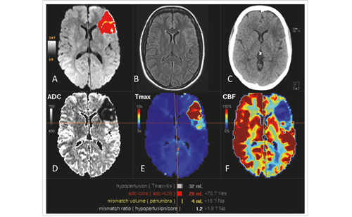
There is considerable interest in the role of MRI, and DWI in particular, in predicting stroke risk after TIA. DWI is a radiological technique that measures the diffusion of water molecules in different tissues in the body and is very sensitive to the early phase of cerebral infarction (see Figure 3). It has therefore been proposed that DWI may identify TIA patients with an active ‘vascular process’ such as a source of emboli or LAA disease, signifying a high risk of further thromboembolism and thus recurrent stroke.
This hypothesis has been tested in a number of studies, although these have been hampered by small size and sometimes retrospective design. For instance, Prabhakaran et al. described a retrospective cohort study of 146 patients with TIA, of whom 37 (25%) had abnormalities on DWI; the presence of these abnormalities was found to be independently associated with a higher risk of in-hospital recurrent TIA or stroke (odds ratio 11.2; p<0.01).37 Purroy et al. described a cohort of 83 consecutive TIA patients attending an ED who were scanned with DWI, with abnormalities identified in 27. The combination of DWI abnormalities and symptoms lasting over an hour was found to be predictive of stroke (hazard ratio [HR] 5.0, 1.4–18.3; p=0.015) or a combined end-point of stroke and other vascular events (HR 3.8, 1.1–13.0; p=0.029).38 Coutts et al. described a cohort of 180 patients with TIA or minor ischaemic stroke presenting to an ED, all of whom had DWI within 12 hours. Stroke risk at 90 days was 18.2% in the 99 patients with DWI abnormalities compared with 2.5% in those without (p<0.001).39
Although the association between DWI and early risk of stroke is clear, it is uncertain what additional prognostic information over and above clinical scores and aetiology it provides. Indeed, focal motor weakness, speech disturbance and symptoms lasting longer than one hour are all associated with DWI lesions in patients with TIA,40 while DWI abnormalities are associated with large-vessel disease. Larger studies are therefore needed to address the interplay between the prognostic information available from clinical features and imaging. ■


