Parkinson’s disease (PD) is the second most common neurodegenerative disorders worldwide1 and is a chronic and progressive condition,2,3 which affects 1 % of the older population (>60 years of age).4 Over the years, major research has been focused on the motor impairments in PD, which has contributed to the development of symptomatic treatments for these patients. Evidence also demonstrates that PD patients have sensory impairments and chronic pain, which reduce their quality of life dramatically. Approximately 43 % of PD patients suffer from pain in general. There is no cure yet available and the number of affected PD patients is increasing, which highlights an existing socioeconomic burden.1
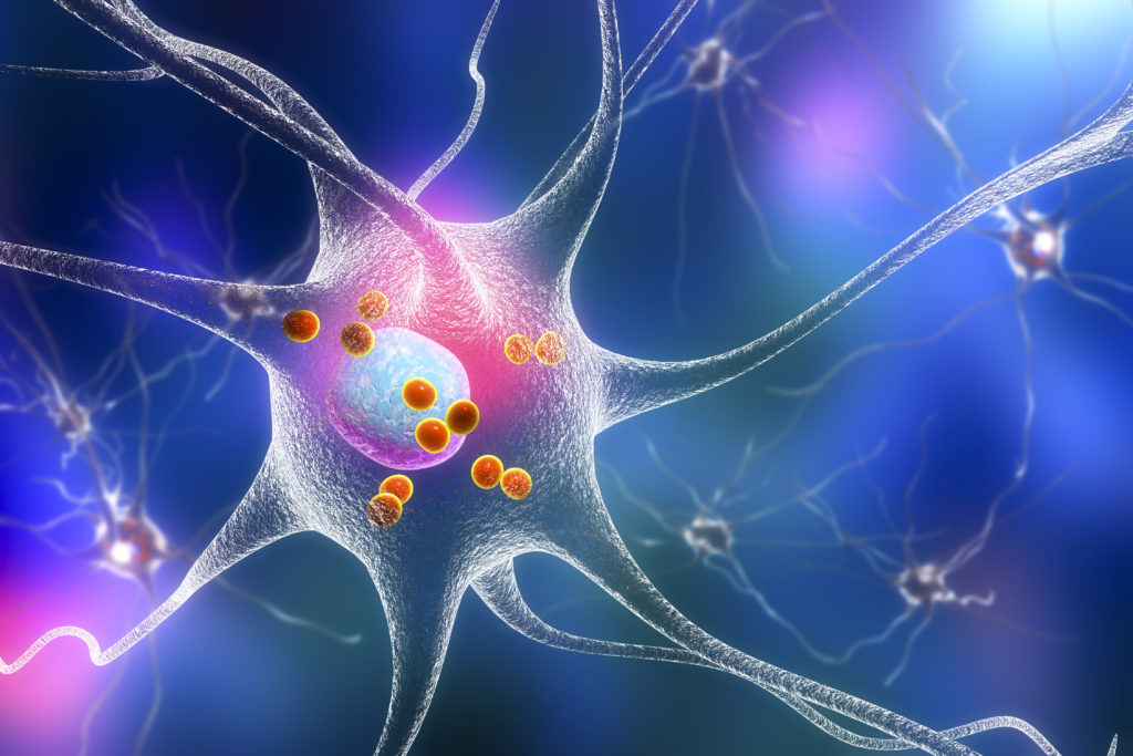
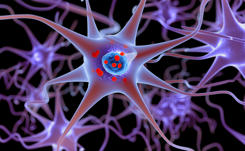
PD is characterized as a primary neurodegenerative disorder5 due to a dysfunction that might occur in the basal ganglia network following degeneration of dopaminergic pigmented neurons in the substantia nigra pars compacta, which gives rise to a significantly reduced dopaminergic deficit in striatum, especially in the putamen part.4 Several studies suggest that an abnormal basal ganglia function in PD can modulate pain directly by increasing or reducing the spread of nociceptive signals or indirectly by changes in affective and cognitive processes related to pain perception.6,7 Over the past decade, it has been gradually revealed that sensory perception in PD patients has been altered8–10 and the putative dysfunction in the basal ganglia is thought to lead to pain and sensory impairment in these patients.
The impairment of the sensory system is a less-explored area in PD. There are only few studies available applying sensory tests in PD patients with conflicting results.11,12 One study showed that PD patients had a lower threshold in both the cold pressure test (CPT) and pressure pain threshold (PPT) test13 and another study showed increased hypersensitivity to cold pain threshold (CPT) in PD patients compared with healthy subjects.6 However, there is a still uncertainty about this alteration in sensory perception as some studies have not been able to demonstrate any significant change.10 It remains to be determined whether PD patients suffer from sensory disturbances in terms of hyposensitivity or hypersensitivity in response to application of a painful or nonpainful stimulus and to what extent. In addition, it is still not clear whether sensory impairment is different in PD patients who suffer from a longterm spontaneous chronic pain, who also often have a poor quality of life, in comparison with those who do not have pain on a daily basis for a long term. It is also not known whether different PD medications have a possible effect on the perception of pain and peripheral sensory input. The primary aim of this study was to investigate whether PD patients have an altered sensory perception that might lead to an increased pain perception in response to noxious and non-noxious stimuli. We also investigated whether different medications taken by the PD patients can have an effect on responsiveness to the applied sensory tests. We proposed that some alterations in pain and sensory perception will be detected in mechanical and thermal perception in PD patients compared with healthy subjects and that drugs that might affect the sensory alterations are most likely levodopa preparation and dopamine agonists.
Materials and Methods
Subjects and Study Design
Twelve PD patients (nine males, three females with the mean age ± standard deviation [SD], 68.67±5.5 years) were recruited through arrangements with the chief physician, Ali Karshenas, Department of Neurology, Aalborg University Hospital, Denmark. Patients were of Caucasian descent, either with PD-related pain or without pain.
Patients >60 years who had been diagnosed ≤5 years with no central or peripheral disorders were included. Patients with pain (except the pain related to PD—defined as pain experienced in PD patients due to no other reason than PD based on the European Parkinson’s Disease Association), on painkillers, psychical disorders such as schizophrenia and dementia, mental retardation, memory impairment, or a Mini Mental State Examination (MMSE) score <24 (see description below) and other disorders of the central nervous system (CNS) or polyneuropathy were excluded from the study. The patients did not take alcohol, caffeinated drinks, or smoke 24 hours before the experiments. In addition, 12 bestmatched healthy volunteers (eight males, four females with the mean age ± SD, 67.5±5.39 years) of Caucasian descent were included as controls. The healthy volunteers were recruited through public notices posted at Aalborg University Hospital, Denmark, and social media. Having pain or taking any painkillers were among the exclusion criteria for healthy volunteers. The experiments took place in the outpatient clinic, Department of Neurology, Aalborg University Hospital, Denmark, and the participants attended one session, which lasted for about 1 hour.
PD patients (with pain and without pain) and healthy subjects were all screened and written informed consent was obtained from all participants before the start of the experiments. The state of patients’ mood (e.g. depression) was not determined through standard questionnaires or test; however, the medications taken by patients were recorded to summarize all medications taken by the patients (e.g. antidepressants). PD medications included: Sifrol®, Sinemet®, Madopar® Quick®, Mylan-Selegiline®, Stalevo®, Exelon®, Requip depot®, Madopar®, Eldepryl®, Requip®, and Azilect®. Other medications were mainly associated with cardiovascular matters and included: Ramipril, Norvasc®, Nifedipine, Corodil®, Cordarone®, Metoprolol, Asasantin®, Simvastatin, Hjertemagnyl®, Marevan®, Centyl®, Diural®, Ancozan®, and Furix®. Some patients were also on the following medications: Tolterodin, Metformin, Kaleorid®, Folimet®, Methotrexate, Euthyrox®, Eltroxin®, Zolpidem, and Gabapentin.
The Ethical Committee of the Region Nordjylland Denmark approved the study protocol (N-20130073) and the experiments were performed in accordance with the Declaration of Helsinki.
Measurements
Evaluation of Cognitive Function MMSE and clock face test were used as screening instruments to assess the cognitive function and investigate cognitive disturbances to ensure that the participants understood the visual analog scale (VAS) (see description below) and the quantitative sensory tests. The MMSE test is a brief standardized method to assess the mental status (score 0–30). Participants with a score <24 were excluded. The clock face test was approved without errors or minimal abnormality in the location of the hands and numbers.
Evaluation of Pain Perception and Daily Life Activities
The McGill pain questionnaire was used for PD patients with pain. This questionnaire was used to get an overview over the location of chronic pain in the PD patients with pain. In the experimental session, the stimuli were given on both forearms (centrally between elbow and wrist) in a supine position, on the dominant hand (few centimetres over the wrist), and on the lumbar part of the back (around vertebrae lumbales LIII on the left side) (see Figure 1). To assess the sensitivity and pain threshold to nonpainful and painful stimuli, a VAS scale was applied after all the mechanical tests and during/after the thermal test. The VAS scale was used in the way that the participants indicated a number from 0 to 10; in which 0 was “no pain” and 10 was the “worst possible pain.” The World Health Organization (WHO) performance status was used to provide an overview of PD patients’ general well-being and activities of daily life.
Dynamic Mechanical Allodynia in Response to Manual Light Brush
Subjects were seated with both forearms rested on the table in a supine position. A standardized brush (SENSELab Brush-05, Somedic, Hörby, Sweden) was used. They were asked to keep their eyes closed during the test. The handheld brush was moved across the skin for five times with a speed of 1–2 cm/second and with an angle of approximately 45˚. The brush was applied alternately from right to left and each stroke was performed from distal to proximal direction (see Figure 1). Each stroke was 5 cm in length over the skin and the test was performed three times for each forearm (interstimulus interval 3–5 second), after which the subjects were asked to rate the pain intensity on a VAS scale (a total three times for each forearm). This test was also performed in the lumbar part (see Figure 1), on the left side, applying the same procedure, while the subjects rested on their stomach on a couch.
Mechanical Pain Sensitivity in Response to Pinprick Stimulation
Seven weight-calibrated pinprick stimulators were applied one by one with one prick at a time in a random order. The handheld pinprick stimulator (Pinprick Stimulator Set, MRC System GmbH, Heidelberg, Germany) consists of seven weighted flat-tipped needles (8mN, 16mN, 32mN, 64mN, 128mN, 256mN, and 512mN) with a contact area of 0.2mm2. This test was carried out in a dotted line in both forearms rested at supine position on the table, while the subjects were seated. The subjects were asked to keep their eyes closed during the stimulations. Each stimulation was repeated three times (interstimulus interval 2–4 seconds) with an angle of approximately 90˚ for each forearm. Stimulators were applied alternately from right to left forearms (see Figure 1) and only one forearm at a time was stimulated with seven weighted needles. During the pinprick test, the subjects were asked to rate their pain intensity to each stimulus on a VAS scale. The pinprick stimulators were also applied on the back (see Figure 1), with the same procedure as the forearm.
Pressure Pain Threshold
A handheld pressure algometer (Somedic, Hörby, Sweden) was applied, which consisted of a gun-shape holder connected to a probe with a circular sensor tip covered with a rubber material with an area on 1 cm2. The pressure rate was set for applied force of 30 kPa/s. The digital display on the pressure algometer showed the pressure (kPa). The subjects pressed a stop key, the first time they felt that the pressure turned to pain. This froze the number corresponding to the pressure on the display, which was noted as the PPT (KPa). The PPT was assessed on both forearms and the lumbar part (see Figure 1). For this test, the subjects were seated with their forearms resting on the desk in a supine position and instructed to use a handheld button to stop the delivered pressure when it reached the point that they felt it uncomfortable. At this point, they were also asked to rate their level of unpleasantness on a VAS scale. This test was performed three times with an angle of approximately 90˚ on each forearm with a resting period of 60 seconds in between each stimulus. The pressure algometer was used with the same procedure on the lumbar part.
Cold Pressor Test
First, the dominant hand was immersed in a bucket of water (30˚C) for 2 minutes in order to provide fairly similar hand temperature. Subsequently, the CPT was carried out and the hand was immersed in a bucket of ice water (5˚C) for maximum of 2 minutes. The hand was immersed in ice water a few centimetres above the wrist (see Figure 1). The subjects were instructed to withdraw their hand when it was uncomfortable or painful. Pain intensity was rated on a VAS scale during and after the experiment. Three measurements were made at 30s, 60s, and at the termination of the test (tolerance time). The subjects were informed that there was a limited maximum possible tolerance time at 2 minutes. Therefore, if subjects removed their hand before this cut-off, the tolerance time was noted; otherwise, 2 minutes was noted for the tolerance time of a subject who kept the hand until to the end. The final pain intensity was also measured on a VAS scale right after the hand removal.
The CPT was then followed by a PPT test. The dominate hand was tested by the pressure algometer before and after the CPT and PPT were noted. The subjects were also asked to rate their unpleasantness on a VAS scale.
Statistical Analysis
All data were first analyzed for normality using the Shapiro-Wilk test. Whenever the data were distributed normally, parametric tests were applied for statistical comparison. Otherwise, nonparametric tests were applied.
An unpaired nonparametric test (Mann-Whitney U test) was used to compare pain intensity (e.g. pain tolerance and pain threshold) in sensory tests. In order to evaluate the most sensitive area in PD patients an unpaired nonparametric test (Kruskal Wallis U test) was used. Furthermore, a paired nonparametric test (Friedman) was applied to test whether there was an exponential curve in the assessment of pain intensity among the participants. Finally, a multiple linear regression analysis was applied to test any correlation in the pinprick test for each stimulator in each area in both groups.
The results are presented as the median and interquartile range (IQR, 25–75th) and the significance level was defined as p<0.05. All data were organized in Excel 2013 and all statistical calculations were performed in SPSS version 18.0 (IBM, Hong Kong) and graphs were created in SigmaPlot 12.0. (Software Inc., Germany).
Result
Subjects
All participants completed the study with no safety concerns or complaints. There was no significant sex (p=0.660) and age (p=0.311) difference between PD patients and healthy controls. There was no statistically significant difference between groups in MMSE score (p=0.060) either.
Five PD patients (41.7 %) had chronic pain and completed the McGill Pain Questionnaire (see Figure 2). All patients had chronic pain in the upper part of the body and 60 % had chronic pain in the lower part. The performance status showed that healthy subjects had normal function (WHO; 0) and the PD patients had a WHO status between 0 and 2, which means that 58.33 % had decreased function in the daily activities. All patients were on the PD medications (dopamine agonists and/or levodopa preparation) and 33.33 % received only one drug and 66.66 % had >1. The two most frequently taken medications were Sifrol® (33.33 %) and Sinemet® (66.66 %).
Dynamic Mechanical Allodynia
The results from brush test showed that 58.33 % of the PD patients suffered from dynamic mechanical allodynia (DMA) (allodynic area: right-, left forearm, and lower back). The median of both groups were calculated for all three locations (right-, left forearm, and lower back) and Mann-Whitney test showed that the PD patients were more sensitive to brush test than the healthy subjects (Pright forearm =0.021, Pleft forearm =0.025, and Plower back=0.002). The test revealed that PD patients were most sensitive in lower back.
Static Mechanical Hyperalgesia
An average for all seven pinprick stimuli was calculated from three areas (right-, left forearm, and lower back) in each subject and the Mann-Whitney test revealed that there was a statistically significant difference between healthy subjects and PD patients by stimulation of the lower back (p<0.001). There was no difference between the groups by pinprick stimulation of the right (p=0.769) and left forearm (p=0.838). The average of each stimulus was calculated for all participants. Mann-Whitney test revealed that PD patients had rated their pain intensity higher than healthy subjects. The perception of pain was significantly increased in PD patients (p for all seven stimulators: <0.001). The three curves showed a clear difference between healthy subjects and PD patients (see Figure 3) following stimulation of right forearm, left forearm, and lower back. Kruskal Wallis test revealed no difference between the two groups in relation to the area that were the most sensitive. The test was also used to calculate the median for all seven stimuli in both groups, which revealed a difference between these two groups, confirming the existence of hyperalgesia in the PD patients (hyperalgesic areas: right-, left forearm, and lower back). Friedman test made it also clear that the higher the stimulus, the higher was the pain intensity (p<0.001). A multiple linear regression analysis did not show a significant result.
Conditioned Pain Modulation
Pressure Pain Threshold
A Mann-Whitney test showed that there was a statistically significant difference in PPT values between PD patients and healthy subjects before (p=0.011) and after (p=0.050) the CPT test (see Figure 4). There was no significant difference in the nondominant hand (p=0.065) or lower back (p=0.106).
Tolerance Time—Cold Pressor Test
All healthy subjects and nine PD patients completed 30 seconds or more in the cold water test. The test showed a difference in pain intensity between these two groups, but the difference was not significant (p=0.183). Only nine healthy subjects and five PD patients completed the test by 60 seconds or more. There was no statistically significant difference between the rated pain intensity in healthy subjects and PD patients (p=0.402). The median of pain on the VAS scale was 6.00 (5.25 to 8.00) and the results showed that the pain intensity was rated higher in PD patients compared with healthy subjects, but not significantly higher (p=0.078). The Mann- Whitney test revealed a significant difference in tolerance time between healthy subjects and PD patients (p=0.016) (see Figure 5). Healthy subjects had a higher tolerance time compared with the PD patients (85.42 versus 52.17 seconds) (see Figure 5). When performing the cold water test it was observed that the PD patients started to shake significantly more than they did before the test. The 30˚ water did not affect the PD patients.
Discussion
The current study investigated the sensory characteristics in PD patients compared with the healthy subjects. There are only few studies available on sensory tests in PD patients with conflicting results.11,12 Our results revealed that PD patients suffered from allodynia to brush and hyperalgesia to prick stimulation in the back and in both forearms. PPT test revealed that the PD patients had lower threshold in the dominant hand both before and after the CPT, but were not different in the nondominant hand and the back. PD patients also had shorter tolerance time in the CPT test. There was no association between sensory impairment and PD medication. It should be highlighted that the present study’s population is limited; a larger population is required to reveal whether the obtained results and conclusions would follow similarities in deviated sensory responses in PD patients.
Spontaneous Pain in Parkinson’s Disease
In the current study, five of the PD patients had PD related pain (41.7 % of the patients had chronic pain), which is in accordance with the results of Chaudhuri et al.,14 who found that approximately 40.0–45.9 % of PD patients suffer from Parkinson-related pain. Furthermore, the PD patients describe their symptoms and marked their pain area on the McGill Pain questionnaire, which is in accordance with the symptoms found by Ford et al.5 and Fil et al.15 One study16 has investigated the association between pain and motor complications in PD patients with and without pain and shown a significant association of pain with motor problems. This finding suggests that in PD pain may occur secondary to motor complications.
Allodynia and Hyperalgesia
We demonstrated here that PD patients had an altered perception in response to the light touch and had increased pain intensity in response to an already painful stimulus compared with healthy subjects. Our results showed that PD patients without pain had the highest pain intensity to light touch in comparison with PD patients with pain. This might indicate that the patients without pain were more sensitive to a nonpainful stimulus than the PD patients with pain, but both were allodynic in comparison with healthy subjects. Based on our knowledge, allodynia has not been tested in PD patients before. Our results showed that 58.33 % of PD patients had discomfort to the nonpainful stimuli by brush. PD patients with pain rated the pain intensity higher than PD patients without pain to a painful stimulus. The stimulus-dependent-response in PD patients showed increased pain intensity to painful stimuli and that the PD patients felt higher pain by larger stimulation compared with healthy subjects. The study also investigated the most sensitive area to pinprick test, the lower back was found to be the most sensitive region, followed by the right forearm, and the lowest pain intensity was in left forearm. However, the result did not show a significant difference between the test areas in the right and left forearms. We could not find similar studies and there would be no point for comparison.
It is not well known that what causes the perception of allodynia and hyperalgesia in PD patients. However, animal studies have shed light on some possible mechanisms that might be involved in development of allodynia in PD. Wisam Dieb et al.17 investigated DMA in a rat model of PD. The rats received an injection of 6-hydroxy dopamine bilaterally to produce a lesion in the nigrostriatal dopaminergic pathways. This study showed significant DMA in the orofacial area, in response to tactile stimulus and the rats received a dopamine 2-receptor agonist (Bromocriptine), a PD medication; the DMA was dramatically reversed compared with control rats that were treated with saline. This study demonstrated that a lesion in the nigrostriatal pathways could result in DMA. Possibly the neuronal loss of dopaminergic neurons results in an abnormal perception of the tactile and nociceptive information or result in central sensitization and presence of allodynia in PD.
Previous studies have shown that CNS disorders can cause altered perception to touch.18 Brush stimulation activates primary sensory neurons encoding signals for low intensity (Aβ-fibers), which under normal conditions should be perceived as sensation of touch.19 Stimulation with pinprick activates Aδ- and C-fibers (encoding for high intensity) and normally should be perceived as pain. However, under pathologic conditions, central sensitization might occur, which is defined as increased response to e.g. light touch.19,20 Our observation suggests that the PD patients might have a state of central sensitization, which leads to increased synaptic ascending transmission and a decrease in the descending inhibition.19,21 Normally, the Aβ-fibers become activated by touch to a light stimulus, such as brushing, but when allodynia occurs it is proposed that the signals from myelinated Aβ-fibers intersect to unmyelinated C-fibers, which causes pain in response to a nonpainful stimulus.19 It is speculated that the PD patients were affected on both pathways and that the interaction between afferent and efferent neurons in the spinal cord has an impact on central sensitization, which possibly reflects on responsiveness of PD patients to painful stimulus.22
This study supports the notion that the PD patients have altered pain perception and this novel observation might contribute to future investigations and extend the literature of allodynic and hyperalgesic conditions in PD patients. In the longer term, increased knowledge on impaired sensory function in PD may lead to a better diagnostic stratification of PD patients, and development of newer medications to help PD patients overcoming sensory disturbances along with motor dysfunction.
Conditioned Pain Modulation—Function of Descending Inhibitory Pain Pathways
PD patients rated higher pain intensity than healthy subjects in response to cold stimulation by immersion of hand in ice water, however, the difference between these two groups were not significant. The drop out and even numbers per group for comparison might have caused insufficient power for statistical analysis. There was however a significant difference in tolerance time between healthy subjects and PD patients, where PD patients had shorter tolerance time than healthy subjects. The result revealed a difference between PD patients with pain that withdrew their hand several minutes before PD patients without pain. The PD patients were more sensitive than healthy subjects, however, a difference between the PD patients with pain and without pain was not observed. The PPT is believed to test the deep pain sensitivity transmitted by Aδ- and C-fibers.23 Our results indicate that these nociceptive fibers were activated in PD patients and in healthy subjects, but the perception of pain occurred faster in PD patients. The PD patients complained of discomfort the first few seconds, after which the pain was initiated. This indicated that the activation of Aδ-fibers were initiated in the beginning of the test and furthermore an activation of C-fibers occurred, when the PD patients felt an uncomfortable pain by performing the CPT.23 The CPT test, however, showed a significant difference between PD patients and healthy subjects, where the PD patients had higher PPT values before the cold test compared with PPTs after the test. After the CPT, PD patients were more sensitive, which might be due to central sensitization present in these patients. There is no similar study that has investigated the PPT before and after the CPT in PD patients.
Vela et al.13 investigated the PPT in PD patients with and without administration of PD medications in comparison with healthy subjects groups and found a significant difference in all four investigated areas (frontal bone, C5-C6 joint, the second metacarpal, and the tibialis anterior muscle). We did not find a significant difference in PPT values between the right and left forearm in PD patients and healthy subjects, but found a tendency that the PD patients had lower pressure in the lower back and the nondominant hand. Due to the lack of data available for other studies on CPT in PD patients, comparison of results obtained here with other similar studies is not possible.
This area needs future investigation to clarify the underlying mechanism, such as the functionality of the descending inhibitory pain pathways and the tolerance to cold water stimulation. Our findings support that the PD patients have altered perception in deep pain sensitivity.
In summary, this pilot study confirmed the existence of sensory and pain disturbances in PD patients in comparison with healthy subjects that might be due to the loss of dopaminergic neurons, which might have an impact on the nigrostriatal pathways leading to an abnormal basal ganglia function and disturbances in afferent and efferent pathways of pain perception. Changes in pain perception might also be due to peripheral pain receptors, structures such as the periaqueductal gray matter, and nondopaminergic neurotransmitter systems. Larger studies are needed to confirm the results obtained in the present study.


