Parkinson’s disease (PD) is the second most common neurodegenerative disease affecting 1 % of the population above the age 65.1 The incidence of PD is expected to increase dramatically worldwide with increased life expectancy.2 PD is manifested by the combination of primary motor disability (bradykinesia, rigidity, tremor, and gait impairment) as well as a spectrum of non-motor symptoms including cognitive, mood, autonomic, and sleep dysfunction.3 The cause of cells degeneration in PD remains unknown. While there is a widespread distribution of neuropathologic changes in the brain responsible for the spectrum of the motor and non-motor manifestations of PD, degeneration of dopamine-producing cells in the substantia nigra pars compacta (SNc) is central to the primary motor signs of the disease.3
Presently, treatment of PD is limited to symptomatic therapy, aimed at mitigating the dopamine deficiency. While treatment can be very effective early in the stages of the disease, it does not impact the progression of the degenerative process. A neuroprotective agent that could slow the progression of the disease has been the holy grail of research in PD. Several agents have been tried, but none have been shown to be effective. The reasons for this failure are unclear, but are likely to stem from the complexity of pathogenesis and the inability to attack the disease process sufficiently early in its course. Another major limitation in clinical trials currently is lack of reliable, objective biomarkers of disease progression. Until such biomarkers are available, the term that should be used in the discussion of the clinical trials outcomes is disease modification, which implies a positive impact on the clinical course of the disease without specifically linking it to pathogenesis. Ultimately, the choice of an agent for neuroprotection in PD should be based on the solid understanding of etiology.
Parkinson’s Disease Etiology
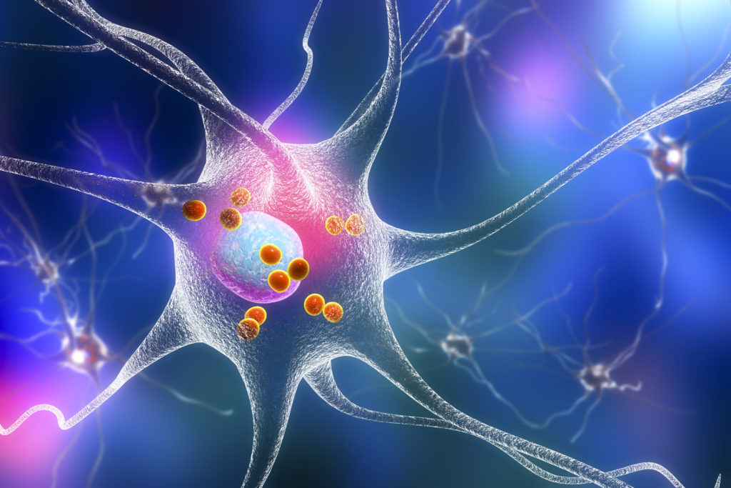
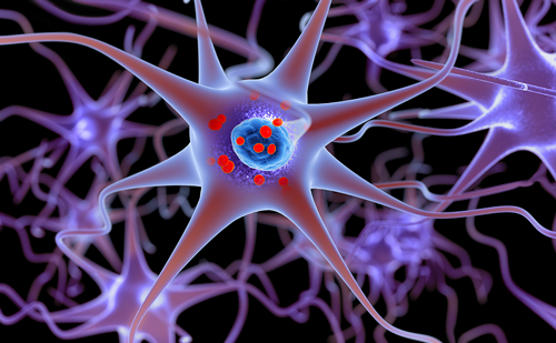
The etiology of PD remains unknown. The pathologic hallmark of PD is the presence of intracytoplasmic eosinophyllic, proteinaceous inclusions, termed Lewy bodies (LB), in surviving neurons. LBs contain the protein α-synuclein,4 which appears to have a role in the release of neurotransmitter from synaptic terminals.5 One proposed mechanism is α-synuclein-induced impairment of the ubiquitin-proteasomal system (UPS), resulting in protein accumulation and leading to cell degeneration.6 LBs are widely distributed in the brains of PD patients, including the SNc, raphe nucleus, locus ceruleus, pedunculopontine nucleus, dorsal motor nucleus of vagus, olfactory bulb, and some cortical structures.7
While the neuropathologic changes in PD are widespread and include a number of neurotransmitter pathways, the cardinal motor manifestations of the disease are clearly linked to the degeneration of the dopamine-producing cells in the SNc. Some of the postulated mechanisms of SNc cells’ death is oxidative stress and complex I mitochondrial dysfunction.8 A mitochondrial linkage is supported by the recent advances in the knowledge of the genetic mutations associated with the rare familial forms of PD. Several of PD genes have a role in mitochondrial function. PINK1, DJ-1, and LRKK2 encode proteins that are localized to the surface or in the mitochondria.9–11 Rotenone, paraquat, and 1-methyl-4-phenyl-1,2,3,6-tetrahydropyridine (MPTP) toxins that produce animal models of parkinsonism are believed to work via mitochondrial complex I inhibition.10 Mitochondrial dysfunction leads to the generation of reactive oxygen species (ROS), that are cytotoxic and can lead to the formation of LB-like intraneuronal filamentous inclusions, containing α-synuclein and ubiquitin.12,13 It is believed that all the above postulated pathogenic factors result in a common pathway of cell degeneration by mechanism of apoptosis.
Based on the postulated mechanisms of cell degeneration and preclinical data a number of clinical trials have been conducted or are underway aiming to attack various levels of the cascade of the neurodegenerative process. Tested agents targeted various potential mechanisms of PD pathogenesis including oxidative stress (eldepril, Vitamin E), mitochondrial dysfunction (CoQ10, ongoing studies of creatine), apoptotic mechanism of cell death (caspase inhibitors), antiexcitatory agents (riluzole), anti-inflammatory agents (minocycline), trophic factors, and others.14–23 So far, none of the agents have demonstrated a positive effect on the course of the disease process. The reasons for negative results of the clinical trials are multifactorial, but one of them is likely a failure to address unique selective vulnerability of the cells in SNc.
The Physiologic Phenotype of Substantia Nigra Pars Compacta Dopaminergic Neurons Potentially Explains Selective Vulnerability
Recent studies demonstrate that dopaminergic (DA) neurons in the SNc, as well as many neurons in other regions affected by PD, have a distinctive physiologic phenotype. They are slow and autonomous pacemakers, with broad action potentials.24–27 Although many neurons in the brain generate autonomous activity, few have this physiologic phenotype. Most pacemakers have very short spikes and limit Ca2+entry to a millisecond period around the spike. In contrast, SNc DA neurons have broad spikes and have membrane potential trajectories that ensure that low threshold Ca2+ channels like those with a Cav1.3 subunit are open virtually all of the time. This continuous Ca2+ influx leads to oscillations in cytosolic Ca2+ concentration.24,25 This Ca2+ influx distinguishes SNc DA neurons from DA neurons in the ventral tegmental area (VTA), which also are slow, broad action potential pacemakers.28 In contrast to SNc DA neurons, VTA DA neurons have a more modest vulnerability in PD.29
Substantia Nigra Pars Compacta Cells Pacemaking and Mitochondrial Oxidant Stress
When neurons generate spikes, the transmembrane ionic gradients that enable this activity are dissipated. These gradients must be restored with ATP-dependent pumps and exchangers. Thus, sustained activity is energetically expensive. Ca2+ ions, because they must be rapidly sequestered or pumped back across the plasma membrane, could pose a particularly significant energetic burden. In neurons, this demand for ATP is met primarily by oxidative phosphorylation in mitochondria.30 Oxidative phosphorylation comes at a cost: the production of potentially damaging superoxide and reactive oxygen species.
Recent studies by our group have shown that Ca2+ entry through L-type channels elevates mitochondrial oxidant stress in SNc DA neurons.25 How this happens is not entirely understood. It does not appear to depend solely upon the energetic burden posed by Ca2+ entry and might involve altered mitochondrial respiratory control. This study also provided an important insight into how this Ca2+-dependent mitochondrial stress and genetic mutations associated with PD might interact to selectively increase the vulnerability of SNc DA neurons. Mutations in DJ-1 are associated with an early onset, recessive form of PD.10 In SNc DA neurons from mice lacking DJ-1, mitochondrial oxidant stress was significantly higher than in wild-type neurons. This was not simply a consequence of losing functional DJ-1 however, as neighbouring VTA DA neurons displayed no measurable stress. As in wild-type neurons, the oxidant stress was reversed by treatment with DHP channel antagonists. These results suggest that DJ-1 is activated only in response to oxidant stress and provide some measure of defense, an idea with broad experimental support.31 These data provide a mechanism by which defects in a widely expressed gene can affect a small population of neurons. Furthermore, while elevated mitochondrial oxidant stress has long been hypothesized to play an important role in the etiology of PD, there has not been a coherent explanation for why SNc DA neurons—in particular—should be stressed. The physiologic phenotype of SNc DA neurons—pacemaking, broad action potentials, sustained opening of L-type Ca2+ channels, and the resulting mitochondrial oxidant stress—provides an explanation for selective vulnerability and, more importantly, establishes the target for potential neuroprotective interventions.
Indeed, antagonizing L-type Ca2+ channels is neuroprotective in toxin models. For example, pre-treatment of mesencephalic brain slices with isradipine, the most potent of the dihydropyridine (DHP) channel antagonists at L-type Ca2+ channels with the Cav1.3 subunit, significantly diminishes the damage to SNc DA neurons caused by the mitochondrial toxin rotenone.24 Moreover, systemic administration of isradipine to mice at doses achieves serum concentrations in the same range as those found in humans following oral administration protects SNc DA neurons in both a chronic MPTP and acute 6-hydroxydopamine (6-OHDA) models.24,32 Although these channels participate in normal pacemaking, they are not essential and antagonizing them with therapeutically relevant concentrations of dihydropyridine has no effect on the rest of the mouse behavior or phenotype.25
Does Physiologic Phenotype Predict the Pattern of Parkinson’s Disease Pathology?
While isradipine might be effective in protecting SNc DA neurons, will it be effective in other regions affected by PD? There are a number of regions of the brain that have cell loss paralleling that of the SNc.7 Although the available data set is fragmented, neurons in the dorsal motor nucleus of the vagus (DMV), locus ceruleus (LC), raphe nuclei (RN), pedunculopontine nucleus (PPN), lateral hypothalamus (LH), tuberomammillary nucleus, basal forebrain (BF), and olfactory bulb all exhibit signs of pathology and all have a physiologic phenotype resembling that of SNc DA neurons. DMV cholinergic neurons, which are thought to be among the earliest neurons with α-synuclein in PD, are spontaneously active;33 this activity is autonomously generated and engages L-type calcium channels (unpublished observations). Serotonergic neurons in the RN have broad spikes and are calcium-dependent autonomous pacemakers.34 This is also true of PPN cholinergic neurons.35 Tuberomammillary neurons are spontaneously active and engage L-type Ca2+ channels.36 DA neurons in the olfactory bulb are calcium-dependent, autonomous pacemakers.37
Perhaps the neurons most affected in PD, other than SNc DA neurons, are LC noradrenergic neurons.7 Like SNc DA neurons, they are autonomous pacemakers (with broad spikes) that engage L-type calcium channels.38 Moreover, these neurons display all the signs of mitochondrial oxidant stress found in SNc DA neurons and this stress is significantly alleviated by isradipine. Taken together, these studies make a compelling case that the DHP class of agents should be broadly effective in slowing the progression of PD.
Epidemiologic Studies
Recent epidemiologic data also supports the potential neuroprotective effect of DHPs in PD. Two studies demonstrated reduced risk of development of PD in subjects treated with calcium channel blockers (CCBs) compared to other antihypertensive agents.39,40 The most recent study assessed the risk of the new diagnosis of PD in a cohort of 1,931 patients with new diagnosis of PD versus 9,651 matched controls. The study demonstrated a 27 % risk reduction (OR=0.73) of a new diagnosis of PD in subjects treated with centrally acting DHP compounds compared to other CCBs or other antihypertensive agents. That study provides strong supporting evidence of channel specific selectivity of the potential neuroprotective effect of CCBs restricted to DHP compounds. The study also provides indirect evidence of sufficient CNS penetration of the subset of DHPs as the risk reduction was seen only with the centrally acting DHPs and not observed with amlodipine that has low CNS penetration.
Isradipine
Currently, there are no Cav1.3 channel selective DHPs. Isradipine is the most potent DHP at these channels and is FDA approved for treatment of hypertension since 1990.41 Isradipine is available in immediate (IR) and controlled release (CR) preparation in 5–20 mg dose range. Isradipine is rapidly and almost completely (90–95 %) adsorbed following oral administration but undergoes extensive first pass metabolism, resulting in bioavailability of 15–24 %. Peak serum levels occur in about 1.5 hours for the IR preparation and 8–10 hours for the CR preparation. Animal studies have demonstrated a neuroprotective effect of isradipine in an intrastriatal 6-OHDA model at the serum concentrations achievable within the dose range approved for human use.32 Isradipine belongs to the group of lipophilic DHPs that have good bioavailability in non-human primates with brain to serum concentration ratios significantly above one.42 Isradipine is among the more lipophilic DHPs, enhancing its brain bioavailability. Recently published epidemiologic data showing a reduced incidence of PD in patients treated with centrally bioavailable DHPs also strongly supports good CNS penetration of DHPs.40
Preliminary Human Data
We have conducted an open-label dose escalation safety and tolerability study of isradipine in patients with early PD. The study demonstrated dose-dependent tolerability of isradipine CR: 94 % for a 5 mg dose; 87 % for a 10 mg dose; 68 % for a 15 mg dose; and 52 % for a 20 mg dose.43 Isardipine had no significant effect on blood pressure or PD motor disability. The two most common reasons for dose reduction were leg edema (seven) and dizziness (three). There was no difference in isradipine tolerability between subjects with and without dopaminergic treatment. That study supports good tolerability of isradipine CR at daily doses up to 10 mg in subjects with early PD.
A pilot Phase II double-blind, placebo-controlled, tolerability- and dosage-finding study of isradipine CR as a disease modifying agent in patients with early PD (STEADY-PD) supported by the Michael J Fox Foundation is ongoing. The objective of the study is to establish safety and tolerability of isradipine CR across the FDA-approved dosing range (5–20 mg) in a larger cohort of patients with early PD and to evaluate the comparative efficacy of three doses of isradipine CR, provided that they are tolerable. The study recruited subjects with early PD not requiring dopaminergic therapy (stable dose of amantadine, anticholinergics, and monoamine oxidase (MAO-B) inhibitors are allowed). The study is designed as a multicenter 52 weeks’ duration, randomized, four-arm double-blind parallel group trial, with 100 subjects randomized to 5, 10, or 20 mg of isradipine CR or matching placebo daily. The dosage that is tolerable and demonstrates preliminary efficacy will be used in the future pivotal efficacy study.44 Tolerability of each active dosage will be compared with the tolerability of placebo. Provided that the dose is tolerable, the choice for the dose selection will be based on efficacy defined as the change in total UPDRS score between the baseline visit and month 12 or the time of sufficient disability to require dopaminergic therapy, whichever occurs first. Comparison will be made between three active treatment arms. The dosage that demonstrates the greatest efficacy will be used in the proposed pivotal study. The study has successfully completed recruitment, with the final data analysis expected to be available in the next six months.
Conclusion
There is solid scientific rationale, preclinical, and epidemiologic data to support the potential benefit of isradipine as a disease modifying agent in early PD. Results of the ongoing Phase II study will be available shortly and will inform the design of the future pivotal efficacy trial.


