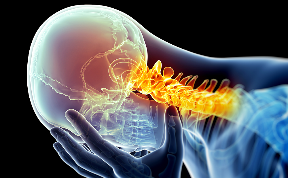
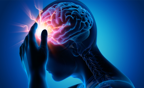
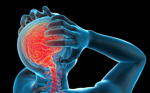
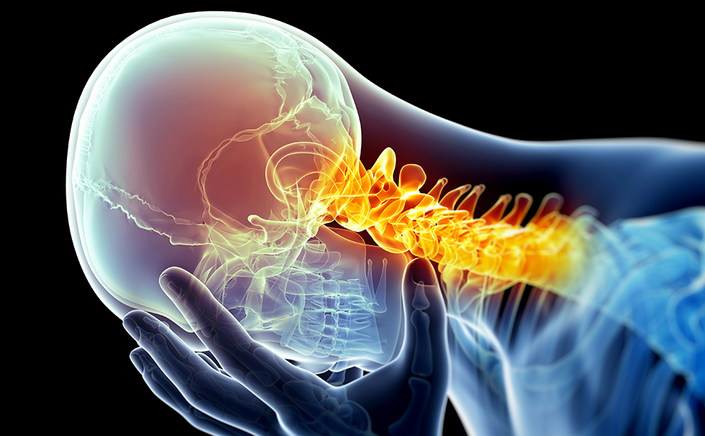
Cluster headache (CH) is, by neurological standards, a relatively common condition that affects about one in 1,000 people,1 although compared with other more common primary headaches such as migraine2 it remains rare in practice. CH has been defined in the second edition of the International Classification of Headache Disorders3 as involving recurrent attacks of severe pain on one side of the head for between 15 and 180 minutes, associated with cranial autonomic features such as lacrimation, conjunctival injection, nasal congestion, or rhinnorhoea (see Table 1).
This condition can be divided into episodic CH (ECH) and chronic CH (CCH) forms. A diagnosis of ECH requires at least two cluster periods lasting from seven days to one year separated by pain-free periods lasting for one month or longer, while a diagnosis of CCH requires attacks to occur for more than one year without remission or with remission lasting for less than one month. CCH affects about 10% of CH patients. General aspects of therapy of the disorder are covered elsewhere.4,5 While it is not directly germane to neuromodulatiuon approaches, an understanding of the broad issues in medical treatment serves as a useful backdrop against which to discuss newer approaches. CH is one of the trigeminal autonomic cephalalgias,6 and their therapies are usefully considered to be background7 as they can also be considered for these newer approaches.
Who Is Suitable for Neuromodulation Approaches?
It seems reasonable to suggest that neuromodulation approaches to the management of CH be employed in patients with medically intractable forms of the condition. While this is currently true, it reflects the relatively primitive state of current interventions. One should observe that as devices become less invasive, the threshold for their use will become lower. For the moment there is a proposed working definition of medically intractable CH.8 The essential components are disabling headache that fails to respond to at least four preventative drugs, including two from the first three of verapamil, lithium, methysergide, melatonin, topiramate, and gabapentin (see Table 2). These considerations are particular to CH and, naturally, generic considerations related to requirements for the devices—such as fitness for anesthesia—are part of the overall assessment of patient suitability.
What Approaches Have Been Tried?
In essence, two classes of neuromodulation have been explored in CH: peripheral and central. Prior to moving to stimulation approaches, the dreadful pain and disability of medically intractable CH lead to a number of destructive procedures. In principle, these seem unlikely to work if one considers CH to fundamentally be a brain condition.9Moreover, they may cause both mortality and significant morbidity, and can induce further pain problems such as anesthesia dolorosa. Previously used approaches have included trigeminal ganglion glycerol injections,10,11 radiofrequency rhizotomy of the Gasserian ganglion,12 or gamma knife aimed at the trigeminal nerve,13,14 microvascular decompression15 and esection or blockade of the N. petrosus superficialis16 or pterygopalatine (sphenopalatine) ganglion.17–19There are case series of trigeminal nerve root section20,21 that illustrate all the issues, including inducing further pain, vision impairment, or indeed death. Moreover, there are also case reports of the complete inefficacy of surgical treatment in CH.21,22 It must also be remembered that annually about 10% of patients with CCH will revert to the episodic form,23 so a procedure should not be less safe than the natural history.
Peripheral Approaches
A number of structures have been suggested as peripheral targets of stimulation in CH. These include the occipital nerve, which will be dealt with in detail below as there are now a number of studies available, the ophthalmic branch of the trigeminal nerve (n=1),24 vagus nerve stimulation (n=6)25 and higher cervical stimulation (n=1).26 Given its relative data and promise, occipital nerve stimulation (ONS) is dealt with in more detail below.
Occipital Nerve Stimulation
Initial interest in the use of ONS to treat headache dates from Weiner and Read,27 who reported a series of cases of intractable occipital neuralgia responding to ONS. Detailed phenotyping of these cases in association with a functional imaging study demonstrated that almost all patients had chronic migraine.28 What was remarkable in the ONS patients studied using functional imaging was that the therapy, while effective in terms of pain, did not seem to alter the brain activation of areas considered to be important in migraine. Instead, it changed thalamic processing. Taken together with experimental data collected by the authors of this paper,29– 31 it was reasoned that ONS may be helpful in selected patients with other primary headache disorders. It seems likely, given that peripheral distribution of the pain does not predict the outcome of stimulation, that ONS has an important central effect on the brain.32 Six out of the eight patients initially undergoing this procedure had sufficient benefit to recommend the procedure to others and to make it an option for other neuromodulation approaches.33 Long-term experience over more than two years demonstrated that device dysfunction almost always led to the return of attacks.34 Thus, it seems unlikely that the useful effect is a prolonged placebo, although much more needs to be done to establish the mechanism of the useful effect.
Central Nervous System Approaches— Deep Brain Stimulation
Recognizing that all invasive treatments bear the risk of severe side effects, international guidelines for patient selection based on expert consensus were published.35 Criteria for the use of deep brain stimulation (DBS) include only considering patients with all of the following: CCH and strictly unilateral attacks without side shift, a normal psychological profile, and no medical/neurological condition contraindicating DBS, such as epilepsy/stroke. Only medically intractable patients should be considered for DBS. Considering that more than 50 patients have been operated on and the results published,36 with an average of 50–70% showing a significant positive response, the question arises of whether it is possible to formulate predictive indicators of which patients will respond to hypothalamic DBS in CH.
Defining the Target Point
The target point for DBS in CH was chosen based on clinical considerations and functional studies, particularly neuroimaging, which revealed the crucial role of the posterior hypothalamic region in CH.37 Neuroimaging with positron-emission tomography (PET) shed light on the genesis of CH, documenting the link between activation in the hypothalamic gray ipsilateral and pain in CH.38 These areas are not simply involved in the response to first-division nociceptive pain impulses but are inherent to each syndrome, probably in some permissive or dysfunctional role.9,39 Furthermore, using high-resolution structural 3D magnetic resonance images and voxel-based morphometry, a significant structural difference in gray matter density of the hypothalamus was found in patients with CH compared with healthy volunteers.40 The co-localization of morphometric and functional changes demonstrates the precise anatomical location for a probable central nervous system lesion in CH. Given that this area is involved in circadian rhythms, sleep–wake cycling,41 and control of the autonomic system,42 the data suggest a crucial involvement of this hypothalamic area, at least in generation of the acute CH attack. Initially, it was thought that the hypothalamic region at the posterior inferior border was activated only in CH. Subsequently, it was shown that this hypothalamic area is also activated during short-lasting, unilateral, neuralgiform headache attacks with conjunctival injection and tearing (SUNCT),43,44 paroxysmal hemicrania,45 and hemicrania continua.46Despite this, a second study found no activation in the hypothalamus in hemicrania continua in a single patient without cranial autonomic features.47 For DBS the electrode is usually implanted stereotactically in the left posterior hypothalamus/anterior periventricular region of the triangle of Sano, according to the co-ordinates published.38 This area does not correspond to a specific anatomical entity and there is no consensus as to whether it is part of the posterior hypothalamus or tegmentum, or even the anterior periventricular gray matter. Two sets of stereotactic co-ordinates have been described:
- those published by Leone and colleagues: x=2mm lateral to the midline, y=6mm behind the mid-commissural point, and z=8mm below the commissural plane;48 and
- the revised co-ordinates published by Franzini et al.:x=2mm lateral, y=3mm posterior, and 5mm inferior to the mid-commissural point.49
Electrophysiology
Several studies have obtained single-unit microrecordings from this region in CH patients. All but one50 were obtained from patients not experiencing an attack and found no particular features in the region. Recently, local field potentials during a CH attack were performed while surgical implantation of a hypothalamic electrode was ongoing. These potentials showed a significant increase in power during the attack.50 This appears to be the first report of neuronal activity during a CH, reinforcing neuroimaging data that implicated hypothalamic activation in cluster attack generation.
Technical Functional Imaging Considerations in Cluster Headache
It is important to note that H2O–PET has a low spatial (4–5mm) and limited temporal (one minute sample time) resolution. Since the changes in individuals are small, group analyses are required. Furthermore, to achieve a statistically significant result in regional cerebral blood flow, a smoothing kernel of at least 10mm is required. It cannot be overstated that the PET study by Leone et al. is the first time that results from functional imaging have been translated into DBS. Likewise, it needs to be pointed out that these patients have been intractable to medical treatment.51 However, a failure rate of up to 50% seems quite considerable for an invasive treatment with the theoretical risk of death.52
Single case studies are possible in principle,53 and it would be easier to define the individual target point for each patient.37 This does not change the fact that hypothalamic DBS should be the last option in CH patients.
Clinical Outcomes from Current Studies
It seems clear that hypothalamic DBS is generally ineffective in ameliorating acute attacks51 and is therefore used to reduce attack frequency with continuous stimulation. To date, 58 intractable CH patients who have been operated on have been reported,36,54 some with a follow-up of more than four years.36 The long-term results are particularly encouraging given that a persistent pain-free state was still present in 10 out of 16 patients (62%), although four of these required preventative medication to control the attacks.51 This is still remarkable given that these patients were not easy to treat and were effectively medically intractable without stimulation.51
Despite the fact that these studies could strengthen the clinical impression of the posterior hypothalamus as a key player in the generation of CH attacks, the responder rate of DBS in this region varies between 50 and 70%55–57 and there are several patients with CH who do not respond.52,55 These differences between patient series55,56 and studies57 cannot simply be attributed to technical details of the devices, operation or target region used as these are more or less identical. It seems clear that the selection of patients and overall care may have a huge impact on the outcome. However, it has to be said that, to date, no predictors for success have been identified.
Open Questions
Given that patients who are operated on using peripheral approaches or DBS are medically intractable and consequently highly disabled, these approaches can substantially restore function. However, there are many issues to be resolved, such as how to design placebo stimuli in ONS and how to better identify the target in DBS. DBS imaging methods seem an obvious way forward, although the authors have been studying a human brain from a patient with CH and it could be contended that imaging pathological reconstructions may be an even better approach. Whatever is said about various techniques, while CH is a devastating disease, primum non nocere.


