The prevalence of Parkinson’s disease (PD) is now estimated to be approximately 1 % in individuals over 60 years of age, with 4.1–4.6 million people affected worldwide,1 a number predicted to more than double by 2030.2 Clinical diagnosis is based upon the recognition of the motor features of tremor, rigidity, and bradykinesia, with other supporting features including asymmetric symptoms and signs, and the absence of signs of other etiologies of Parkinsonism. However, improved diagnostic tools are desirable for several reasons. By the time that motor symptoms are recognized, the PD pathologic process may already be advanced in the central and peripheral nervous systems. In addition, symptoms and signs may overlap with other neurodegenerative disorders, leading to the risk for misdiagnosis. This is particularly true in early PD, when only subtle motor signs are present, leading also to delayed diagnosis. Therefore, in order to identify PD earlier and more accurately, a number of approaches are being tested. One focus is the well-described non-motor symptoms of PD, as it is now clear that they may occur prior to motor symptoms in some cases, and might therefore define a ‘pre-motor’ phase of PD. Biomarker development is also being intensively pursued as an aid to diagnosis, with potential markers including neuroimaging as well as sophisticated biochemical and molecular techniques. Advances in understanding the genetic contribution to PD are expanding the number of gene loci that are not only causes of heritable PD but are also identified in genome-wide association studies (GWAS) as PD risk loci. Improved diagnosis would not only impact upon clinical management, but such approaches are also desirable in order to identify disease-modifying treatments.
Where Does Parkinson’s Disease Pathology Begin?
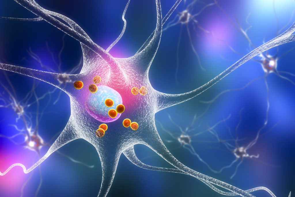
PD has long been attributed to pathology in the substantia nigra (SN), with loss of pigmented dopaminergic neurons observed at autopsy. Surviving SN neurons often have intracytoplasmic proteinaceous inclusions, termed Lewy bodies, containing aggregates of a misfolded pre-synaptic protein, α-synuclein.3 The α-synuclein protein is well recognized as key to PD pathogenesis, with Lewy bodies, as well as dystrophic Lewy neurites containing concentrations of α-synuclein, having been widely described in other tissues. PD pathology is therefore detected in, and may in some other tissues. PD pathology is therefore detected in, and may in some cases begin at, sites outside the central nervous system (CNS). For example, α-synuclein pathology is seen in autonomic nerves innervating the heart,4 gut,5 and prostate,6 and immunoreactivity in skin tissue is observed in autopsy-identified cases with Lewy body disease.7
A PD pathology staging system has been proposed based on more than 125 autopsy cases.8 Six neuropathological stages have been proposed, each characterized by α-synuclein pathology, beginning in non-dopaminergic structures of the brainstem and olfactory bulb with progression to more caudal regions of the CNS. The six stages involve: 1) the caudal medulla, including the dorsal motor nucleus of the vagus, and the olfactory bulb; 2) the medulla oblongata and adjoining portions of the pontine tegmentum, including the locus ceruleus and other ‘gain setting nuclei’; 3) the SN; 4) the forebrain and temporal mesocortex; and 5) and 6) widespread regions of the cerebral cortex.8 It is proposed that cardinal motor signs of PD begin to occur at the third stage. Although this study did not set out to provide clinicopathologic correlation, certain non-motor symptoms, such as olfactory dysfunction and sleep disorders, may occur prior to motor symptoms (see below), and the Braak staging suggests precise pathological underpinnings.
This hypothesis has provided a springboard for critical analysis of the pattern of α-synuclein pathology and its relationship to clinical features.9,10 For example, in the Baltimore longitudinal study of aging, of 117 brains examined at autopsy, α-synuclein pathology was observed in 100 % of seven cases of PD, in 31.5 % of 56 cases with Alzheimer’s disease pathology, and in 8.3 % of 36 older control brains.11 Another consideration is that the presence of Lewy bodies and Lewy neurites may not be necessary for cellular dysfunction and may not therefore fully define regions of PD cellular dysfunction. There is accumulating evidence that axonal loss may occur earlier and as a distinct process from cell body degeneration in PD.12 For instance, examination of α-synuclein pathology and tyrosine hydroxylase immunostaining in cardiac sympathetic axons and ganglia in patients with incidental Lewy body pathology, PD, and controls, suggests that the disease process begins in the distal axon and proceeds in a retrograde fashion.13 Moreover, the existence of distinct mechanisms mediating cell soma and axonal degeneration is supported by the protection of dopamine neuron cell bodies, but not axons, by jnk2 and jnk3 null mutations in the 6-hydroxydopamine rodent model of Parkinsonism.14 This is important, since axonal loss, occurring as a distinct process prior to neuronal cell body loss in PD, has implications regarding target choices for neuroprotection. Although preservation of neuron cell bodies is essential for the function and prolonged survival of axons, focusing exclusively upon interventions aimed at protecting cell bodies against programmed cell death may be missing important therapeutic targets.12,15
How Does Parkinson’s Disease Present Clinically?
The variety of structures in which PD pathology has been observed suggests that a focus upon predicted clinical correlates may yield measures that could aid in early diagnosis. Langston in 2006 described Parkinsonism as the ‘tip of the iceberg’ of PD, with non-motor symptoms including mood disorders, cognitive dysfunction, sleep disorders, autonomic dysfunction, and pain or sensory syndromes coexisting alongside motor symptoms.16 It is now increasingly recognized that non-motor symptoms might in some cases arise prior to the characteristic motor symptoms, possibly forming a pre-motor syndrome. The term ‘Parkinson’s associated risk syndrome’ (PARS) has been coined to describe a combination of features identifying individuals at risk for developing PD (see Table 1).17
Rapid Eye Movement Sleep Behavior Disorder
Rapid eye movement sleep behavior disorder (RBD) is characterized by recurrent episodes of sudden, abnormally vigorous body, head, or limb movements during rapid eye movement (REM) sleep, and has been reported in 15–47 % of patients with PD,18 occurring up to 20 years before motor symptoms arise.19 There is a high risk for conversion to PD, in addition to conversion to dementia with Lewy bodies (DLB) and multiple system atrophy (MSA), in multiple studies. For example, in a retrospective study of 44 patients with idiopathic RBD, 45 % went on to be diagnosed with PD or DLB a mean of 5.1 years after RBD diagnosis, and 11.5 years after the onset of RBD symptoms.20 In a careful prospective study of 67 patients with idiopathic RBD, six went on to develop PD and 11 went on to develop dementia with DLB characteristics at five years, and 52 % developed a neurodegenerative disorder at 12 years.21
Olfactory Dysfunction
In addition to RBD, olfactory dysfunction is one of the most well-known pre-motor symptoms, and multiple studies suggest it is an early sign of PD,22 consistent with proposed involvement of the olfactory bulb in Braak stage I. In a recent analysis of individuals in the Honolulu-Asia aging study, those individuals who fell into the lowest quartile on odor identification scores had an approximately five-fold increase in PD risk compared with those in the top two quartiles.23 Neuroimaging studies also support a strong link between hyposmia or dysosmia and PD. One study found that the degree of olfactory sense impairment, reflected by University of Pennsylvania Smell Identification Test (UPSIT) scores, correlated with striatal dopamine transporter density, as measured by [99mTc]TRODAT-1 single photon emission computed tomography (SPECT) in 24 individuals with early PD.24 Magnetic resonance imaging (MRI) diffusion tensor imaging (DTI) revealed significant differences in the anterior olfactory region and SN in 14 newly diagnosed patients with PD compared with controls.25 In addition to sporadic PD, hyposmia is evident in some genetic forms of PD, such as that associated with phosphatase and tensin homolog (PTEN)-induced putative kinase-1 (PINK1), and, of note, a milder deficit has been demonstrated in asymptomatic heterozygotes.26
Constipation
Constipation is another symptom that often predates clinical PD and has been attributed to α-synuclein pathology in the myenteric and submucosal plexus.27 In the Honolulu Heart Program, a prospective study of more than 8,000 Japanese-American men followed with periodic health surveys, men who reported less than one bowel movement per day were more than four times more likely to develop PD than those who reported two or more bowel movements per day.28 Gastrointestinal dysfunction in PD is not limited to constipation, but includes delayed gastric emptying, dysphagia, and anorectal dysfunction.29
Cardiac Autonomic Dysfunction
More than 40 studies on cardiac autonomic dysfunction in PD raise the possibility that neurophysiologic measures could contribute to earlier PD diagnosis. Sympathetic denervation has been detected in those with early PD, and, in one patient, denervation was observed four years before the onset of motor symptoms was recognized.30,31 131I-metaiodobenzylguanidine (131I-MIBG) is taken up by catecholaminergic neurons, and reduced 131I-MIBG uptake has been demonstrated in PDpatients.4 It may also be possible to differentiate PD from MSA using this technique. In a study of 246 individuals, PD could be distinguished from MSA with 89.7 % sensitivity and 94 % specificity.32 The role of a more accessible and cheaper test is now under examination; decreased heart rate variability (HRV), which may be detected by electrocardiogram (EKG) recordings, has now been observed in patients with PD in a number of studies, including during polysomnographic studies.33,34
Neurobehavioral Features
Symptoms including depression, anxiety, and cognitive changes are more commonly observed after PD diagnosis, but may be present up to 20 years prior to the diagnosis of motor symptoms. A general practice-based registry study found that the lifetime incidence of depressive disorder prior to PD onset was 9.2 % compared with 4.0 % in controls from the same register (odds ratio 2.4).35 In a case-control study, odds ratios of 2.2 and 1.9 were determined for increased anxiety and depressive disorders, respectively, prior to PD onset.36 Although cognitive dysfunction is more typically recognized clinically in advanced PD, careful testing reveals early changes that could also be of use in diagnosis. In early PD, almost 20 % of individuals were classified with mild cognitive impairment compared with controls (relative risk 2.1) in a recent study of PD in a Norwegian cohort.37
Possibilities for Earlier Diagnosis of Parkinson’s Disease
The PARS hierarchy, as proposed by Stern and Siderowf,17 divides individuals who are at risk for PD into pre-physiologic, preclinical, pre-motor, and pre-diagnostic stages (see Figure 1). The pre-physiologic stage corresponds to a genetic predisposition to PD, and possibly to exposures that might increase PD risk. The preclinical stage is characterized by neuroimaging abnormalities, while in the pre-motor stage clinical symptoms and signs of PD have appeared that are non-motor in nature. The pre-diagnostic stage constitutes subtle neurological findings not yet diagnosed by clinical practitioners.17
Clinical Detection of Pre-motor Parkinson’s Disease
What are the possibilities for identifying individuals in the pre-motor stage of PD and pushing back diagnosis even before subtle motor signs are evident (see Figure 2)? Hitherto, individual clinical markers have not been specific; for instance, the severe olfactory dysfunction that can occur in PD also occurs in certain other neurodegenerative conditions, including MSA, corticobasal degeneration (CBD), progressive supranuclear palsy (PSP), and other disorders.38 The most reliable method of pre-motor PD detection will therefore most likely rely on a combinatorial approach. In a recent study, non-motor signs across six different domains were explored in patients with early motor PD.39 Tests addressed visual deficits tested by color discrimination, contrast sensitivity, and spatial discrimination; olfactory function; sleep abnormalities by Parkinson’s Disease Sleep Scale rating and documentation of loss of muscle atonia during REM sleep on polysomnography; dysautonomia as reported on two self-completed questionnaires; executive dysfunction; and depression. Thirty patients with motor signs of PD for less than three years were compared with a control group in each of these domains. Across all domains, patients with PD performed worse than the control group, with statistically significant differences in visual deficits, sleep abnormalities, hyposmia, dysautonomia, and depression; the most marked distinction between the test and control groups emerged in visual deficits, hyposmia, and dysautonomia.39 By combining the results of the Farnsworth–Munsell color discrimination test and the non-motor symptom questionnaire (NMSQuest), the sensitivity and specificity for detection of PD peaked at 0.77, suggesting a relatively simple model for predicting PD.
Testing performance characteristics of combinatorial markers in PD is useful, but ultimately needs to be performed in at-risk individuals. It is therefore important that clustering of non-motor features has also been observed in studies of individuals at risk for PD. For example, in a small study of 20 patients with RBD, HRV as determined by EKG was compared with that of control subjects with primary insomnia.40 In those subjects with RBD, HRV was decreased compared with controls.
Neuroimaging as a Diagnostic Tool of Pre-motor Phase Parkinson’s Disease
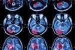
PD neuroimaging biomarker development has focused primarily on ligands whose uptake reflects the integrity of the nigrostriatal tract, as detected by SPECT or positron emission tomography (PET). These show promise in facilitating earlier detection of PD and improving diagnostic accuracy.41–43 For example, a large prospective study examined the correlation of initial clinical diagnosis in 118 patients with clinically uncertain Parkinsonian syndromes (CUPS) with SPECT imaging using the 123I-ioflupane ligand, which binds to the dopamine transporter. In 36 % of those with a suspected diagnosis of a Parkinsonian syndrome, 123I-ioflupane binding was normal, while 54 % with a suspected diagnosis of non-Parkinsonian syndrome had an abnormal scan.44 Significantly, after a two-year follow-up, clinical diagnosis agreed with the initial scan in 90 % of patients in whom a specific diagnosis was established.45
Valuable information on the use of neuroimaging in the pre-motor stage is anticipated from the PARS study, which follows a large cohort of individuals using a combination of olfactory testing and 2β-carbomethoxy-3β-(4-iodophenyl)-tropane (β-CIT) SPECT imaging.42 Dopaminergic imaging, however, has not been helpful in differentiating PD from atypical Parkinsonian syndromes including MSA, CBD, and PSP, as all of these syndromes are associated with pre-synaptic dopaminergic deficiency. However, 18F-fluorodeoxyglucose PET imaging has shown promise in identifying disease-specific metabolic brain patterns in MSA and PSP, which may be useful in distinguishing them from PD.46
A distinct and more accessible imaging technique, ultrasound, now appears promising as a PD biomarker, and demonstrates hyperechogenicity of the SN in PD. An ultrasound study of 102 subjects with PD, 34 with MSA, and 21 with PSP, found that the combination of hyperechogenicity in the SN plus regular echogenicity in the lentiform nucleus versus moderate or less SN echogenicity plus hyperechogenicity in the lentiform nucleus yielded a positive predictive value of 0.91 for PD versus atypical Parkinsonian syndromes.47
Genetic, Molecular, and Biochemical Biomarker Development in Parkinson’s Disease
Whereas these markers might potentially detect pre-physiologic PD, testing has so far been limited to motor PD to allow development and validation of specific testing modalities.
Genomic analysis is useful as a ‘trait’ rather than ‘state’ biomarker and therefore may be most useful as a marker to stratify individuals in terms of PD risk. Multiple genes have now been identified that lead to Mendelian inheritance of PD and Parkinsonism being reviewed.48,49 In general, how best to use genetic testing as a predictor of PD remains to be established, and genes leading to heritable PD may only be useful in a minority of cases, with the exception of leucine-rich repeat kinase-2 (LRRK2) in specific populations. However, identification of genetic risk loci and the use of GWAS holds the promise of risk stratification,50 with the possibility that significant PD risk might be accounted for on the basis of particular alleles of genes including α-synuclein, microtubule-associated protein tau (MAPT), the human leukocyte antigen (HLA) locus, HLA-DRB5, and others.51
Neuropathological studies and advances in genetic causes of PD have identified pathways that are potentially impaired in PD and have therefore provided potential markers. For example, a recent study in over 100 PD subjects determined values of 92 % sensitivity and 58 % specificity for low cerebrospinal fluid (CSF) concentrations of α-synuclein in differentiating PD versus control subjects. In the same study, 90 % sensitivity and 70 % specificity for decreased DJ-1 protein concentrations in CSF were determined.52 One question is whether using this measure in combination with other biomarkers could result in enhanced tools for diagnosis. Recently, a panel of seven proteins (α-synuclein, DJ-1, tau, hyperphosphorylated tau, fractalkine, Flt3 ligand, and Aβ1–42) was able to differentiate CSF samples from PD patients versus normal controls, individuals with Alzheimer’s disease, or those with MSA.53 Finally, increased understanding of the pathogenesis of PD has opened other avenues that are now being examined, for example oxidative stress markers54 and transcriptomic and metabolomic analysis.55,56
Conclusions
The worldwide increase in PD that is currently forecast makes efforts to improve its management imperative, and there is a critical need to develop improved clinical tests and biomarkers for PD to improve diagnostic accuracy and timeliness. The use of clinical batteries incorporating non-motor PD features and development of sophisticated biomarker techniques now provides hope for earlier and more accurate identification of individuals in whom pathogenesis has already begun. Even earlier identification of individuals at risk for this pathogenetic process is now possible through genetic testing, and in the future genomic profiling will conceivably expand the practical clinical use of DNA studies. Another application of clinical batteries and molecular biomarkers is to define PD subtypes or endophenotypes of relevance to prognosis and treatment. For example, it may be possible to define PD subtypes based on pathogenetic mechanisms such as oxidative stress.57 Definition of such possible PD subtypes could then allow rational development and implementation of individualized medicine in the future, and this could have significant impact upon clinical trial design. How far are we toward the goal of earlier and more accurate diagnosis? There have been dramatic advances in understanding the clinical spectrum of PD as well as the underlying pathology. By recognizing a ‘Parkinson’s associated risk syndrome,’ it is possible that PD may be detected earlier in the course of disease, and appropriate interventions employed. The advantage of clinical batteries is that many are employed easily in the clinic, for example EKG to measure HRV. However, a major limitation is lack of specificity. On the other hand, neuroimaging and other biomarkers may provide more specificity, but in the near future will be largely limited to specialist centers, and are expensive.58 It is unlikely that one specific clinical test or biomarker will serve all purposes. A constellation of clinical batteries and biomarkers, incorporating clinical symptom measurement, molecular genetics, and markers based upon PD pathophysiology may eventually provide better and more predictable risk profiling than single markers. Indeed, the Alzheimer’s disease neuroimaging initiative (ADNI), an international study obtaining longitudinal multimodal imaging data in individuals with Alzheimer’s disease, mild cognitive impairment, and controls (www.adni-info.org), is examining such a combinatorial approach.59 New initiatives in PD are now testing how a collaborative approach for comparison and combination of markers may be applied to biomarker development, for example the Parkinson’s progression markers initiative (PPMI: NCT01141023).
Finally, more accurate diagnosis is predicted to enhance patient care significantly, and will certainly remove much of the initial uncertainty in practice as patients are newly diagnosed. However, can it be shown that earlier diagnosis of PD will have a significant impact upon long-term patient outcomes? If neurologists become inclined to increase effort in this area to speed initiation of treatment, will this result in health or social/economic benefit? As we develop technologies that put these goals within our reach, and provide a window of opportunity to redefine treatment paradigms, it will be important to pay attention to measures of long-term outcomes and impact upon overall health and social function.